This website uses cookies to ensure you get the best experience on our website.
- Table of Contents
2 Citations
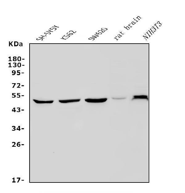
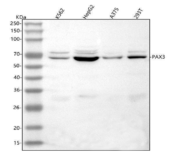
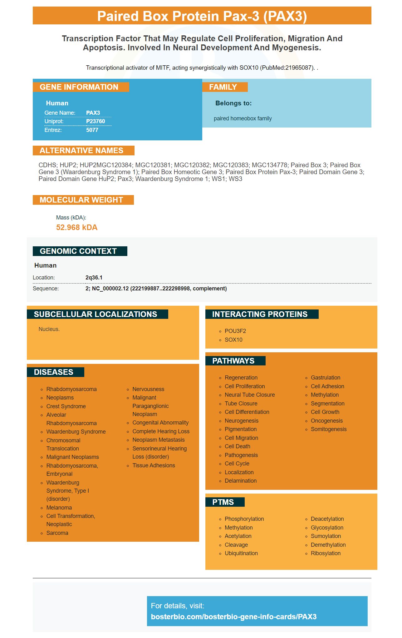
Facts about Paired box protein Pax-3.

Transcriptional activator of MITF, acting synergistically with SOX10 (PubMed:21965087). .
| Human | |
|---|---|
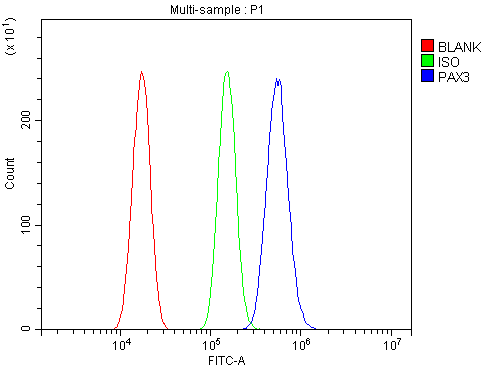
| Gene Name: | PAX3 |
| Uniprot: | P23760 |
| Entrez: | 5077 |

| Belongs to: |
|---|
| paired homeobox family |

CDHS; HUP2; HUP2MGC120384; MGC120381; MGC120382; MGC120383; MGC134778; paired box 3; paired box gene 3 (Waardenburg syndrome 1); paired box homeotic gene 3; paired box protein Pax-3; paired domain gene 3; paired domain gene HuP2; Pax3; Waardenburg syndrome 1; WS1; WS3
Mass (kDA):
52.968 kDA

| Human | |
|---|---|
| Location: | 2q36.1 |
| Sequence: | 2; NC_000002.12 (222199887..222298998, complement) |
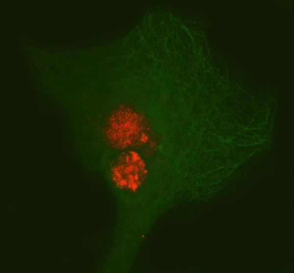
Nucleus.


PMID: 14639621 by Parker C.J., et al. Expression of PAX 3 alternatively spliced transcripts and identification of two new isoforms in human tumors of neural crest origin.
PMID: 7782066 by Macina R.A., et al. Genomic organization of the human PAX3 gene: DNA sequence analysis of the region disrupted in alveolar rhabdomyosarcoma.
*More publications can be found for each product on its corresponding product page