PCK1 / Phosphoenolpyruvate carboxykinase, cytosolic [GTP] · IHC design guide

Design Immunohistochemistry for PCK1

Plan chromogenic PCK1 IHC in paraffin sections using kidney tubules as a high-staining reference (HPA tissue IHC). This guide covers fixation consistency, cytoplasmic scoring (HPA tissue IHC), and interpretation when staining and RNA data disagree (HPA tissue IHC).

Evidence assembled Oct 2026 · For research use; verify linked source records and product datasheet before use
Immunohistochemistry protocol sheet for PCK1 (IHC for PCK1): expected localisation Cytoplasmic tissue staining (HPA tissue IHC), antibody A02022-3, validated IHC image, and IHC protocol steps
Printable PCK1 IHC protocol sheet — expected localisation Cytoplasmic tissue staining (HPA tissue IHC), antibody A02022-3, controls and protocol steps. Open the full PCK1 IHC guide →

PCK1 Immunohistochemistry Experimental Design Guide

Expected localisation, validated protocols, controls and antibodies — the at-a-glance facts below, then the full design guide.

Must know before staining
Expected localisation Cytoplasmic tissue staining (HPA tissue IHC)
Staining pattern High in kidney tubules; medium in gut glands, cytoplasmic (HPA tissue IHC)
Antigen retrieval EDTA pH 8.0 HIER, heat-mediated (datasheet A02022-3)
Positive control ⓘ Kidney+4 more · see all
Negative control ⓘ Adrenal gland+4 more · see all
Important caveats
Reasons your staining may differ from the expected pattern.
Fixation Keep paraffin-section fixation consistent across samples. (standard IHC practice; not target-specific)
Caveat Antibody staining has low consistency with RNA data (HPA tissue IHC)
Regulation Memory CD8+ rise (by similarity) (UniProt)
Isoform / epitope 2 isoforms; check epitope coverage for each (UniProt)
Section 1

Recommended PCK1 IHC & IF Protocols

The catalog antibody’s IHC-P protocol is supplemented by published PCK1 staining methods for xenograft, kidney, and liver paraffin sections (PMC11053060; PMC12128133; PMC6304441).

Recommended immunohistochemistry (IHC-P) protocol parameters
SampleParaffin-embedded mouse kidney tissue; fixative not specified (datasheet A02022-3)
FixationImage fixative and duration unreported (datasheet A02022-3); verify before use.
Sectioning4–5 µm sections on charged slides (standard)
DeparaffinisationXylene, graded ethanol series to water (standard)
Antigen retrievalHeat retrieval: EDTA pH 8.0 (datasheet A02022-3); 20 min, 95–100 °C (standard)
Peroxidase block3% H2O2, 10 min, room temperature (standard)
Blocking10% goat serum (datasheet A02022-3)
Primary antibodyRabbit anti-PCK1, 0.5-1μg/ml (datasheet A02022-3)
Primary incubationOvernight at 4 °C (datasheet A02022-3)
DetectionStreptavidin-biotin complex (SABC), DAB chromogen (datasheet A02022-3)
CounterstainHematoxylin, blue, dehydrate and mount (standard)
Expected resultPCK1-positive staining in cells in tubules of kidney (HPA tissue IHC: High). HPA tissue profile: Cytoplasmic expression in several different tissue types, including kidney and gastrointestinal tract. No signal in the no-primary control.
💡Decision noteStart with heat-mediated EDTA at pH 8.0 for the catalog antibody (datasheet A02022-3); citrate retrieval is a published kidney-section alternative (PMC12128133).
Section 2

What Is the Expected PCK1 Staining Pattern?

PCK1 should appear chiefly as cytoplasmic staining in kidney tubular cells (High) and gastrointestinal glandular cells (Medium) (HPA tissue IHC). A cytosolic protein without a transmembrane segment, it can also move to the endoplasmic reticulum after Ser-90 phosphorylation (UniProt P35558). HPA rates the tissue IHC evidence Approved but reports low consistency between antibody staining and RNA expression (HPA tissue IHC).

What am I looking at on my slide?
Strong cytoplasmic staining in kidney tubular cells, with less intense staining in intestinal glands (HPA tissue IHC).This matches the reported High kidney tubular and Medium gastrointestinal glandular staining (HPA tissue IHC). Assess the cell type and compartment together: staining elsewhere on the section does not by itself establish PCK1 specificity.
Predominantly nuclear, surface, or extracellular staining in cells expected to be positive.The expected compartment is cytoplasmic (HPA tissue IHC; UniProt P35558). Treat a dominant signal in another compartment as suspect and review morphology and controls. UniProt describes conditional movement to the endoplasmic reticulum, but that does not establish a routine nuclear or surface IHC pattern.
Strong staining in bronchial respiratory epithelial cells, reported as Not detected (HPA tissue IHC).Consider cross-reactivity or endogenous chromogen detection activity, then compare with a control lacking primary antibody. The HPA result concerns the named cell population; it does not mean every cell in bronchus must be unstained.
Diffuse chromogen across cells and tissue spaces, without a clear cytoplasmic cell pattern.Broad background can reflect nonspecific binding or detection activity (general IHC practice). Judge it against a control lacking primary antibody and tissue morphology. Legitimate PCK1 may fill the cytoplasm, so distribution across an entire section is the concern.
No staining in kidney tubular cells despite interpretable tissue morphology.These cells are a useful positive reference because HPA reports High staining (HPA tissue IHC). Check antibody suitability for IHC-P, the documented dilution and retrieval conditions, and detection controls before calling the sample negative; HPA does not establish a PCK1-specific fixation effect.
💡Expected PCK1 appearanceCall a result consistent with PCK1 when kidney tubular cytoplasm stains strongly and gastrointestinal glandular cytoplasm stains moderately; dominant nuclear, surface, or tissue-wide chromogen is suspect (HPA tissue IHC; UniProt P35558).
How each factor affects the staining
Compartment and topologyPCK1 is annotated in the cytosol and has no transmembrane segment (UniProt P35558). Score cytoplasmic staining by cell type; a membrane outline alone is not the expected distribution. Ser-90 phosphorylation can promote endoplasmic reticulum translocation, but these data do not define its frequency or appearance in routine IHC sections.
Choice of reference tissueKidney tubular cells provide a High staining reference; appendix, colon, duodenum, rectum, and small-intestinal glandular cells are reported at Medium (HPA tissue IHC). Liver is RNA enhanced, while the supplied liver IHC entry specifies Low staining in cholangiocytes (HPA tissue IHC); do not infer a hepatocyte IHC score from it.
Strength of tissue evidenceThree listed antibodies have Approved IHC status, and HPA reports low consistency between antibody staining and RNA expression (HPA antibodies; HPA tissue IHC). Use the observed tissue pattern as a guide, and interpret an unexpected result with controls rather than treating an RNA category as a slide-level staining prediction.
Isoforms and epitope coverageUniProt lists 2 PCK1 isoforms and a full-length 1–622 chain, with no signal peptide or propeptide annotated (UniProt P35558). The supplied evidence gives no antibody epitope or isoform coverage; a difference between antibodies cannot be assigned to isoform recognition from these records alone.
IF/ICC expected pattern?A cytoplasmic pattern is the biologically supported expectation (UniProt P35558). HPA supplies no ICC-IF location call, image-bearing cell line, or ICC validation status for the listed antibodies (HPA subcellular; HPA antibodies). Treat this as an expectation, not a verified IF/ICC result; consult the separate IF/ICC guide for assay design.
Why is my staining missing, weak or wrong?
SituationLikely causeNext action
Kidney tubules are blank or much weaker than expected (HPA tissue IHC).The IHC-P assay or detection chain may have failed; the supplied sources do not identify a PCK1-specific fixation cause.Confirm the antibody's IHC-P validation and its documented conditions; inspect a suitable positive section and detection controls before interpreting the specimen as negative (general IHC practice).
Nuclei dominate the signal in otherwise recognizable kidney tubules.A dominant nuclear pattern conflicts with cytoplasmic localisation (HPA tissue IHC; UniProt P35558).Review the counterstain and a control lacking primary antibody, then reassess localisation at the cell level; do not score nuclear color as the expected PCK1 pattern (general IHC practice).
Bronchial respiratory epithelium stains strongly despite its Not detected HPA entry.Cross-reactivity or endogenous detection activity is possible; the HPA entry alone cannot identify the cause (HPA tissue IHC).Compare a control lacking primary antibody and, for enzyme-based chromogenic detection, check the relevant endogenous activity block (general IHC practice).
Chromogen covers tissue spaces and obscures cell borders.Nonspecific binding or detection background may prevent a reliable cytoplasmic read (general IHC practice).Inspect controls, wash and blocking steps, and the documented primary antibody dilution; score only cells whose boundaries and staining compartment can be resolved (general IHC practice).
Staining differs between two IHC antibodies.Their epitope and isoform coverage are unspecified here; PCK1 has 2 annotated isoforms (UniProt P35558).Compare the antibodies on the same reference tissue and review each antibody's IHC-P documentation. Do not attribute the difference to an isoform or modification without direct evidence.
IF/ICC gives punctate or perinuclear signal instead of a broad cytoplasmic pattern.Ser-90 phosphorylation can promote endoplasmic reticulum translocation (UniProt P35558), but HPA provides no ICC-IF images or location call to validate this appearance (HPA subcellular).Record the observed compartment and assess IF/ICC controls using the separate guide. Do not treat a perinuclear pattern alone as proof of phosphorylation or endoplasmic reticulum localisation.

Sample controls for PCK1 IHC & IF

🧪Run kidney first: cells in tubules should stain (HPA: High in kidney tubules). Use adrenal glandular cells as a negative tissue (HPA: Not detected), and assess adjacent nontubular cells on the kidney slide for background staining rather than assuming they are PCK1-negative (HPA: kidney row specifies tubule cells only).
Positive control tissue: Kidney (Cells in tubules, HPA High)
Negative control tissue: Adrenal gland (HPA Not detected)
ICC-IF cell lines (HPA subcellular resource): HPA carries no ICC-IF cell line for PCK1; derive a cell-line control from the positive tissue's cell type (Cells in tubules) and confirm it by RNA or western blot first.
Technical controls: Include a no-primary, secondary-only control and a species- and class-matched rabbit IgG isotype control; the primary is rabbit, but its clonality is unreported (caption: rabbit primary; standard IHC practice). Use PCK1 knockout tissue as a biological negative and check endogenous peroxidase and biotin background in kidney, especially with the caption’s biotin-based DAB detection (standard IHC practice; caption: biotinylated secondary, SABC and DAB).
⚠️Feasibility: A target-specific fixation window or fixation effect is unreported, and the selected A02022-3 tissue-IHC caption does not state the fixative (caption: fixative unreported). The caption uses heat retrieval in EDTA at pH 8.0, but does not establish that retrieval is required; the supplied evidence does not show whether frozen sections or IF are easier (caption: paraffin-section IHC; HPA: no ICC-IF image-bearing cell lines). Kidney background from endogenous peroxidase or biotin may complicate chromogenic scoring (standard IHC practice; caption: SABC/DAB detection).

HPA tissue IHC evidence for PCK1

Comprehensive Human Protein Atlas IHC scoring per tissue (reliability: Approved — Low consistency between antibody staining and RNA expression data.). Rows are taken directly from the HPA tissue chart — click any row's HPA link to view the source.

Positive expression · recommended positive controls

TissueCell typeLevelEvidenceSource
Kidney Cells in tubules High Protein (IHC) HPA →
Appendix Glandular cells Medium Protein (IHC) HPA →
Colon Glandular cells Medium Protein (IHC) HPA →
Duodenum Glandular cells Medium Protein (IHC) HPA →
Rectum Glandular cells Medium Protein (IHC) HPA →

Undetected expression · recommended negative controls

TissueCell typeLevelEvidenceSource
Adrenal gland Glandular cells Not detected Protein (IHC) HPA →
Bone marrow Hematopoietic cells Not detected Protein (IHC) HPA →
Bronchus Respiratory epithelial cells Not detected Protein (IHC) HPA →
Caudate Glial cells Not detected Protein (IHC) HPA →
Cerebellum Cells in granular layer Not detected Protein (IHC) HPA →
Section 3

Advanced PCK1 IHC Tips

Troubleshoot PCK1 staining in paraffin-section chromogenic IHC using the catalog antibody’s kidney example and expected cytoplasmic pattern.

Which retrieval conditions should I try first for weak PCK1 staining?
Start with heat-mediated EDTA at pH 8.0 for paraffin sections stained with A02022-3 (datasheet A02022-3). Use the mouse kidney example as a benchmark: it reports a 10% goat-serum block, 1 μg/mL primary antibody overnight at 4°C, and DAB detection (datasheet A02022-3). If staining is weak, compare heating and cooling conditions on adjacent sections while keeping the buffer and detection settings constant (standard IHC practice). Check for cytoplasmic staining in tubule cells and run a no-primary control before trying another retrieval buffer as a fallback (HPA: high in kidney tubule cells; standard IHC practice).
How should I investigate staining differences between fixation batches?
The mouse kidney example documents paraffin embedding but does not state the fixative, so target-specific fixation sensitivity is unknown (datasheet A02022-3). Record the fixative, fixation duration, and delay before fixation for each batch, then compare sections retrieved in EDTA at pH 8.0 under matched conditions (datasheet A02022-3; standard IHC practice). Keep the primary antibody at the documented 1 μg/mL and inspect tissue morphology alongside staining intensity (datasheet A02022-3; standard IHC practice). Include a similarly processed kidney control before assigning a weak result to fixation, because tubule cells provide an expected positive population (HPA: high in kidney tubule cells; standard IHC practice).
What PCK1 staining pattern is credible in kidney sections?
Assess cytoplasmic staining in kidney tubule cells first: PCK1 is assigned to the cytosol, and HPA reports high staining in those cells (UniProt P35558: localisation; HPA: high in kidney tubule cells). HPA also describes cytoplasmic expression across several tissue types, although its tissue staining has low consistency with RNA data (HPA: tissue profile and reliability). PCK1 can relocate to the endoplasmic reticulum after Ser-90 phosphorylation, so a perinuclear pattern is a hypothesis to test rather than proof of that modification (UniProt P35558: Ser-90 localisation). Investigate predominantly nuclear or cell-surface staining with controls, since neither is an annotated primary location (UniProt P35558: localisation; standard IHC practice).
Can this stain distinguish PCK1 isoforms or modified epitopes?
The record lists 2 PCK1 isoforms, while the supplied A02022-3 tissue caption does not map its antibody epitope (UniProt P35558: isoforms; datasheet A02022-3). Consequently, chromogenic staining alone cannot assign a positive cell to either isoform; obtain epitope information and compare it with both sequences before making that claim (UniProt P35558: isoforms; standard IHC practice). Reported modifications include acetylation at Lys-70 and Lys-71 and phosphorylation at Ser-90, but the caption supplies no evidence that they alter antibody binding (UniProt P35558: modified residues; datasheet A02022-3). If modification state matters, validate a suitable modification-specific assay with appropriate controls (standard IHC practice).
How should I adapt the PCK1 question to multiplex immunofluorescence?
Establish IF suitability separately: the supplied A02022-3 example documents paraffin-section DAB IHC and leaves its fixative unreported (datasheet A02022-3). In kidney sections, multiplex PCK1 with a validated tubular epithelial marker and assess signal within marker-positive cells, since HPA scores tubule cells high (HPA: high in kidney tubule cells; standard IF practice). Image unstained tissue first, then choose separated fluorophores in channels with relatively low autofluorescence and include a secondary-only control (standard IF practice). Because PCK1 is cytosolic and lacks a transmembrane segment, permeabilize for intracellular epitope access; test conditions carefully when assessing possible endoplasmic-reticulum redistribution after Ser-90 phosphorylation (UniProt P35558: topology and localisation; standard IF practice).
How can I reduce diffuse brown signal in PCK1 IHC?
The documented kidney workflow uses a biotinylated secondary antibody, streptavidin-biotin complex, and DAB, so examine each detection step when brown signal appears outside cells (datasheet A02022-3; standard IHC practice). Run a no-primary control and apply an endogenous peroxidase block before DAB development; these are general chromogenic IHC controls (standard IHC practice). If background remains with biotin-based detection, assess endogenous biotin and compare an appropriate biotin-independent detection system (standard IHC practice). The example uses 10% goat serum for blocking; retain a matched block while optimizing washes and detection so changes remain interpretable (datasheet A02022-3; standard IHC practice).
How should I score PCK1 staining across kidney sections? ⚠ ANSWER MARKED FOR VERIFICATION
Define comparable viable tubule-cell regions before scoring, because kidney tubule cells are the HPA high-staining population (HPA: high in kidney tubule cells; standard IHC practice). Report the percentage of positive tubule cells and, when intensity matters, an H-score using 0–3 intensity grades weighted by cell percentages, yielding 0–300 (standard IHC practice). For spatial comparisons, report positive-cell density per mm² of viable tubule area and normalize counts to the same cell population or measured area (standard IHC practice). Keep retrieval, DAB development, imaging, and scoring thresholds consistent; interpret comparisons cautiously because HPA notes low consistency between staining and RNA data (standard IHC practice; HPA: reliability description).
How do I separate credible PCK1 staining from artefact?
A credible pattern is intracellular cytoplasmic staining in kidney tubule cells, supported by PCK1’s cytosolic annotation and HPA’s high tubule-cell signal (UniProt P35558: localisation; HPA: high in kidney tubule cells). Evaluate a perinuclear shift cautiously: Ser-90 phosphorylation can promote endoplasmic-reticulum translocation, but routine chromogenic staining does not establish phosphorylation state (UniProt P35558: Ser-90 localisation; standard IHC practice). Exclude section edges, necrotic areas, and diffuse deposits, then check no-primary and detection controls for endogenous peroxidase or biotin signal (standard IHC practice). Compare cell type and compartment as well as intensity, and avoid treating staining alone as a quantitative proxy for RNA expression (HPA: low staining–RNA consistency; standard IHC practice).
Boster reagents

Best PCK1 / Phosphoenolpyruvate carboxykinase, cytosolic [GTP] IHC Antibodies

Explore anti-PCK1 antibodies for tissue IHC and cell IF: the catalog includes mouse and rat kidney paraffin IHC images, a HeLa cell IF image, and a human-reactive IHC option (catalog captions; applications/reactivity).

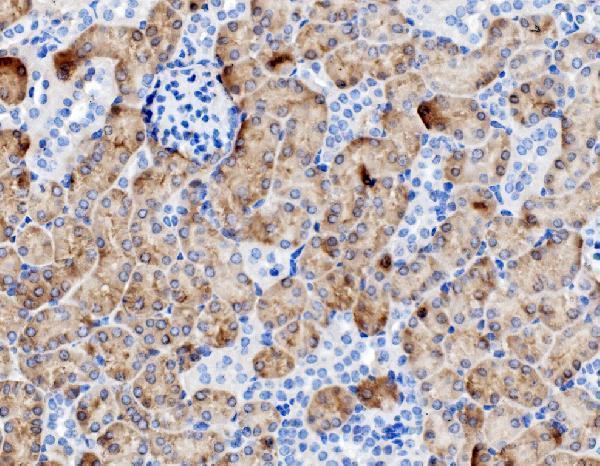
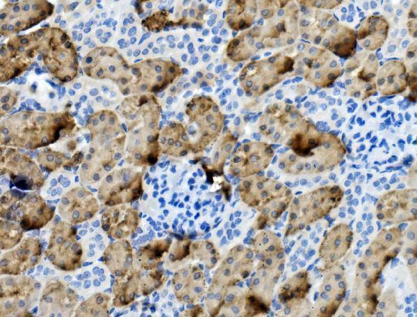
Real IHC data IHC analysis of PCK1/PEPC using anti-PCK1/PEPC antibody (A02022-3). PCK1/PEPC was detected in paraffin-embedded section of mouse kidney tissue. Heat mediated antigen retrieval was performed in EDTA buffer (pH8.0, epitope retrieval solution). The tissue section was blocked with 10% goat serum. The tissue section was then incubated with 1μg/ml rabbit anti-PCK1/PEPC Antibody (A02022-3) overnight at 4°C. Biotinylated goat anti-rabbit IgG was used as secondary antibody and incubated for 30 minutes at 37°C. The tissue section was developed using Strepavidin-Biotin-Complex (SABC) (Catalog # SA1022) with DAB as the chromogen.
Anti-PCK1/PEPC Antibody ®
Cat # A02022-3

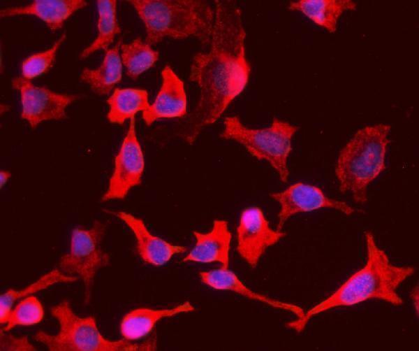
A02022-3 has IHC images from mouse and rat kidney paraffin sections and an ICC/IF image from HeLa cells (A02022-3 image captions). M02022-1 lists human reactivity and IHC use, but has no supplied IHC image (M02022-1 catalog applications/reactivity; image inventory).

Which to pick: Choose A02022-3 for tissue IHC in mouse or rat: its own captions document kidney paraffin sections, and the catalog also lists human reactivity (A02022-3 IHC captions; catalog reactivity). Choose A02022-3 for IF/ICC because its HeLa cell caption documents IF at 4 μg/mL (A02022-3 IF caption). M02022-1 is a rabbit monoclonal option listed for human IHC, but has no tissue image or processing details; the fixative is unreported for both products (M02022-1 catalog; A02022-3 IHC captions).

Each figure is that product's own IHC / IF validation image from its datasheet.

References

  1. UniProt Consortium. UniProt entry P35558 (PCKGC_HUMAN, Phosphoenolpyruvate carboxykinase, cytosolic [GTP]).
  2. Human Protein Atlas. PCK1 tissue IHC expression (reliability: Approved).
  3. Human Protein Atlas. PCK1 subcellular location (ICC-IF): Highest expression in Hep-G2: 2.2 nTPM.
  4. Human Protein Atlas. PCK1 antibody validation summary (3 antibodies).
  5. Expression and functional significance of phosphoenolpyruvate carboxykinase 1 in uveal melanoma. Cell death discovery 2024 — PMC11053060.
  6. Protective effect of phosphoenolpyruvate carboxykinase 1 on inflammation and fibrotic progression of IgA nephropathy. Renal failure 2025 — PMC12128133.
  7. Pharmacologic Antagonization of Cannabinoid Receptor 1 Improves Cholestasis in Abcb4(-/-) Mice. Cellular and molecular gastroenterology and hepatology 2022 — PMC8873597.
  8. PCK1 Downregulation Promotes TXNRD1 Expression and Hepatoma Cell Growth via the Nrf2/Keap1 Pathway. Frontiers in oncology 2018 — PMC6304441.
  9. PubMed PMID:8490617 — UniProt-cited evidence.
  10. PubMed PMID:8325643 — UniProt-cited evidence.
  11. PubMed PMID:14702039 — UniProt-cited evidence.