This website uses cookies to ensure you get the best experience on our website.
- Table of Contents
1 Citations
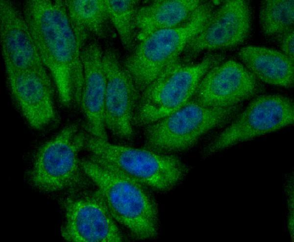
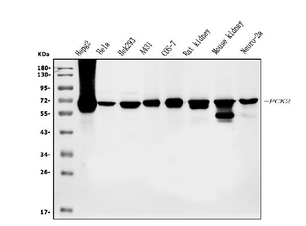
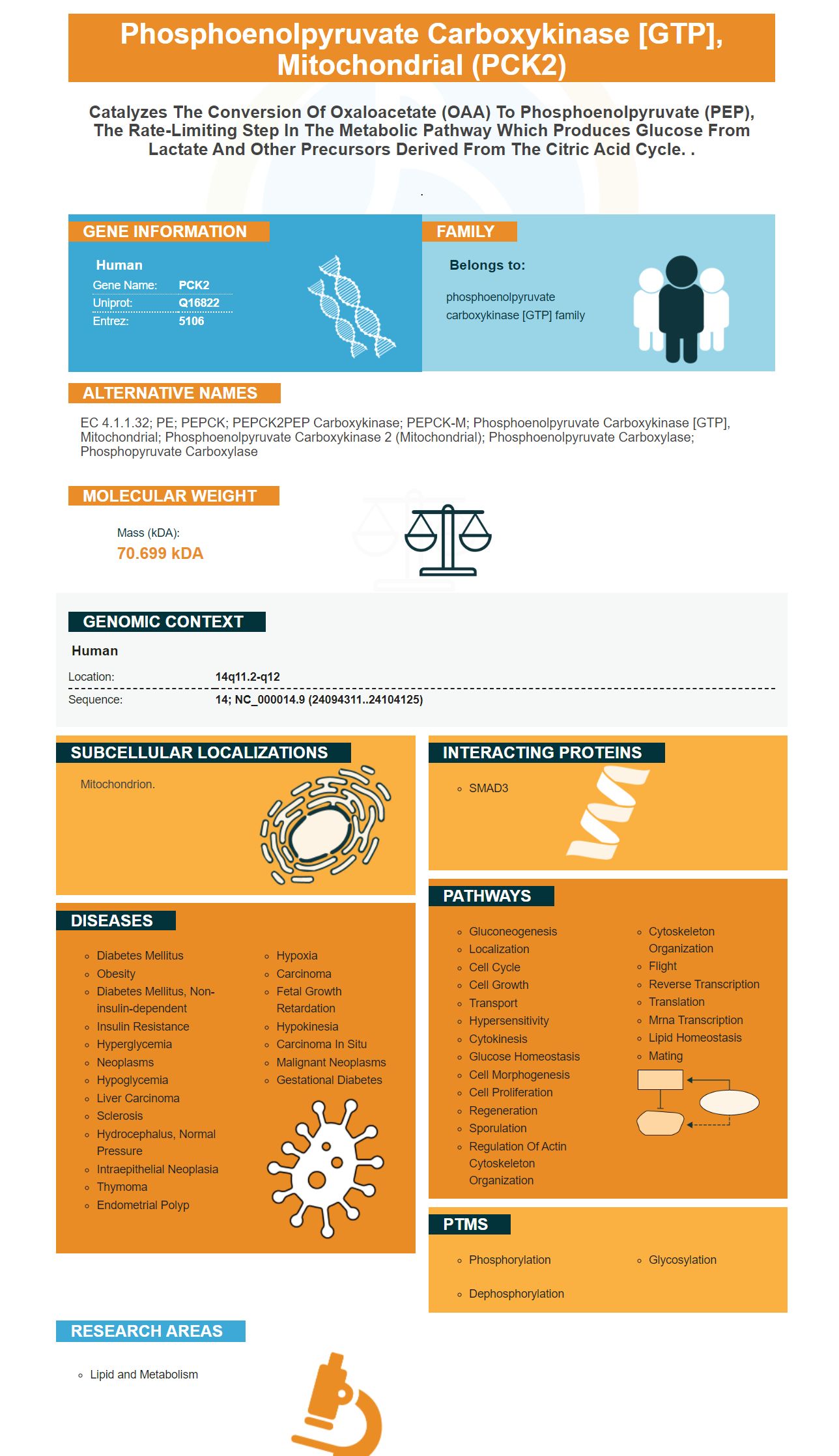
Facts about Phosphoenolpyruvate carboxykinase [GTP], mitochondrial.

.
| Human | |
|---|---|
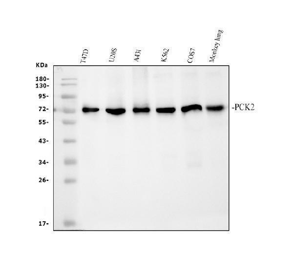
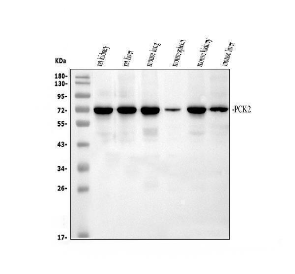
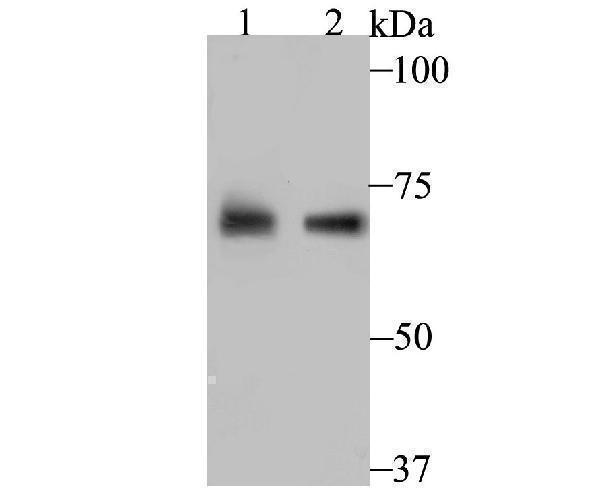
| Gene Name: | PCK2 |
| Uniprot: | Q16822 |
| Entrez: | 5106 |

| Belongs to: |
|---|
| phosphoenolpyruvate carboxykinase [GTP] family |

EC 4.1.1.32; PE; PEPCK; PEPCK2PEP carboxykinase; PEPCK-M; phosphoenolpyruvate carboxykinase [GTP], mitochondrial; phosphoenolpyruvate carboxykinase 2 (mitochondrial); Phosphoenolpyruvate carboxylase; phosphopyruvate carboxylase
Mass (kDA):
70.699 kDA

| Human | |
|---|---|
| Location: | 14q11.2-q12 |
| Sequence: | 14; NC_000014.9 (24094311..24104125) |
Mitochondrion.


PMID: 8645161 by Modaressi S., et al. Molecular cloning, sequencing and expression of the cDNA of the mitochondrial form of phosphoenolpyruvate carboxykinase from human liver.
PMID: 9657976 by Modaressi S., et al. Human mitochondrial phosphoenolpyruvate carboxykinase 2 gene. Structure, chromosomal localization and tissue-specific expression.