PCK2 / Phosphoenolpyruvate carboxykinase [GTP], mitochondrial · IHC design guide

Design Immunohistochemistry for PCK2

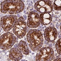
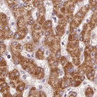
Plan PCK2 staining in paraffin sections using an IHC-validated antibody. Look for granular cytoplasmic staining in intestinal cells, hepatocytes and renal tubules (HPA tissue IHC), and include a kidney proximal tubule positive control (HPA tissue IHC).

Evidence assembled Oct 2026 · For research use; verify linked source records and product datasheet before use
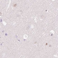
Immunohistochemistry protocol sheet for PCK2 (IHC for PCK2): expected localisation Granular cytoplasm (HPA tissue IHC); mitochondrion (UniProt), antibody M04772-2, validated IHC image, and IHC protocol steps
Printable PCK2 IHC protocol sheet — expected localisation Granular cytoplasm (HPA tissue IHC); mitochondrion (UniProt), antibody M04772-2, controls and protocol steps. Open the full PCK2 IHC guide →

PCK2 Immunohistochemistry Experimental Design Guide

Expected localisation, validated protocols, controls and antibodies — the at-a-glance facts below, then the full design guide.

Must know before staining
Expected localisation Granular cytoplasm (HPA tissue IHC); mitochondrion (UniProt)
Staining pattern Granular cytoplasm in renal tubules and intestinal endocrine cells (HPA tissue IHC)
Antigen retrieval EDTA pH 8.0 HIER, heat-mediated (datasheet M04772-2)
Positive control ⓘ Appendix+4 more · see all
Negative control ⓘ Adipose tissue+4 more · see all
Important caveats
Reasons your staining may differ from the expected pattern.
Fixation Keep fixation consistent across paraffin sections. (standard IHC practice; not target-specific)
Caveat Presumed off-target binding can confound staining (HPA tissue IHC)
Regulation Tissue-dependent staining intensity (HPA tissue IHC)
Isoform / epitope 3 isoforms; mature chain starts at residue 33—check epitope coverage (UniProt)
Section 1

Recommended PCK2 IHC & IF Protocols

Compare the catalog antibody’s IHC-P protocol (datasheet M04772-2) with published PCK2 IHC protocols for medulloblastoma (PMC12935851), lymphoma (PMC12642796), renal biopsies (PMC7710132), and glioma (PMC11685687).

Recommended immunohistochemistry (IHC-P) protocol parameters
SampleParaffin-embedded human prostate cancer tissue; fixative not specified (datasheet M04772-2)
FixationImage fixative and duration unreported (datasheet M04772-2); verify before use.
Sectioning4–5 µm sections on charged slides (standard)
DeparaffinisationXylene, graded ethanol series to water (standard)
Antigen retrievalHeat retrieval: EDTA pH 8.0 (datasheet M04772-2); 20 min, 95–100 °C (standard)
Peroxidase block3% H2O2, 10 min, room temperature (standard)
Blocking10% goat serum (datasheet M04772-2)
Primary antibodyMouse monoclonal (clone 3F7) anti-PCK2, 2μg/ml (datasheet M04772-2)
Primary incubationOvernight at 4 °C (datasheet M04772-2)
DetectionStreptavidin-biotin complex (SABC), DAB chromogen (datasheet M04772-2)
CounterstainHematoxylin, blue, dehydrate and mount (standard)
Expected resultPCK2-positive staining in endocrine cells of appendix (HPA tissue IHC: High). HPA tissue profile: Distinct granular cytoplasmic expression, mainly in the small intestine, hepatocytes and renal tubules. No signal in the no-primary control.
💡Decision noteStart with heat-mediated EDTA pH 8.0 retrieval (datasheet M04772-2). If optimization is needed, compare the published citrate retrieval conditions (PMC12642796; PMC7710132).
Section 2

What Is the Expected PCK2 Staining Pattern?

PCK2 localizes to mitochondria and has no transmembrane segment (UniProt Q16822). In paraffin sections, expect granular cytoplasmic staining in intestinal cells, hepatocytes and renal tubules (HPA tissue IHC). HPA reports high staining in intestinal endocrine cells and kidney proximal tubules; its tissue IHC reliability is Enhanced, although presumed off target binding was observed and disregarded (HPA tissue IHC).

What am I looking at on my slide?
Distinct cytoplasmic granules in kidney proximal tubules or intestinal endocrine cells, with little nuclear signal.This fits mitochondrial localization (UniProt Q16822; HPA subcellular) and the high staining reported for those cells (HPA tissue IHC). Compare the pattern across cells within the section; a brown deposit alone does not establish that it is PCK2.
Predominantly nuclear, membrane rim or extracellular staining replaces the granular cytoplasmic pattern.The dominant compartment conflicts with mitochondrial PCK2 (UniProt Q16822; HPA subcellular). Treat it as a suspected artefact and review morphology, counterstain and detection controls before scoring it as positive.
Strong signal appears in adipocytes or another cell population scored as not detected by HPA.HPA reports adipocytes as not detected in adipose tissue and breast (HPA tissue IHC). Unexpected staining raises concern for antibody cross reactivity or endogenous detection activity; an HPA absence call is a comparator, not proof that every specimen must be negative.
Broad, diffuse colour obscures cell boundaries and overwhelms discrete cytoplasmic granules.Diffuse background prevents a reliable localization call. In general IHC practice, review antibody concentration, blocking, washing and chromogen development, then judge signal against a section processed without primary antibody.
No signal is visible in an adequately preserved kidney proximal tubule control.HPA reports high staining in proximal tubules (HPA tissue IHC), so a blank control calls the assay run into question. Check section identity and the retrieval, primary antibody and detection steps before interpreting other blank sections.
💡Expected PCK2 appearanceCall a section positive when it shows distinct granular cytoplasmic staining in expected cells, especially proximal tubules or intestinal endocrine cells with high HPA staining; dominant nuclear staining or uniform colour over unrelated cells is suspect (UniProt Q16822; HPA tissue IHC and subcellular).
How each factor affects the staining
CompartmentPCK2 is mitochondrial and has no transmembrane segment (UniProt Q16822); HPA describes granular cytoplasmic tissue staining and enhanced mitochondrial localization by ICC-IF (HPA tissue IHC; HPA subcellular).
Tissue and cell selectionHPA reports high staining in kidney proximal tubules and intestinal endocrine cells, and a granular pattern in hepatocytes (HPA tissue IHC). Adipocytes are reported as not detected, making them a useful pattern comparator (HPA tissue IHC).
Strength of validationHPA classifies tissue IHC reliability as Enhanced and lists Enhanced IHC validation for HPA051162, HPA053502 and CAB018734 (HPA tissue IHC; HPA antibodies). Presumed off target binding was also observed and disregarded (HPA tissue IHC).
Processing and isoformsUniProt lists three isoforms and a mature chain spanning residues 33–640 (UniProt Q16822). The supplied evidence gives no antibody epitopes, so it cannot establish which isoforms or processed forms a given stain detects.
Antigen retrieval and fixationTarget specific fixation sensitivity and retrieval conditions are unreported in the supplied sources. As general paraffin IHC practice, follow the chosen antibody's validated retrieval instructions and keep conditions consistent across comparisons.
IF/ICC cross check?Yes: HPA reports enhanced mitochondrial localization by ICC-IF (HPA subcellular). Use that observation to assess whether a cytoplasmic IHC pattern is plausible; it does not supply an IF/ICC protocol for this IHC guide.
Why is my staining missing, weak or wrong?
SituationLikely causeNext action
Kidney proximal tubules show no stain.The run may have failed, or the sampled section may lack an interpretable positive region; HPA reports high proximal tubule staining (HPA tissue IHC).Confirm tubule identity and tissue integrity, then check the catalog antibody's IHC retrieval, dilution and detection instructions. Reassess the run with a known positive section.
A blank or weak section is being called PCK2 negative.Without an internal or parallel positive control, staining failure cannot be separated from low expression. PCK2 is widely expressed (UniProt Q16822), but HPA levels vary by cell type (HPA tissue IHC).Include a section containing an HPA high cell population, such as kidney proximal tubules (HPA tissue IHC), and report the test section as uninterpretable until that control works.
Nuclei stain more strongly than cytoplasm.That distribution conflicts with mitochondrial PCK2 (UniProt Q16822; HPA subcellular); excess chromogen, background or nonspecific antibody binding may obscure localization.Inspect a section processed without primary antibody, shorten chromogen development if overdeveloped, and score only signal whose compartment can be resolved.
Adipocytes or many unrelated cells stain strongly.HPA reports adipocytes as not detected (HPA tissue IHC). Cross reactivity or endogenous detection activity is possible, especially when colour also appears in the no primary control.Run that control alongside a kidney or intestinal positive section, review the blocking step and compare the cellular pattern before attributing the stain to PCK2.
Diffuse brown haze hides the expected granules.In general chromogenic IHC practice, overly concentrated primary antibody, incomplete washing or excessive chromogen development can raise background; the payload does not identify a PCK2 specific cause.Optimize those steps using the chosen antibody's instructions, keep exposure and counterstaining comparable, and accept only discrete cytoplasmic signal above control background.
IHC looks diffuse while an IF image appears mitochondrial.HPA reports mitochondrial ICC-IF localization and granular tissue IHC staining (HPA subcellular; HPA tissue IHC). The imaging methods resolve detail differently, so appearance alone cannot identify the cause.Recheck IHC focus, section thickness and counterstain; compare expected cells and a no primary control. Use IF as a localization cross check, without treating its image as an IHC protocol.

Sample controls for PCK2 IHC & IF

🧪Run kidney first: proximal tubule cell bodies must stain strongly (HPA: High in kidney proximal tubules). Use adipose tissue adipocytes as the negative comparison (HPA: Not detected in adipocytes); cells outside the proximal tubules on the kidney slide should provide low-background internal contrast, but their absence of PCK2 is not established by the supplied HPA rows.
Positive control tissue: Appendix (Endocrine cells, HPA High)
Negative control tissue: Adipose tissue (HPA Not detected)
ICC-IF cell lines (HPA subcellular resource): HPA ICC-IF images show PCK2 in A-431, MCF-7, U2OS, Hep-G2, U2OS, siRNA 2 (10x), U2OS, scrambled (10x), HeLa BAC 6291, HeLa , NIH 3T3, with annotated localisation: Mitochondria (enhanced) (HPA subcellular).
Technical controls: Include a no-primary, secondary-only control; a mouse IgG control matched to the primary’s isotype and clonality where known (selected-SKU caption: mouse primary); and PCK2-knockout tissue or cells as a biological negative. In kidney, block endogenous peroxidase and, if using the caption’s biotin-based detection, endogenous biotin to limit false chromogenic signal (selected-SKU caption: biotinylated secondary and DAB).
⚠️Feasibility: A PCK2-specific fixation window or fixation effect is unreported, and the selected-SKU paraffin-section caption does not state the fixative (selected-SKU caption: fixative not stated). Heat-mediated EDTA retrieval at pH 8.0 was used in the reported IHC example, but a retrieval requirement has not been established (selected-SKU caption: EDTA retrieval). Frozen-section IHC has no supplied comparison with paraffin IHC; IF can assess the expected mitochondrial pattern in cells, while kidney proximal tubules require attention to endogenous biotin during chromogenic detection (HPA subcellular: mitochondria; selected-SKU caption: biotin-based detection).

HPA tissue IHC evidence for PCK2

Comprehensive Human Protein Atlas IHC scoring per tissue (reliability: Enhanced — High consistency between antibody staining and RNA expression data. Presumed off target binding observed and disregarded.). Rows are taken directly from the HPA tissue chart — click any row's HPA link to view the source.

Positive expression · recommended positive controls

TissueCell typeLevelEvidenceSource
Appendix Endocrine cells High Protein (IHC) HPA →
Colon Endocrine cells High Protein (IHC) HPA →
Duodenum Endocrine cells High Protein (IHC) HPA →
Kidney Proximal tubules (cell body) High Protein (IHC) HPA →
Lung Alveolar cells type II High Protein (IHC) HPA →

Undetected expression · recommended negative controls

TissueCell typeLevelEvidenceSource
Adipose tissue Adipocytes Not detected Protein (IHC) HPA →
Adrenal gland Glandular cells Not detected Protein (IHC) HPA →
Bone marrow Hematopoietic cells Not detected Protein (IHC) HPA →
Breast Adipocytes Not detected Protein (IHC) HPA →
Caudate Glial cells Not detected Protein (IHC) HPA →
Section 3

Advanced PCK2 IHC Tips

Troubleshoot PCK2 staining in paraffin sections by checking retrieval, assay controls, cellular pattern and cell type before comparing chromogenic signal.

What retrieval should I try first when PCK2 staining is weak in paraffin sections?
Start with heat-mediated antigen retrieval in EDTA at pH 8.0 for paraffin-section IHC (datasheet M04772-2). The selected PCK2 image used that retrieval before incubation with 2 µg/ml primary antibody overnight at 4°C (datasheet M04772-2). If signal is weak, compare heating and cooling durations on matched sections while holding antibody concentration and detection constant; insufficient retrieval and tissue damage can both obscure a granular pattern (standard IHC practice). Record the retrieval conditions for every slide, because the caption reports neither a heating duration nor a fixative (datasheet M04772-2).
Could fixation explain inconsistent PCK2 staining across paraffin blocks?
Target-specific fixation sensitivity is unknown: the selected paraffin-section caption does not state its fixative or fixation duration (datasheet M04772-2). Record each block’s fixative, time before fixation and fixation duration, then compare matched sections using EDTA at pH 8.0 and the same detection run (datasheet M04772-2; standard IHC practice). If those conditions vary, treat differences in staining as unresolved until a controlled comparison separates tissue processing from biological variation (standard IHC practice). Do not infer a PCK2-specific fixation effect from its mitochondrial location, modified residues or reported tissue distribution (UniProt Q16822; HPA tissue IHC).
What PCK2 staining pattern should I expect within positive cells?
Expect a granular cytoplasmic pattern consistent with mitochondrial localisation, rather than a dominant nuclear or surface outline (HPA tissue IHC; HPA subcellular; UniProt Q16822). HPA describes distinct granular cytoplasmic expression, mainly in small intestine, hepatocytes and renal tubules, and reports high staining in kidney proximal tubule cell bodies (HPA tissue IHC). PCK2 has no annotated transmembrane segment, but that fact alone does not establish which mitochondrial compartment an antibody can reach (UniProt Q16822 topology). Compare suspicious regions with cell morphology and a no-primary control before assigning diffuse chromogen deposits to PCK2 (standard IHC practice).
How could isoforms or epitope placement affect PCK2 IHC?
PCK2 has 3 annotated isoforms, while the listed processed chain spans residues 33–640 (UniProt Q16822). The supplied antibody evidence does not map its epitope, so staining cannot establish which isoforms it detects or whether an amino-terminal epitope is retained (datasheet M04772-2; UniProt Q16822). Annotated modifications include phosphothreonine at residue 42 and phosphoserine at 115; their effect on this antibody’s staining is untested here (UniProt Q16822). For discrepant cases, seek epitope documentation and compare staining with an independent PCK2 measurement or a separately mapped antibody on matched material (standard IHC practice).
How should I check PCK2 localisation by IF alongside tissue IHC?
For a matched IF experiment, pair PCK2 with a validated marker of the expected cell type, such as kidney proximal tubule cells, and assess mitochondrial pattern within those cells (HPA tissue IHC; HPA subcellular). Choose a fluorophore away from strong tissue autofluorescence, often in the far-red range, and include single-channel controls to check bleed-through (standard IF practice). Permeabilisation must give the antibody access to its epitope; because its mitochondrial side is not mapped here, compare mild and stronger permeabilisation conditions without assuming an orientation (UniProt Q16822 topology; standard IF practice). HPA reports mitochondrial IF localisation, but the supplied evidence gives no IF fixation or permeabilisation settings to transfer into a protocol (HPA subcellular).
How do I separate PCK2 signal from chromogenic background?
Run a no-primary control and inspect whether brown deposits persist in the same compartments or collect at section edges (standard IHC practice). The selected assay used 10% goat serum, a biotinylated secondary antibody, a streptavidin–biotin complex and DAB, so check nonspecific secondary binding and endogenous biotin alongside routine peroxidase blocking (datasheet M04772-2; standard IHC practice). Keep DAB development comparable across slides when judging subtle granular signal, because longer development can amplify background (standard IHC practice). HPA rates tissue staining as enhanced while noting presumed off-target binding that was disregarded; an isolated unexpected pattern therefore needs independent confirmation (HPA tissue IHC).
How should I score PCK2 staining across different tissue regions? ⚠ ANSWER MARKED FOR VERIFICATION
Define the cell population first, then score viable cells for staining intensity from 0–3 and calculate an H-score as the sum of intensity multiplied by percent of cells at each intensity (standard IHC practice). Report the percentage positive separately, with a consistent positivity threshold set before comparing groups (standard IHC practice). Normalise counts or signal area to the number of evaluable cells or viable tissue area in mm², and record which cell type was assessed (standard IHC practice). This matters for PCK2 because HPA reports cell-specific staining, including high signal in kidney proximal tubule cell bodies (HPA tissue IHC).
When is apparent PCK2 positivity more likely to be artefact?
A credible positive shows granular cytoplasmic staining compatible with mitochondrial PCK2 in an identifiable cell population (HPA tissue IHC; HPA subcellular; UniProt Q16822). Strong nuclear staining, a cell-surface outline or uniform extracellular DAB should prompt review before it is assigned to PCK2 (UniProt Q16822 topology; standard IHC practice). Compare the finding with expected cell patterns, including high proximal tubule staining and undetected adipocyte staining in the reported HPA sections, without treating either as a universal rule (HPA tissue IHC). Edge-restricted signal, necrotic deposits or staining retained in a no-primary control can reflect processing or endogenous enzyme activity; confirm unexpected positives independently (standard IHC practice).
Boster reagents

Best PCK2 / Phosphoenolpyruvate carboxykinase [GTP], mitochondrial IHC Antibodies

M04772-2 has IHC images from paraffin sections of human prostate, breast and rectal cancer tissue, plus an IF/ICC image from HEPG2 cells (M04772-2 image captions).

Real IHC data IHC analysis of PCK2 using anti-PCK2 antibody (M04772-2). PCK2 was detected in paraffin-embedded section of human prostate cancer tissue. Heat mediated antigen retrieval was performed in EDTA buffer (pH8.0, epitope retrieval solution). The tissue section was blocked with 10% goat serum. The tissue section was then incubated with 2μg/ml mouse anti-PCK2 Antibody (M04772-2) overnight at 4°C. Biotinylated goat anti-mouse IgG was used as secondary antibody and incubated for 30 minutes at 37°C. The tissue section was developed using Strepavidin-Biotin-Complex (SABC) (Catalog # SA1021) with DAB as the chromogen.
Anti-PCK2 Antibody ® (monoclonal, 3F7)
Cat # M04772-2

M04772-2 will render with its IHC figure from a paraffin section of human prostate cancer tissue (M04772-2 IHC caption). Its IF/ICC image shows HEPG2 cells, and the catalog lists Human, Mouse, Rat and Monkey reactivity (M04772-2 IF caption; M04772-2 catalog reactivity).

Which to pick: For tissue IHC, choose mouse monoclonal M04772-2, clone 3F7: its own paraffin-section caption documents EDTA retrieval at pH 8.0 and 2 μg/ml primary antibody overnight; the fixative is unreported (M04772-2 catalog host/clone; M04772-2 IHC caption). For IF/ICC, A04772-1 has a human MCF-7 cell image and a listed 5 μg/ml IF dilution (A04772-1 IF caption; A04772-1 catalog dilution). For cross-species paraffin IHC, A04772-1 lists Human, Mouse and Rat for that application, while its IHC images document human tissue (A04772-1 catalog dilution; A04772-1 IHC captions).

Each figure is that product's own IHC / IF validation image from its datasheet.

References

  1. UniProt Consortium. UniProt entry Q16822 (PCKGM_HUMAN, Phosphoenolpyruvate carboxykinase [GTP], mitochondrial).
  2. Human Protein Atlas. PCK2 tissue IHC expression (reliability: Enhanced).
  3. Human Protein Atlas. PCK2 subcellular location (ICC-IF): Localized to the mitochondria..
  4. Human Protein Atlas. PCK2 antibody validation summary (3 antibodies).
  5. Phosphoenolpyruvate carboxykinase 2 as a prognostic biomarker: expression and clinical significance in Group 3 and Group 4 medulloblastoma. Journal of neuro-oncology 2026 — PMC12935851.
  6. PCK2, a SASP-Associated Gene, Serves as an Independent Prognostic Indicator in Diffuse Large B-Cell Lymphoma. Journal of inflammation research 2025 — PMC12642796.
  7. The proteomic landscape of small urinary extracellular vesicles during kidney transplantation. Journal of extracellular vesicles 2020 — PMC7710132.
  8. Phosphoenolpyruvate carboxykinase 2 is a promising prognostic biomarker that correlates with peritumoral dendritic cell infiltration in glioblastoma. Journal of Cancer 2025 — PMC11685687.
  9. PubMed PMID:8645161 — UniProt-cited evidence.
  10. PubMed PMID:9657976 — UniProt-cited evidence.
  11. PubMed PMID:14702039 — UniProt-cited evidence.