This website uses cookies to ensure you get the best experience on our website.


- Table of Contents
Plan PCOLCE staining in paraffin sections using breast glandular cells as a high-staining reference (HPA tissue IHC). The catalog antibody’s illustrated workflow uses 2 μg/ml primary antibody overnight at 4°C, followed by HRP/DAB detection (datasheet A06428-2).

Expected localisation, validated protocols, controls and antibodies — the at-a-glance facts below, then the full design guide.
| Expected localisation | Extracellular and cytoplasmic staining (HPA tissue IHC) | |
| Staining pattern | Extracellular signal and cytoplasmic staining in several tissues (HPA tissue IHC) | |
| Antigen retrieval | EDTA pH 8.0 HIER, heat-mediated (datasheet A06428-2) | |
| Positive control | Breast+4 more · see all | |
| Negative control | Appendix+4 more · see all |
| Fixation | Keep fixation consistent across sections (standard IHC practice; not target-specific) | |
| Caveat | Secreted PCOLCE may stain beyond producing cells (UniProt) | |
| Regulation | Specific expression regulation is not annotated (UniProt) | |
| Isoform / epitope | No annotated isoforms; signal peptide is cleaved (UniProt) |
The catalog antibody’s IHC protocol uses EDTA pH 8.0 retrieval (datasheet). Published PCOLCE IHC protocols describe glioma and renal carcinoma tissue (PMC11733849; PMC8740509).
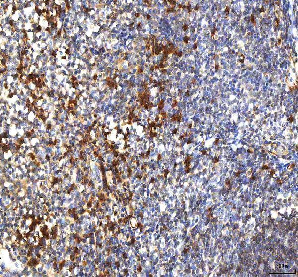
| Sample | Paraffin-embedded human tonsil tissue; fixative not specified (datasheet A06428-2) |
| Fixation | Image fixative and duration unreported (datasheet A06428-2); verify before use. |
| Sectioning | 4–5 µm sections on charged slides (standard) |
| Deparaffinisation | Xylene, graded ethanol series to water (standard) |
| Antigen retrieval | Heat retrieval: EDTA pH 8.0 (datasheet A06428-2); 20 min, 95–100 °C (standard) |
| Peroxidase block | 3% H2O2, 10 min, room temperature (standard) |
| Blocking | 10% goat serum (datasheet A06428-2) |
| Primary antibody | Rabbit anti-PCOLCE, 2-5 μg/ml (datasheet A06428-2) |
| Primary incubation | Overnight at 4 °C (datasheet A06428-2) |
| Detection | HRP-conjugated secondary, DAB chromogen (datasheet A06428-2) |
| Counterstain | Hematoxylin, blue, dehydrate and mount (standard) |
| Expected result | PCOLCE-positive staining in glandular cells of breast (HPA tissue IHC: High). HPA tissue profile: Extracellular positivity and cytoplasmic expression in several tissues. No signal in the no-primary control. |
PCOLCE is secreted and has no transmembrane segment (UniProt Q15113 topology). In paraffin-section IHC, expect extracellular positivity and cytoplasmic staining in several tissues (HPA tissue IHC: Approved). Breast glandular cells provide a high-staining example; endometrial stromal cells and bronchial respiratory epithelial cells are medium-staining examples (HPA tissue IHC). Interpret cellular and extracellular signal together, since tissue RNA and protein locations can differ for this secreted protein (HPA reliability note).
| Extracellular staining accompanies glandular or stromal cytoplasmic staining. | This fits a secreted protein (UniProt Q15113 topology) and HPA's observed extracellular and cytoplasmic IHC profile (HPA tissue IHC). Judge each compartment against the tissue and cell type: breast glandular cells stain high, while endometrial stromal cells stain medium (HPA tissue IHC). |
| A crisp nuclear-only or membrane-rim pattern dominates the section. | Neither is the reported PCOLCE IHC pattern (HPA tissue IHC), and PCOLCE has no transmembrane segment (UniProt Q15113 topology). Treat the pattern as suspect; compare its distribution with the extracellular and cytoplasmic reference pattern before assigning specificity. |
| The strongest signal occurs in a cell population expected to be unstained. | For example, appendix glandular cells are not detected in HPA tissue IHC. Cross-reactivity or endogenous detection activity is plausible (general IHC practice). Check whether the signal persists when primary antibody is omitted, and compare it with a known-positive section. |
| Color spreads uniformly across cells, stroma, and empty-looking spaces. | Uniform haze cannot distinguish the extracellular positivity reported for PCOLCE from background (HPA tissue IHC; general IHC practice). Examine morphology, a primary-omission control, and a positive tissue processed in the same run before scoring extracellular staining. |
| Breast glandular cells show no detectable signal. | High staining is reported for these cells (HPA tissue IHC), so the negative result warrants a run check. Review tissue preservation, retrieval, antibody dilution, detection reagents, and the concurrent positive control as general IHC workflow checks; no PCOLCE-specific fixation sensitivity is established here. |
| Secretion and maturation | PCOLCE has a signal peptide at residues 1–25 and a mature chain at 26–449 (UniProt Q15113 processing). Its extracellular IHC signal can be spatially separate from stained cells (HPA tissue IHC). Do not infer the producing cell solely from extracellular deposits. |
| Tissue and cell baseline | HPA reports high breast glandular staining, medium endometrial stromal staining, and no detection in appendix glandular cells (HPA tissue IHC). These are reference observations for interpreting a run, not a claim that every section or cell must have identical intensity. |
| Antibody validation | The HPA tissue profile is Approved, and CAB017623 has Approved IHC status (HPA tissue IHC; HPA antibodies). Approval supports use of the reported pattern as a reference; it does not establish specificity for every specimen or detection system. |
| Epitope and retrieval limits | The supplied UniProt record identifies CUB and NTR domains and glycosylation sites at residues 29 and 431 (UniProt Q15113). It gives no antibody epitope or target-specific retrieval response, so choose retrieval conditions from the IHC-validated antibody's instructions and validate the resulting pattern (general IHC practice). |
| IF/ICC cross-check? | HPA reports approved Golgi and vesicle localization in ICC-IF, with U2OS images (HPA subcellular ICC-IF). This intracellular view can coexist with secretion and the extracellular tissue IHC profile (UniProt Q15113 topology; HPA tissue IHC). It does not define an IF protocol for this IHC guide. |
| Situation | Likely cause | Next action |
|---|---|---|
| No staining in the concurrent breast glandular positive section. | A run failure or insufficient assay signal is possible; breast glandular staining is high in the HPA reference (HPA tissue IHC). | Check antibody preparation and dilution, retrieval, detection reagents, and counterstain against the IHC antibody instructions (general IHC practice). Repeat with a concurrent positive section before interpreting test sections. |
| Only nuclei or sharply outlined cell membranes stain. | The location conflicts with extracellular and cytoplasmic PCOLCE tissue staining (HPA tissue IHC) and its lack of a transmembrane segment (UniProt Q15113 topology). | Review morphology and controls; reassess antibody specificity and detection background before scoring the signal as PCOLCE (general IHC practice). |
| Appendix glandular cells stain strongly. | Those cells are not detected in the HPA reference (HPA tissue IHC). Cross-reactivity or endogenous detection activity may explain the discrepancy (general IHC practice). | Run a primary-omission control and inspect the detection system's endogenous-activity blocking step (general IHC practice). Compare the result with breast glandular cells in the same run (HPA tissue IHC). |
| Extracellular staining is diffuse and hard to separate from haze. | Extracellular positivity is expected (HPA tissue IHC), but widespread nonspecific color can obscure its boundaries (general IHC practice). | Compare signal with the primary-omission control, tissue morphology, and expected stained cells. Review blocking, washes, and detection development as general IHC checks; do not score uniform haze alone. |
| A positive tissue stains, but the test tissue is weak or negative. | PCOLCE tissue levels vary: breast glandular cells are high, whereas lung alveolar cells are low (HPA tissue IHC). A working positive control does not establish expression in the test cell population. | Identify the exact tissue and cell population, then compare its HPA reference level. Score the observed compartment and intensity without extending the positive control's expected intensity to another tissue. |
| An ICC-IF image shows Golgi or vesicles while tissue IHC shows extracellular signal. | Both locations are reported: approved Golgi and vesicle ICC-IF localization (HPA subcellular ICC-IF) and extracellular tissue positivity (HPA tissue IHC); PCOLCE is secreted (UniProt Q15113 topology). | Interpret each application against its own HPA reference. For paraffin-section IHC, assess extracellular and cytoplasmic staining in the relevant tissue cells (HPA tissue IHC); use the separate IF/ICC guide for that application's setup. |
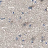


Comprehensive Human Protein Atlas IHC scoring per tissue (reliability: Approved — Secreted protein, tissue location of RNA and protein is expected to differ.). Rows are taken directly from the HPA tissue chart — click any row's HPA link to view the source.
| Tissue | Cell type | Level | Evidence | Source |
|---|---|---|---|---|
| Breast | Glandular cells | High | Protein (IHC) | HPA → |
| Adrenal gland | Glandular cells | Medium | Protein (IHC) | HPA → |
| Bronchus | Respiratory epithelial cells | Medium | Protein (IHC) | HPA → |
| Endometrium | Cells in endometrial stroma | Medium | Protein (IHC) | HPA → |
| Esophagus | Squamous epithelial cells | Medium | Protein (IHC) | HPA → |
| Tissue | Cell type | Level | Evidence | Source |
|---|---|---|---|---|
| Appendix | Glandular cells | Not detected | Protein (IHC) | HPA → |
| Cerebellum | Cells in granular layer | Not detected | Protein (IHC) | HPA → |
| Epididymis | Glandular cells | Not detected | Protein (IHC) | HPA → |
| Fallopian tube | Glandular cells | Not detected | Protein (IHC) | HPA → |
| Hippocampus | Glial cells | Not detected | Protein (IHC) | HPA → |
Troubleshoot PCOLCE staining by assessing retrieval, compartment, and detection alongside matched tissue controls (UniProt Q15113; HPA tissue IHC).
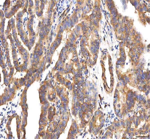
The catalog contains one human-reactive anti-PCOLCE antibody with paraffin-section IHC images from human tonsil and thyroid papillary carcinoma; no IF image is supplied (catalog: A06428-2 image captions).
A06428-2 is listed for human IHC (catalog: A06428-2 applications and reactivity). Its image captions show staining in paraffin sections of human tonsil and thyroid papillary carcinoma (catalog: A06428-2 IHC image captions).
Which to pick: Choose A06428-2 for human paraffin-section IHC; its caption documents EDTA retrieval at pH 8.0 and 2 μg/ml primary antibody (catalog: A06428-2 tonsil IHC caption). Its application list provides no IF/ICC validation, and its reactivity is limited to human, so the payload supports neither an IF/ICC nor a cross-species recommendation (catalog: A06428-2 applications and reactivity). The caption identifies paraffin sections but does not report the fixative (catalog: A06428-2 IHC image captions).