This website uses cookies to ensure you get the best experience on our website.


- Table of Contents
Plan paraffin-section PCYOX1 IHC around cytoplasmic staining in most tissues, including high staining in hepatocytes and kidney tubule cells (HPA tissue IHC). Use the catalog antibody’s IHC-P conditions (datasheet A11180-1), and interpret staining with HPA’s reported low consistency between antibody staining and RNA expression (HPA tissue IHC).

Expected localisation, validated protocols, controls and antibodies — the at-a-glance facts below, then the full design guide.
| Expected localisation | Cytoplasmic tissue staining (HPA tissue IHC); lysosomal protein (UniProt) | |
| Staining pattern | Cytoplasmic staining in many cell types across most tissues (HPA tissue IHC) | |
| Antigen retrieval | EDTA pH 8.0 HIER, heat-mediated (datasheet A11180-1) | |
| Positive control | Heart muscle+4 more · see all | |
| Negative control | Bone marrow+4 more · see all |
| Fixation | Matched tissue-IHC evidence does not establish the fixation claim. Validate the specimen-specific method before use. (selected-SKU IHC image A11180-1) | |
| Caveat | Staining may diverge from RNA expression (HPA tissue IHC) | |
| Regulation | Widely expressed; no regulator specified (UniProt) | |
| Isoform / epitope | Two isoforms; signal peptide cleaved; epitope impact unknown (UniProt) |
The catalog antibody’s IHC-P protocol is paired with a published protocol for human atherosclerotic lesions (PMC8455616).
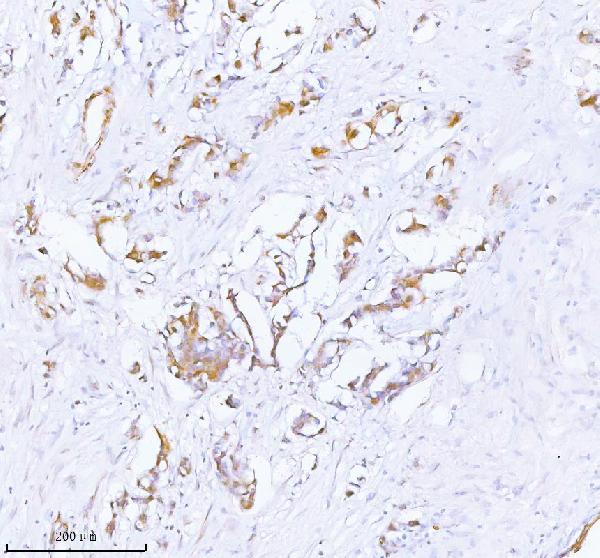
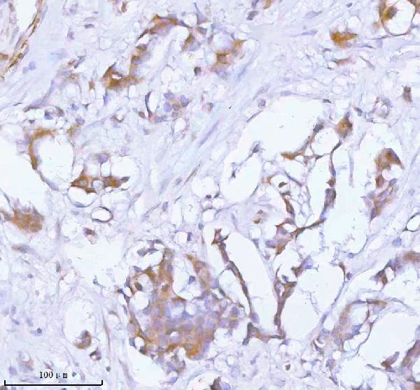
| Sample | Paraffin-embedded human breast cancer tissue; fixative not specified (datasheet A11180-1) |
| Fixation | Image fixative and duration unreported (datasheet A11180-1); verify before use. |
| Sectioning | 4–5 µm sections on charged slides (standard) |
| Deparaffinisation | Xylene, graded ethanol series to water (standard) |
| Antigen retrieval | Heat retrieval: EDTA pH 8.0 (datasheet A11180-1); 20 min, 95–100 °C (standard) |
| Peroxidase block | 3% H2O2, 10 min, room temperature (standard) |
| Blocking | 10% goat serum (datasheet A11180-1) |
| Primary antibody | Rabbit anti-PCYOX1, 2-5 μg/ml (datasheet A11180-1) |
| Primary incubation | Overnight at 4 °C (datasheet A11180-1) |
| Detection | HRP-conjugated secondary, DAB chromogen (datasheet A11180-1) |
| Counterstain | Hematoxylin, blue, dehydrate and mount (standard) |
| Expected result | PCYOX1-positive staining in cardiomyocytes of heart muscle (HPA tissue IHC: High). HPA tissue profile: Cytoplasmic expression in most tissues. No signal in the no-primary control. |
PCYOX1 is lysosomal and has no transmembrane segment (UniProt Q9UHG3). In paraffin sections, expect cytoplasmic staining in the relevant cells, including hepatocytes, kidney tubule cells and cardiomyocytes, which stain High in HPA tissue IHC (HPA: tissue IHC). HPA describes cytoplasmic expression in most tissues but rates the staining Approved with low consistency between antibody staining and RNA expression; interpret unexpected patterns cautiously (HPA: tissue IHC).
| Cytoplasmic signal in hepatocytes, kidney tubule cells or cardiomyocytes, with visible cell boundaries. | These are useful positive reference populations: each is rated High in HPA tissue IHC (HPA: liver, kidney and heart muscle). A lysosomal protein can give intracellular staining (UniProt Q9UHG3); HPA reports a broad cytoplasmic pattern and does not establish that every positive cell must show discrete puncta (HPA: tissue IHC). |
| Predominantly nuclear staining or a sharp outline confined to the plasma membrane. | That distribution does not match the reported cytoplasmic tissue pattern or lysosomal location (HPA: tissue IHC; UniProt Q9UHG3). Treat it as suspect until it is reproduced with a suitable control. A compartment mismatch alone cannot identify whether the cause is nonspecific binding, detection background or an interpretation error. |
| Strong signal in hematopoietic cells of bone marrow or germinal center cells of tonsil. | Those particular cell populations are Not detected in HPA tissue IHC (HPA: bone marrow; HPA: tonsil). Check cell identity and a no-primary control. Persistent staining could reflect antibody cross-reactivity or endogenous detection activity; a discordant slide alone cannot establish which explanation applies. |
| Diffuse chromogen across cells and extracellular spaces, obscuring intracellular detail. | The result is hard to score as PCYOX1 because HPA describes cytoplasmic expression in most tissues (HPA: tissue IHC). General IHC practice: compare a no-primary control and background in adjacent tissue, then review blocking, washes and detection chemistry. Widespread color without recognizable cell-specific staining is insufficient evidence of a positive result. |
| No visible signal in hepatocytes, kidney tubule cells or cardiomyocytes. | Absence in one of these High populations conflicts with the HPA reference pattern (HPA: liver, kidney and heart muscle). First confirm that the expected cell population is present and that the run's positive control worked. If both checks pass, review the antibody and detection workflow before calling the specimen negative. |
| Cell population and tissue | PCYOX1 is widely expressed (UniProt Q9UHG3), yet HPA levels vary by cell population: Leydig cells are High, while ovarian stroma cells are Not detected (HPA: testis; HPA: ovary). Score the identified cells, not the whole section. |
| Antibody evidence | HPA035193 is a rabbit polyclonal antibody with Approved IHC status (HPA: antibodies). HPA also reports low consistency between staining and RNA expression (HPA: tissue IHC), so the rating is not independent proof of every observed signal. |
| Protein processing and glycosylation | The annotated signal peptide spans residues 1–27; the mature chain spans 28–505, with three glycosylation sites (UniProt Q9UHG3). Epitope position is unspecified here, so these annotations do not predict retrieval needs or staining intensity. |
| Isoforms | Two isoforms are listed (UniProt Q9UHG3). The supplied HPA IHC observations do not distinguish them (HPA: tissue IHC); an IHC-positive cell therefore cannot be assigned to isoform 1 or 2 from these data. |
| IF/ICC Q&A: Is there a confirmed cellular image pattern? | No HPA ICC-IF image, main location or ICC validation entry is supplied (HPA: subcellular; HPA: antibodies). Lysosomal localization is an expectation from UniProt Q9UHG3, not an HPA-confirmed IF pattern; use the separate IF/ICC guide for that application. |
| Situation | Likely cause | Next action |
|---|---|---|
| Expected positive tissue is blank. | The reference cell type may be absent, or the staining run may have failed; hepatocytes, kidney tubule cells and cardiomyocytes are High references (HPA: tissue IHC). | Verify the cells on the section and compare an independent positive control. General IHC practice: check reagent sequence, detection and the validated retrieval conditions before interpreting a negative result. |
| Signal is much weaker than the reference image. | HPA's High, Medium and Not detected calls apply to specified cell populations (HPA: tissue IHC). A different population or scoring threshold can make a section appear weaker. | Compare like cells at similar magnification and document the proportion and intensity of stained cells. General IHC practice: assess the run control before changing staining conditions. |
| Nuclear or membrane-only color dominates. | This conflicts with HPA's cytoplasmic tissue pattern and UniProt's lysosomal location (HPA: tissue IHC; UniProt Q9UHG3). The supplied sources do not identify a PCYOX1-specific cause. | Check the no-primary control, tissue morphology and antibody localization in a High reference population (HPA: tissue IHC). Reassess the pattern only after background has been excluded. |
| Bone marrow hematopoietic cells or tonsil germinal centers stain strongly. | Both populations are Not detected in HPA tissue IHC (HPA: bone marrow; HPA: tonsil). Cross-reactivity or endogenous chromogenic activity is possible, but neither is established by the slide alone. | Confirm the cell type and compare a no-primary control. General IHC practice: if enzyme-based detection is used, check the appropriate endogenous-activity control. |
| Color covers most of the section and masks cell detail. | Diffuse background can obscure the cytoplasmic pattern reported by HPA (HPA: tissue IHC). The supplied evidence does not establish a target-specific fixation or retrieval effect. | General IHC practice: inspect a no-primary control, blocking, washes and detection exposure; then repeat with a positive reference tissue in the same run. |
| An IF/ICC image seems to show a different compartment. | HPA supplies no ICC-IF images or main subcellular location for PCYOX1 (HPA: subcellular). UniProt assigns PCYOX1 to lysosomes (UniProt Q9UHG3), but that does not validate the image. | Treat the image as unconfirmed for IF/ICC and consult that application's separate guide. Do not transfer an IHC-P staining pattern into an IF/ICC validation claim. |


Comprehensive Human Protein Atlas IHC scoring per tissue (reliability: Approved — Low consistency between antibody staining and RNA expression data.). Rows are taken directly from the HPA tissue chart — click any row's HPA link to view the source.
| Tissue | Cell type | Level | Evidence | Source |
|---|---|---|---|---|
| Heart muscle | Cardiomyocytes | High | Protein (IHC) | HPA → |
| Kidney | Cells in tubules | High | Protein (IHC) | HPA → |
| Liver | Hepatocytes | High | Protein (IHC) | HPA → |
| Salivary gland | Glandular cells | High | Protein (IHC) | HPA → |
| Testis | Leydig cells | High | Protein (IHC) | HPA → |
| Tissue | Cell type | Level | Evidence | Source |
|---|---|---|---|---|
| Bone marrow | Hematopoietic cells | Not detected | Protein (IHC) | HPA → |
| Endometrium | Cells in endometrial stroma | Not detected | Protein (IHC) | HPA → |
| Fallopian tube | Glandular cells | Not detected | Protein (IHC) | HPA → |
| Ovary | Ovarian stroma cells | Not detected | Protein (IHC) | HPA → |
| Parathyroid gland | Glandular cells | Not detected | Protein (IHC) | HPA → |
Troubleshoot PCYOX1 staining by checking retrieval, cellular pattern, and controls before scoring signal (datasheet A11180-1; UniProt Q9UHG3; standard IHC practice).
A11180-1 has real IHC images from human paraffin sections of breast cancer and testicular seminoma (A11180-1 image captions). No IF/ICC data are listed (catalog applications and image payload).
A11180-1 is listed for human IHC and is shown staining paraffin sections of human breast cancer (catalog applications and reactivity; A11180-1 image captions). The same SKU is shown staining paraffin sections of human testicular seminoma (A11180-1 image captions).
Which to pick: For human paraffin-section IHC, choose A11180-1: its captions document EDTA retrieval at pH 8.0 and primary antibody at 2 μg/ml (A11180-1 image captions), within the listed 2–5 μg/ml IHC range (catalog dilution). No listed SKU has IF/ICC validation or an IF image (catalog applications and image payload). No cross-species choice is supported because A11180-1 lists human reactivity only; its captions specify paraffin sections but do not report the fixative (catalog reactivity; A11180-1 image captions).