This website uses cookies to ensure you get the best experience on our website.


- Table of Contents
Plan chromogenic PDCD2L IHC in paraffin sections with the catalog antibody at 1:50–1:200 (datasheet). Compare strongly stained breast glandular cells with oral mucosal squamous cells reported as undetected, and score cytoplasmic signal (HPA tissue IHC).

Expected localisation, validated protocols, controls and antibodies — the at-a-glance facts below, then the full design guide.
| Expected localisation | Cytoplasmic staining in tissue sections (HPA tissue IHC) | |
| Staining pattern | Cytoplasmic signal across tissues; high in glandular cells (HPA tissue IHC) | |
| Antigen retrieval | Tris-EDTA pH 9.0 HIER, 95–98 °C, 20 min (rule: nuclear antigen) | |
| Positive control | Adrenal gland+4 more · see all | |
| Negative control | Oral mucosa+1 more · see all |
| Fixation | Matched tissue-IHC evidence does not establish the fixation claim. Validate the specimen-specific method before use. (selected-SKU IHC image A14105) | |
| Caveat | Staining and RNA show medium consistency; verify specificity (HPA tissue IHC) | |
| Regulation | Expression regulation is unreported (UniProt) | |
| Isoform / epitope | No isoforms annotated; mature chain spans residues 2–358 (UniProt) |
The catalog antibody’s IHC-P protocol (datasheet) is followed by one published PDCD2L paraffin-section workflow (PMC13085924).
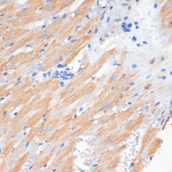
| Sample | Paraffin-embedded rat heart tissue; fixative not specified (datasheet A14105) |
| Fixation | Image fixative and duration unreported (datasheet A14105); verify before use. |
| Sectioning | 4–5 µm sections on charged slides (standard) |
| Deparaffinisation | Xylene, graded ethanol series to water (standard) |
| Antigen retrieval | Heat-induced epitope retrieval in Tris-EDTA buffer, pH 9.0, 20 min at 95–98 °C (standard rule: nuclear antigen) |
| Peroxidase block | 3% H2O2, 10 min, room temperature (standard) |
| Blocking | 10% normal serum of the secondary host, 30 min, room temperature (standard) |
| Primary antibody | Rabbit anti-PDCD2L, 1:50-1:200 (datasheet A14105) |
| Primary incubation | Overnight at 4 °C (standard) |
| Detection | HRP-polymer secondary, DAB chromogen 5–10 min (standard) |
| Counterstain | Hematoxylin, blue, dehydrate and mount (standard) |
| Expected result | PDCD2L-positive staining in glandular cells of adrenal gland (HPA tissue IHC: High). HPA tissue profile: Cytoplasmic expression in all tissues. No signal in the no-primary control. |
PDCD2L has a cytoplasmic tissue IHC profile (HPA tissue IHC), while UniProt annotates nucleolar, nuclear and cytosolic localisation and no transmembrane segment (UniProt Q9BRP1). High staining is reported in selected glandular, respiratory epithelial and neuronal cells (HPA tissue IHC). HPA rates the tissue staining Approved, with medium consistency against RNA data and external verification pending (HPA tissue IHC).
| Clear cytoplasmic staining in breast or cervical glandular cells, bronchial respiratory epithelial cells, or caudate neurons. | This matches cell types scored High in tissue IHC (HPA tissue IHC). Assess the stained cells and compartment together; a strong chromogen signal alone does not establish the expected pattern (HPA tissue IHC). |
| Predominantly nuclear, membranous or sharply organelle-like staining in an IHC section, with little cytoplasmic signal. | This differs from the reported cytoplasmic tissue IHC profile (HPA tissue IHC). Nuclear localisation is annotated separately (UniProt Q9BRP1), so review the controls and staining distribution before calling a nuclear result artefactual. A membrane-only result also conflicts with the lack of a transmembrane segment (UniProt Q9BRP1). |
| Strong staining in oral or vaginal squamous epithelial cells. | Those cells are scored Not detected (HPA tissue IHC). Consider cross-reactivity or endogenous detection activity, then compare matched control sections and the expected cytoplasmic pattern (HPA tissue IHC; general IHC practice). Do not treat one unexpected cell population as proof of PDCD2L expression. |
| Diffuse chromogen across tissue, extracellular spaces or the whole section, without cell boundaries. | This cannot be scored as the reported cellular cytoplasmic pattern (HPA tissue IHC). Check the detection-only control and background handling; widespread colour may reflect nonspecific binding or endogenous detection activity (general IHC practice). |
| No signal in a section containing breast glandular or bronchial respiratory epithelial cells. | Both cell populations are scored High (HPA tissue IHC), making the result a useful reason to review section quality, antigen retrieval, antibody incubation and detection controls (general IHC practice). A single negative section does not overturn the HPA pattern, whose external verification remains pending (HPA tissue IHC). |
| Tissue and cell selection | HPA scores adrenal, breast, cervical, duodenal, endometrial and fallopian glandular cells High; bronchial respiratory epithelial and caudate neuronal cells are also High (HPA tissue IHC). Oral and vaginal squamous cells are Not detected, while esophageal squamous cells, skeletal myocytes and adipocytes are Low (HPA tissue IHC). Interpret intensity by cell population. |
| Compartment and assay | Tissue IHC is described as cytoplasmic (HPA tissue IHC). UniProt also annotates the nucleus and nucleolus alongside cytosol (UniProt Q9BRP1). These records support checking assay context when a nuclear component appears; they do not establish a required nuclear IHC signal. |
| IF/ICC Q: What localisation should be considered? | A: HPA reports approved mitochondrial localisation in ICC-IF images from A-431, A-549 and U2OS (HPA subcellular). This is separate from the cytoplasmic tissue IHC profile (HPA tissue IHC) and the nuclear, nucleolar and cytosolic UniProt annotation (UniProt Q9BRP1). Interpret the assays in their own context. |
| Evidence strength | The listed HPA052181 antibody is Approved for IHC and ICC (HPA antibodies). Tissue IHC has medium consistency with RNA data and awaits external verification (HPA tissue IHC). Approval supports using the published pattern as a reference, while ambiguous or unexpected staining still needs controls. |
| RNA versus protein staining | HPA calls RNA tissue enhanced in skeletal muscle, yet scores skeletal myocytes Low by tissue IHC (HPA tissue IHC). UniProt lists higher expression in lung, colon, mammary gland, cervix, stomach and small intestine (UniProt Q9BRP1). Use the cell-level IHC observations to choose staining comparators; RNA enrichment alone does not predict a strong chromogen result. |
| Topology and processing | UniProt annotates no transmembrane segment or signal peptide and lists a PDCD2L chain spanning residues 2–358 (UniProt Q9BRP1). These annotations give no basis to expect a membrane-restricted pattern. They do not specify antibody epitope position or target-specific antigen retrieval conditions (UniProt Q9BRP1). |
| Situation | Likely cause | Next action |
|---|---|---|
| Expected positive cells are blank. | Section or detection failure is possible; the chosen cells may also differ from an HPA High population (HPA tissue IHC; general IHC practice). | Confirm cell identity against the HPA tissue IHC entries, then review retrieval, antibody incubation and detection controls in the established IHC workflow (HPA tissue IHC; general IHC practice). No PDCD2L-specific retrieval condition is supplied. |
| Oral or vaginal squamous cells stain strongly. | That conflicts with their Not detected scores (HPA tissue IHC); nonspecific binding or endogenous detection activity is possible (general IHC practice). | Review a detection-only control and the staining distribution, then compare with a High cell population on a suitable section (HPA tissue IHC; general IHC practice). |
| Colour covers the section uniformly. | Acellular or uniform colour does not match the cell-associated cytoplasmic tissue profile (HPA tissue IHC); background from binding or detection is possible (general IHC practice). | Inspect tissue edges and empty areas, assess the detection-only control, and review blocking, washes and chromogen development (general IHC practice). |
| Only nuclei appear positive in IHC. | Nuclear localisation is annotated (UniProt Q9BRP1), but the HPA tissue IHC profile is cytoplasmic (HPA tissue IHC). | Check whether cytoplasmic signal is present in HPA High cells and compare control sections before deciding whether the nuclear-only result is specific (HPA tissue IHC; general IHC practice). |
| A membrane outline dominates the IHC result. | A membrane-restricted pattern is unsupported by the reported cytoplasmic staining and lack of a transmembrane segment (HPA tissue IHC; UniProt Q9BRP1). | Reassess cellular localisation at higher magnification and inspect detection controls for edge staining or background (general IHC practice). |
| Skeletal muscle is weak despite the RNA enrichment label. | HPA labels RNA tissue enhanced in skeletal muscle while scoring myocytes Low by IHC (HPA tissue IHC). | Score the observed myocyte staining against the protein IHC entry and use an HPA High cell population to assess whether the staining run produced an interpretable signal (HPA tissue IHC; general IHC practice). |


Comprehensive Human Protein Atlas IHC scoring per tissue (reliability: Approved — Medium consistency between antibody staining and RNA expression data. Pending external verification.). Rows are taken directly from the HPA tissue chart — click any row's HPA link to view the source.
| Tissue | Cell type | Level | Evidence | Source |
|---|---|---|---|---|
| Adrenal gland | Glandular cells | High | Protein (IHC) | HPA → |
| Breast | Glandular cells | High | Protein (IHC) | HPA → |
| Bronchus | Respiratory epithelial cells | High | Protein (IHC) | HPA → |
| Caudate | Neuronal cells | High | Protein (IHC) | HPA → |
| Cervix | Glandular cells | High | Protein (IHC) | HPA → |
Troubleshoot PDCD2L staining in paraffin sections by checking retrieval, compartment, controls and scoring before interpreting biological differences.
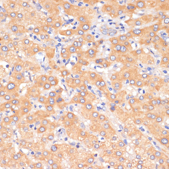
A14105 has IHC images from paraffin-embedded rat heart and human liver (catalog image captions). No IF images or IF/ICC application are listed (catalog applications and image captions).
A14105 is listed for IHC and has images of paraffin-embedded rat heart and human liver stained at 1:100 (catalog applications and image captions). Its listed reactivity is human, mouse and rat; the supplied IHC images cover human and rat only (catalog reactivity and image captions).
Which to pick: Choose A14105 for paraffin-section IHC: its captions document microwave antigen retrieval in 10 mM PBS, pH 7.2, and the fixative is unreported (catalog image captions). For cross-species work, A14105 lists human, mouse and rat reactivity, with IHC images for human and rat; its host is rabbit and clonality is unreported (catalog reactivity, image captions, host and clone fields). No IF/ICC antibody can be selected from this payload because A14105 has no listed IF/ICC application or IF image (catalog applications and image captions).