This website uses cookies to ensure you get the best experience on our website.


- Table of Contents
Plan PDE4D staining in paraffin sections using the IHC-validated antibody A01111-1 at 0.5–1 μg/ml (datasheet: IHC-P dilution). Compare the cytoplasmic pattern with tissue controls while accounting for uncertain staining reliability (HPA tissue IHC).

Expected localisation, validated protocols, controls and antibodies — the at-a-glance facts below, then the full design guide.
| Expected localisation | Ubiquitous cytoplasmic tissue staining (HPA tissue IHC) | |
| Staining pattern | Cytoplasmic staining across many cell types (HPA tissue IHC) | |
| Antigen retrieval | Citrate pH 6 HIER, heat-mediated (datasheet A01111-1) | |
| Positive control | Appendix+4 more · see all | |
| Negative control | None in HPA (detected in all 45 tissues); use no-primary + isotype controls |
| Fixation | Keep fixation consistent across sections. (standard IHC practice; not target-specific) | |
| Caveat | Staining has only medium consistency with RNA expression (HPA tissue IHC) | |
| Regulation | Tissue-enhanced RNA: marrow, muscle (HPA tissue RNA) | |
| Isoform / epitope | 12 isoforms; N-terminal epitopes may vary; no TM span (UniProt) |
The catalog antibody’s IHC-P protocol is accompanied by four published PDE4D IHC protocols covering liver, aorta, rat cortex, and pancreatic tumor sections (PMC12375944; PMC8776755; PMC7655962; PMC6856734).
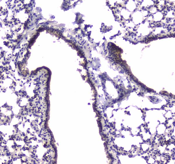
| Sample | Paraffin-embedded mouse lung tissue; fixative not specified (datasheet A01111-1) |
| Fixation | Image fixative and duration unreported (datasheet A01111-1); verify before use. |
| Sectioning | 4–5 µm sections on charged slides (standard) |
| Deparaffinisation | Xylene, graded ethanol series to water (standard) |
| Antigen retrieval | Heat retrieval: Citrate pH 6, 20 min (datasheet A01111-1) |
| Peroxidase block | 3% H2O2, 10 min, room temperature (standard) |
| Blocking | 10% goat serum (datasheet A01111-1) |
| Primary antibody | Rabbit anti-PDE4D, 0.5-1μg/ml (datasheet A01111-1) |
| Primary incubation | Overnight at 4 °C (datasheet A01111-1) |
| Detection | Streptavidin-biotin complex (SABC), DAB chromogen (datasheet A01111-1) |
| Counterstain | Hematoxylin, blue, dehydrate and mount (standard) |
| Expected result | PDE4D-positive staining in glandular cells of appendix (HPA tissue IHC: High). HPA tissue profile: Ubiquitous cytoplasmic expression. No signal in the no-primary control. |
Expect PDE4D mainly in the cytoplasm of many cell types, with possible membrane-associated staining, including the apical membrane of colonic crypt cells (HPA: ubiquitous cytoplasmic expression; UniProt Q08499: subcellular location). Colon glandular cells and heart cardiomyocytes are among the reported high-staining cells (HPA: tissue IHC). Treat intensity and compartment calls cautiously: the tissue IHC profile has uncertain reliability (HPA: tissue IHC). PDE4D has no transmembrane segment (UniProt Q08499: topology).
| Cytoplasmic staining in colon glandular cells, with possible apical accentuation. | This fits the reported high staining of colon glandular cells (HPA: tissue IHC) and PDE4D's cytoplasmic location and apical association in colonic crypt cells (UniProt Q08499: subcellular location). Assess the glandular cells themselves; a stained section alone does not establish that every coloured structure is PDE4D (general IHC practice). |
| Strong staining confined to nuclei, with little cytoplasmic or membrane signal. | A nuclear-only IHC pattern differs from the reported ubiquitous cytoplasmic tissue pattern (HPA: tissue IHC). Nucleoplasm is only an additional, uncertain ICC-IF location (HPA: subcellular). Check the counterstain, detection background and antibody controls before calling the nuclear signal specific (general IHC practice). |
| Signal mainly in cells other than the expected glandular cells in a colon section. | Colon glandular cells are reported as high staining (HPA: tissue IHC). An unexpected cell distribution can reflect off-target antibody binding or endogenous detection activity (general IHC practice); it cannot be assigned to PDE4D solely from colour. Compare cellular morphology and appropriate detection controls (general IHC practice). |
| Weak, diffuse colour across tissue and blank areas, with poor cell boundaries. | A broadly diffuse deposit is difficult to reconcile with a readable cellular pattern (general IHC practice). HPA reports cytoplasmic expression, but its tissue IHC reliability is uncertain (HPA: tissue IHC). Inspect a no-primary control and optimise blocking, washing or detection conditions before scoring (general IHC practice). |
| No signal in colon glandular cells or heart cardiomyocytes. | Both are listed as high-staining cell populations (HPA: tissue IHC), so a blank run warrants a technical check. Verify that tissue is present and interpretable, then review retrieval and detection controls (general IHC practice). Because the HPA tissue IHC profile is uncertain, absence alone does not prove biological loss of PDE4D (HPA: tissue IHC). |
| Cell and tissue choice (HPA: tissue IHC) | Colon, appendix, duodenum, epididymis and fallopian tube glandular cells; cerebellar granular-layer cells; endometrial stromal cells; and cardiomyocytes are listed as high (HPA: tissue IHC). Parathyroid glandular cells, vaginal squamous cells and soft-tissue fibroblasts are listed as low, not negative (HPA: tissue IHC). |
| Location and topology (UniProt Q08499: subcellular location and topology) | PDE4D is reported in cytoplasm, membranes and cytoskeletal compartments, with apical colonic crypt localisation (UniProt Q08499: subcellular location). It has no transmembrane segment (UniProt Q08499: topology); membrane-associated staining therefore need not resemble an integral membrane protein (general interpretation of topology). |
| Strength of the tissue evidence (HPA: tissue IHC and antibody validation) | The tissue profile is described as ubiquitous cytoplasmic expression, but antibody staining has only medium consistency with RNA data and is rated uncertain (HPA: tissue IHC). The listed antibody, HPA045895, has uncertain IHC validation (HPA: antibody validation). Treat a matching pattern as supportive, not definitive. |
| Isoform coverage (UniProt Q08499: isoforms) | UniProt lists 12 PDE4D isoforms (UniProt Q08499: isoforms). Without an epitope assignment for the antibody in the supplied evidence, the observed stain cannot be assigned to a particular isoform or assumed to cover all isoforms. Compare like tissue compartments when interpreting differences (general IHC practice). |
| Situation | Likely cause | Next action |
|---|---|---|
| Expected high-staining cells are blank. | A failed retrieval, primary-antibody or detection step is possible (general IHC practice); high staining is reported for colon glandular cells and cardiomyocytes (HPA: tissue IHC). | Run a suitable positive section with the same IHC workflow; check reagent delivery, retrieval conditions and the detection control (general IHC practice). Avoid calling biological absence from a failed control, especially given uncertain tissue IHC reliability (HPA: tissue IHC). |
| Colour spreads through tissue with little cellular definition. | Excess detection deposit, inadequate washing or nonspecific antibody binding can obscure cellular localisation (general IHC practice). | Review a no-primary control, blocking and wash steps; adjust antibody or detection conditions using the catalog IHC-P protocol as the starting point (general IHC practice). Score only a resolved cellular pattern, considering the uncertain HPA IHC profile (HPA: tissue IHC). |
| Nuclear staining dominates the section. | The IHC tissue summary is cytoplasmic (HPA: tissue IHC); nucleoplasm is an uncertain additional ICC-IF location (HPA: subcellular). Nuclear colour alone has limited support. | Compare the counterstain and no-primary control, then seek cytoplasmic staining in an expected cell population before interpreting the result (general IHC practice; HPA: tissue IHC). |
| A low-staining comparison tissue appears negative. | Parathyroid glandular cells, vaginal squamous cells and soft-tissue fibroblasts are described as low, not absent (HPA: tissue IHC). A low signal may be hard to resolve in a particular run (general IHC practice). | Use a reported high-staining cell population to verify the run; record the comparison as below detection if appropriate, rather than asserting PDE4D absence (HPA: tissue IHC; general IHC practice). |
| Unexpected cells stain more strongly than the expected population. | Off-target binding or endogenous detection activity is possible (general IHC practice). The reported HPA levels identify cell populations to compare, but have uncertain tissue IHC reliability (HPA: tissue IHC). | Confirm cell identity by morphology and counterstain; inspect no-primary and detection controls, then repeat under optimised IHC conditions if needed (general IHC practice). |
| IF/ICC shows membrane, cytosolic or nuclear signal: how should it be read? | Plasma membrane is supported; cytosol, nucleoplasm, nuclear membrane and primary cilium are uncertain ICC-IF locations (HPA: subcellular). | Interpret the IF/ICC image using its own localisation evidence and controls (HPA: subcellular; general IF practice). Do not use an ICC-IF compartment call alone to validate an IHC tissue stain, whose reliability is uncertain (HPA: tissue IHC). |
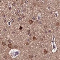
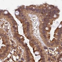

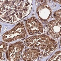


Comprehensive Human Protein Atlas IHC scoring per tissue (reliability: Uncertain — Medium consistency between antibody staining and RNA expression data.). Rows are taken directly from the HPA tissue chart — click any row's HPA link to view the source.
| Tissue | Cell type | Level | Evidence | Source |
|---|---|---|---|---|
| Appendix | Glandular cells | High | Protein (IHC) | HPA → |
| Cerebellum | Cells in granular layer | High | Protein (IHC) | HPA → |
| Colon | Glandular cells | High | Protein (IHC) | HPA → |
| Duodenum | Glandular cells | High | Protein (IHC) | HPA → |
| Endometrium | Cells in endometrial stroma | High | Protein (IHC) | HPA → |
| Tissue | Cell type | Level | Evidence | Source |
|---|---|---|---|---|
| None in HPA: PDE4D is detected in all 45 scored tissues. Use a no-primary (secondary-only) and an isotype control instead. | ||||
Troubleshoot PDE4D staining in paraffin sections by checking retrieval, compartment, background and scoring against the available tissue evidence.
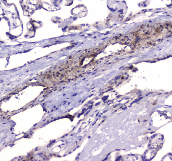
A01111-1 has IHC images from paraffin sections of mouse lung, human placenta and rat lung (catalog image captions). ICC is listed as an application; no IF image is supplied (catalog applications and images).
A01111-1 will render with its mouse lung IHC figure (card image caption). Its catalog also shows paraffin-section IHC in human placenta and rat lung and lists human, mouse and rat reactivity plus ICC (catalog image captions and reactivity/application lists).
Which to pick: For tissue IHC, choose A01111-1: its own caption documents citrate retrieval at pH 6 for 20 minutes and antibody at 1 μg/ml on a paraffin mouse lung section (card image caption). For IF/ICC, A01111-1 lists ICC and 0.5–1 μg/ml, but supplies no IF image; for cross-species IHC, its captions show human, mouse and rat tissues (catalog applications, dilution and image captions). Clonality and fixative are unreported (catalog clone field and image captions).