This website uses cookies to ensure you get the best experience on our website.


- Table of Contents
Use this IHC-P guide to assess PDE6A staining in retinal photoreceptor rod segments (HPA tissue IHC). It covers the expected outer segment membrane location (UniProt) and paraffin-section staining with the catalog antibody (datasheet A07800-1).

Expected localisation, validated protocols, controls and antibodies — the at-a-glance facts below, then the full design guide.
| Expected localisation | Rod outer segment membrane expected (UniProt) | |
| Staining pattern | Strong staining in photoreceptor rod segments (HPA tissue IHC) | |
| Antigen retrieval | EDTA pH 8.0 HIER, heat-mediated (datasheet A07800-1) | |
| Positive control | Retina | |
| Negative control | Adipose tissue+4 more · see all |
| Fixation | Keep fixation consistent across matched paraffin sections. (selected-SKU IHC image A07800-1) | |
| Caveat | Sections missing rod outer segments may lack expected signal (HPA tissue IHC) | |
| Regulation | No expression regulator annotated (UniProt) | |
| Isoform / epitope | No isoforms annotated; mature chain spans residues 2–857 (UniProt) |
The catalog antibody has an IHC-P protocol (datasheet A07800-1). Published PDE6A IHC examples use FFPE tissue arrays (PMC3297615) and canine retinal sections (PMC3720143).
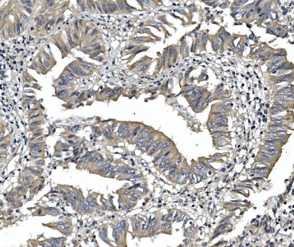
| Sample | Paraffin-embedded human rectal cancer tissue; fixative not specified (datasheet A07800-1) |
| Fixation | Image fixative and duration unreported (datasheet A07800-1); verify before use. |
| Sectioning | 4–5 µm sections on charged slides (standard) |
| Deparaffinisation | Xylene, graded ethanol series to water (standard) |
| Antigen retrieval | Heat retrieval: EDTA pH 8.0 (datasheet A07800-1); 20 min, 95–100 °C (standard) |
| Peroxidase block | 3% H2O2, 10 min, room temperature (standard) |
| Blocking | 10% goat serum (datasheet A07800-1) |
| Primary antibody | Rabbit anti-PDE6A, 0.5-1μg/ml (datasheet A07800-1) |
| Primary incubation | Overnight at 4 °C (datasheet A07800-1) |
| Detection | Streptavidin-biotin complex (SABC), DAB chromogen (datasheet A07800-1) |
| Counterstain | Hematoxylin, blue, dehydrate and mount (standard) |
| Expected result | PDE6A-positive staining in photoreceptor cells of retina (HPA tissue IHC: High). HPA tissue profile: Expression in rods segments. No signal in the no-primary control. |
PDE6A is a rod phototransduction protein associated with the photoreceptor outer segment membrane; UniProt reports no transmembrane segment (UniProt P16499 topology and function). In paraffin retina sections, expect staining in photoreceptor segments, consistent with HPA's high photoreceptor staining and rod-segment profile (HPA: Retina, High; HPA: expression in rods segments). HPA rates its tissue IHC pattern Enhanced, with external verification pending (HPA: reliability).
| Strong staining follows the photoreceptor segment layer in retina (HPA: Retina photoreceptor cells, High). | This matches HPA's rod-segment profile and UniProt's photoreceptor outer segment membrane location (HPA: tissue profile; UniProt P16499 subcellular location). Judge the distribution across the retinal layers as well as staining strength. |
| Signal is mainly nuclear or concentrated away from photoreceptor segments (HPA: tissue profile). | That distribution conflicts with the reported outer segment location (UniProt P16499 subcellular location). Check retinal orientation, counterstain and staining controls before interpreting the signal as PDE6A; intensity alone cannot establish localisation. |
| Prominent signal appears in other cell types or in an HPA negative tissue, such as adrenal glandular cells (HPA: adrenal gland, Not detected). | Investigate antibody cross-reactivity or chromogen generated by endogenous detection activity (general IHC practice). HPA's negative call applies to the specified tissue and cell type; it does not prove that every cell in every specimen must be negative. |
| Colour covers several retinal layers or the section broadly, without a defined segment pattern (HPA: tissue profile). | Diffuse staining obscures the expected rod-segment distribution (HPA: expression in rods segments). Review blocking, antibody concentration, washes and the detection-only control for background (general IHC practice). Do not score diffuse colour as a positive compartment. |
| Photoreceptor segments show no convincing signal in an intact retina section (HPA: Retina photoreceptor cells, High). | A negative result in the expected positive tissue needs technical checks before a biological conclusion (HPA: Retina photoreceptor cells, High; general IHC practice). Verify section orientation, tissue preservation, antibody performance and detection controls. |
| Compartment and topology (UniProt P16499 subcellular location and topology) | PDE6A is associated with the photoreceptor outer segment membrane and has no transmembrane segment (UniProt P16499). Interpret a segment-associated pattern without requiring a crisp outline of each cell membrane; section geometry can affect its appearance (general IHC practice). |
| Tissue expression and validation (HPA: Retina, High; reliability Enhanced) | HPA reports high photoreceptor staining, a rod-segment profile and retina-enriched RNA (HPA: tissue IHC). Its Enhanced rating reflects staining and RNA consistency; external verification remains pending (HPA: reliability description). Use these as expectations, not a guarantee for every specimen. |
| Antibody-specific evidence (HPA: HPA016970 and HPA074677, IHC Enhanced) | Both listed rabbit polyclonal antibodies have Enhanced IHC status (HPA: antibody validation). That supports the reported tissue pattern, but does not establish that an unspecified catalog antibody shares their performance or that either has ICC-IF validation. |
| Protein processing (UniProt P16499 processing and modified residues) | UniProt lists a chain spanning residues 2–857, a short C-terminal propeptide and modified residues at positions 2 and 857 (UniProt P16499). Those annotations alone cannot predict epitope access or antigen retrieval needs without the antibody's epitope information. |
| Situation | Likely cause | Next action |
|---|---|---|
| Retinal photoreceptor segments are unstained despite an intact expected positive area (HPA: Retina, High). | Antibody, retrieval or detection conditions may have failed; the supplied sources do not establish PDE6A-specific fixation sensitivity (general IHC practice; HPA/UniProt scope). | Check the catalog antibody's IHC-P instructions, reagent performance, section orientation and a known positive control. Adjust retrieval only as a general IHC optimisation, then compare the segment pattern (general IHC practice; HPA: tissue profile). |
| Strong nuclear or unrelated-layer staining dominates the retina section (UniProt P16499 subcellular location). | The distribution is inconsistent with the reported outer segment location; nonspecific binding or misread anatomy may explain it (UniProt P16499; general IHC practice). | Use the counterstain to identify retinal layers, compare an adjacent section and review antibody concentration and controls before assigning PDE6A positivity (general IHC practice; HPA: rod-segment profile). |
| Adrenal glandular cells stain strongly (HPA: adrenal gland glandular cells, Not detected). | This conflicts with HPA's observed negative pattern and warrants investigation of cross-reactivity or detection background (HPA: adrenal gland; general IHC practice). | Check an antibody-omission control and the detection chemistry. If the method uses peroxidase, assess endogenous peroxidase blocking (general IHC practice); retain HPA's cell-specific negative call when comparing sections. |
| A broad haze makes photoreceptor segments hard to distinguish (HPA: rod-segment profile). | Excess antibody or insufficient blocking or washing can raise background in chromogenic IHC (general IHC practice). | Review the catalog IHC-P dilution and incubation guidance, then optimise blocking and washes with controls (general IHC practice). Score only a resolved segment pattern against HPA's expected distribution (HPA: tissue profile). |
| The chromogen obscures the boundary between photoreceptor segments and neighbouring layers (HPA: rod-segment profile). | Overdevelopment or an overly dark counterstain can limit localisation assessment (general IHC practice). | Review development time and counterstain intensity, then reassess the segment layer with the same anatomical landmarks across sections (general IHC practice; HPA: expression in rods segments). |
| Can an IF/ICC pattern establish the expected PDE6A location? | HPA provides no main ICC-IF location or cell-line images, and lists no ICC validation for the two antibodies (HPA: subcellular record; HPA: antibody validation). | Use the outer segment location as a biological expectation, not as evidence of an established ICC-IF staining pattern (UniProt P16499 subcellular location; HPA: subcellular record). Consult the separate IF/ICC guide for that application. |


Comprehensive Human Protein Atlas IHC scoring per tissue (reliability: Enhanced — High consistency between antibody staining and RNA expression data. Pending external verification.). Rows are taken directly from the HPA tissue chart — click any row's HPA link to view the source.
| Tissue | Cell type | Level | Evidence | Source |
|---|---|---|---|---|
| Retina | Photoreceptor cells | High | Protein (IHC) | HPA → |
| Tissue | Cell type | Level | Evidence | Source |
|---|---|---|---|---|
| Adipose tissue | Adipocytes | Not detected | Protein (IHC) | HPA → |
| Adrenal gland | Glandular cells | Not detected | Protein (IHC) | HPA → |
| Appendix | Endocrine cells | Not detected | Protein (IHC) | HPA → |
| Bone marrow | Hematopoietic cells | Not detected | Protein (IHC) | HPA → |
| Breast | Adipocytes | Not detected | Protein (IHC) | HPA → |
Troubleshoot PDE6A staining in paraffin section IHC using the catalog antibody’s documented workflow and retinal rod localisation as reference points.
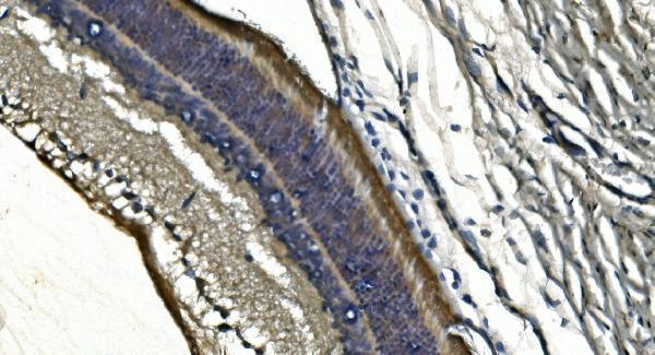
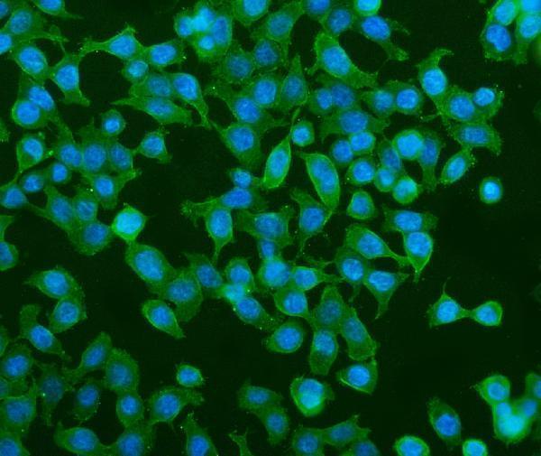
A07800-1 has IHC images from human rectal cancer and rat retina paraffin sections, plus an IF image from CACO-2 cells (catalog image captions).
A07800-1 has IHC images from human rectal cancer and rat retina paraffin sections (catalog IHC captions). The same SKU has an IF image from CACO-2 cells and lists human, mouse and rat reactivity (catalog IF caption; catalog reactivity).
Which to pick: For tissue IHC, choose A07800-1: its paraffin-section images use 1 μg/ml antibody after heat retrieval in EDTA at pH 8.0; the fixative is unreported (catalog IHC captions). For IF/ICC, A07800-1 also has a CACO-2 cell image at 4 μg/ml (catalog IF caption). For cross-species work, A07800-1 lists human, mouse and rat reactivity, with IHC images supplied for human and rat (catalog reactivity; catalog IHC captions).