This website uses cookies to ensure you get the best experience on our website.


- Table of Contents
Plan chromogenic FFPE IHC for PDGFRB using testis peritubular cells as a positive control and adipocytes as a cell-level negative reference (HPA tissue IHC). Interpret staining alongside the receptor’s membrane location and ligand-dependent internalization (UniProt).

Expected localisation, validated protocols, controls and antibodies — the at-a-glance facts below, then the full design guide.
| Expected localisation | Cell membrane; cytoplasmic vesicles and lysosome lumen (UniProt) | |
| Staining pattern | Endothelial, decidual and fibroblast staining; compartment unspecified (HPA tissue IHC) | |
| Antigen retrieval | Citrate pH 6.0 HIER, 95–98 °C, 20 min (rule: cytoplasmic / membrane antigen) | |
| Positive control | Testis+4 more · see all | |
| Negative control | Adipose tissue+4 more · see all |
| Fixation | Matched tissue-IHC evidence does not establish the fixation claim. Validate the specimen-specific method before use. (selected-SKU IHC image M00096-6) | |
| Caveat | Ligand binding can shift receptor signal from membrane to vesicles (UniProt) | |
| Regulation | Ligand binding promotes degradation (UniProt) | |
| Isoform / epitope | 2 isoforms; epitope differences are unspecified—map extracellular versus cytoplasmic domains (UniProt) |
The catalog antibody’s IHC-P protocol is accompanied by 4 published PDGFRB IHC protocols covering glomus tumors, colorectal lesions, human FFPE sections, and mouse tissues (PMC7554116; PMC9497196; PMC3244048; PMC2848338).
| Sample | Tissue sections; selected-image fixative not specified (standard IHC workflow) |
| Fixation | Image fixative and duration unreported (datasheet M00096-6); verify before use. |
| Sectioning | 4–5 µm sections on charged slides (standard) |
| Deparaffinisation | Xylene, graded ethanol series to water (standard) |
| Antigen retrieval | Heat-induced epitope retrieval in citrate buffer, pH 6.0, 20 min at 95–98 °C (standard rule: cytoplasmic / membrane antigen) |
| Peroxidase block | 3% H2O2, 10 min, room temperature (standard) |
| Blocking | 10% normal serum of the secondary host, 30 min, room temperature (standard) |
| Primary antibody | Rabbit anti-PDGFRB, 1:200-1:1000 (datasheet M00096-6) |
| Primary incubation | Overnight at 4 °C (standard) |
| Detection | HRP-polymer secondary, DAB chromogen 5–10 min (standard) |
| Counterstain | Hematoxylin, blue, dehydrate and mount (standard) |
| Expected result | PDGFRB-positive staining in peritubular cells of testis (HPA tissue IHC: High). HPA tissue profile: High expression in endothelial cells, decidual cells and fibroblasts with distinct positivity in connective tissue. No signal in the no-primary control. |
PDGFRB is a single-pass receptor with an extracellular region and a cytoplasmic kinase region; UniProt places it at the cell membrane and in cytoplasmic vesicles (UniProt P09619 topology; subcellular location). In tissue IHC, assess staining in the relevant cells, including fibroblasts, endothelial cells and decidual cells (HPA tissue IHC). HPA rates the tissue pattern “Enhanced,” while reporting medium consistency between staining and RNA data (HPA tissue IHC).
| Cell-associated staining in connective tissue, including fibroblasts or endothelial cells; strong staining of testis peritubular cells. | This fits the reported tissue distribution: HPA describes distinct connective-tissue positivity and high expression in fibroblasts and endothelial cells, while grading testis peritubular cells High (HPA tissue IHC). Judge the stained cell population as well as intensity; a positive-looking region alone does not identify which cells carry the signal. |
| Membrane-associated staining, with possible cytoplasmic puncta, in an expected cell population. | A membrane component agrees with the receptor’s single transmembrane segment; puncta can agree with its reported cytoplasmic vesicle location and internalization after ligand binding (UniProt P09619 topology; subcellular location). Chromogenic IHC may not resolve small vesicles individually (general IHC practice), so interpret puncta alongside cell identity and the overall pattern. |
| A dominant nuclear-only pattern, without convincing cell-surface or vesicular staining. | Treat this as a localization mismatch rather than a characteristic PDGFRB result: the supplied locations are membrane, cytoplasmic vesicles and lysosome lumen, with no nuclear location listed (UniProt P09619 subcellular location). Review morphology, counterstain and controls before assigning the chromogen to the nucleus (general IHC practice). |
| Strong staining mainly in adipocytes or glandular cells expected to be unstained in the chosen reference tissue. | HPA reports adipocytes as Not detected in adipose tissue and glandular cells as Not detected in several listed tissues (HPA tissue IHC). Check the exact tissue and cell type before calling cross-reactivity: other cells in the same section may legitimately stain, and HPA’s tissue assessment has medium staining–RNA consistency (HPA tissue IHC). |
| Diffuse chromogen across cells and spaces, or no signal in testis peritubular cells. | Neither is an interpretable positive pattern. Diffuse signal can reflect nonspecific background or endogenous detection activity (general IHC practice). Complete absence in a well-preserved reference area conflicts with HPA’s High peritubular-cell result, but does not alone distinguish a failed stain from sample variation (HPA tissue IHC; general IHC practice). |
| Topology and unknown antibody epitope | The receptor spans residues 533–553, with extracellular residues 33–532 and cytoplasmic residues 554–1106 (UniProt P09619 topology). The supplied record does not identify the IHC antibody’s epitope, so topology cannot predict which region that antibody recognizes. |
| Ligand-dependent trafficking | After ligand binding, autophosphorylated PDGFRB is ubiquitinated and internalized, then degraded (UniProt P09619 subcellular location). Membrane and intracellular patterns can therefore both fit its biology; a vesicular pattern alone does not establish ligand exposure or activation state in a fixed section. |
| Reference-pattern confidence | HPA labels tissue IHC reliability “Enhanced” and lists two IHC antibodies with Enhanced validation, CAB003842 and CAB018144 (HPA tissue IHC; HPA antibodies). HPA also reports only medium consistency with RNA expression, so use its cell-specific observations as references rather than absolute pass–fail thresholds. |
| IF/ICC: what localization should I expect? | HPA reports mainly vesicles, with additional Golgi localization, in ICC-IF; its ICC-supported antibody is HPA028499 (HPA subcellular; HPA antibodies). This answers the localization question for the separate IF/ICC guide. It does not establish a staining protocol or transfer an ICC validation claim to IHC. |
| Situation | Likely cause | Next action |
|---|---|---|
| No staining in testis peritubular cells | HPA grades these cells High, so absence may indicate an assay or section problem; the observation alone cannot identify which one (HPA tissue IHC; general IHC practice). | Confirm the peritubular cells are present and preserved, then review antibody identity, controls, retrieval conditions and detection steps against the validated IHC procedure (general IHC practice). |
| Uniform brown haze obscures cell boundaries | Nonspecific reagent binding, incomplete blocking or endogenous detection activity can produce diffuse chromogenic background (general IHC practice). | Inspect appropriate negative controls and reagent-only areas; review blocking, washes and endogenous-enzyme quenching for the detection system used (general IHC practice). |
| Apparent signal is confined to nuclei | That compartment conflicts with the membrane and vesicle locations supplied for PDGFRB (UniProt P09619 subcellular location). | Compare the chromogen with the counterstain and cell borders, then check control sections before scoring nuclear signal as PDGFRB (general IHC practice). |
| Adipocytes appear strongly positive | HPA reports adipocytes as Not detected in adipose tissue; signal may instead arise from adjacent connective-tissue cells or background (HPA tissue IHC; general IHC practice). | Identify the stained cell boundaries and compare with a negative control; score adipocytes separately from nearby stromal cells (general IHC practice). |
| A glandular region stains despite an expected negative reference | HPA lists glandular cells as Not detected in several specified tissues, but its observations must be matched to the actual tissue and cell type (HPA tissue IHC). | Verify that the reference tissue matches an HPA-listed negative, then inspect morphology and detection controls before treating the signal as cross-reactivity (HPA tissue IHC; general IHC practice). |
| Membrane staining is faint while cytoplasmic puncta remain | PDGFRB can occupy cytoplasmic vesicles and undergo internalization after ligand binding; intensity alone cannot establish why a particular section looks this way (UniProt P09619 subcellular location). | Assess whether the puncta are cell-associated in an HPA-supported cell population, and compare positive and negative controls before changing the interpretation (HPA tissue IHC; general IHC practice). |
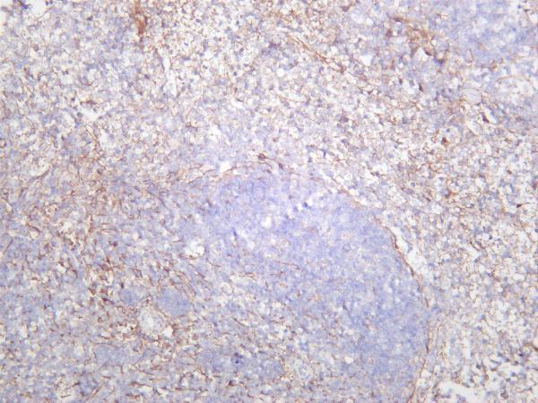
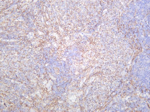

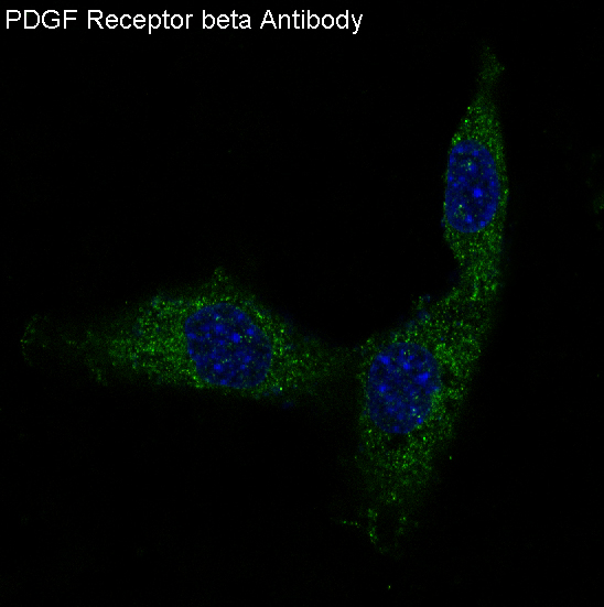


Comprehensive Human Protein Atlas IHC scoring per tissue (reliability: Enhanced — Medium consistency between antibody staining and RNA expression data.). Rows are taken directly from the HPA tissue chart — click any row's HPA link to view the source.
| Tissue | Cell type | Level | Evidence | Source |
|---|---|---|---|---|
| Testis | Peritubular cells | High | Protein (IHC) | HPA → |
| Breast | Myoepithelial cells | Medium | Protein (IHC) | HPA → |
| Endometrium | Cells in endometrial stroma | Medium | Protein (IHC) | HPA → |
| Kidney | Cells in glomeruli | Medium | Protein (IHC) | HPA → |
| Ovary | Ovarian stroma cells | Medium | Protein (IHC) | HPA → |
| Tissue | Cell type | Level | Evidence | Source |
|---|---|---|---|---|
| Adipose tissue | Adipocytes | Not detected | Protein (IHC) | HPA → |
| Adrenal gland | Glandular cells | Not detected | Protein (IHC) | HPA → |
| Appendix | Glandular cells | Not detected | Protein (IHC) | HPA → |
| Bone marrow | Hematopoietic cells | Not detected | Protein (IHC) | HPA → |
| Bronchus | Respiratory epithelial cells | Not detected | Protein (IHC) | HPA → |
Troubleshoot PDGFRB staining by checking retrieval, cellular location, controls, and scoring against the expected vascular and stromal pattern.
Two anti-PDGFRB antibodies support IHC in human, mouse, and rat samples (catalog: applications and reactivity); the catalog also provides an IF image from 3T3 cells (M00096-1: IF image caption).
M00096-6 has IHC images from mouse spleen, rat spleen, and human kidney, and lists IF among its applications (M00096-6: IHC image captions; catalog: applications). M00096-1 has an IHC image from paraffin-embedded human uterus and an IF image from 3T3 cells; its listed reactivity is human, mouse, and rat (M00096-1: image captions; catalog: reactivity).
Which to pick: For paraffin-section IHC, choose M00096-1 when the human uterus example is relevant; its caption reports paraffin embedding but does not report the fixative (M00096-1: IHC image caption). For IF/ICC, choose M00096-1 because ICC and IF are listed applications and it has a 3T3 IF image (M00096-1: applications; IF image caption). For IHC across species, consider M00096-6 because its own IHC images show mouse spleen, rat spleen, and human kidney; both SKUs are rabbit monoclonals with human, mouse, and rat listed as reactive species (M00096-6: IHC image captions; catalog: antibody titles and reactivity).