PDIA5 / Protein disulfide-isomerase A5 · IHC design guide

Design Immunohistochemistry for PDIA5

Plan PDIA5 paraffin IHC using the cytoplasmic tissue pattern and high staining in placental syncytiotrophoblasts (HPA tissue IHC). The guide pairs tissue controls with the catalog antibody’s IHC-P conditions (datasheet A09898).

Evidence assembled Oct 2026 · For research use; verify linked source records and product datasheet before use
Immunohistochemistry protocol sheet for PDIA5 (IHC for PDIA5): expected localisation Cytoplasmic staining in most tissues (HPA tissue IHC), antibody A09898, validated IHC image, and IHC protocol steps
Printable PDIA5 IHC protocol sheet — expected localisation Cytoplasmic staining in most tissues (HPA tissue IHC), antibody A09898, controls and protocol steps. Open the full PDIA5 IHC guide →

PDIA5 Immunohistochemistry Experimental Design Guide

Expected localisation, validated protocols, controls and antibodies — the at-a-glance facts below, then the full design guide.

Must know before staining
Expected localisation Cytoplasmic staining in most tissues (HPA tissue IHC)
Staining pattern Cytoplasmic signal in placental syncytiotrophoblasts (HPA tissue IHC)
Antigen retrieval EDTA pH 8.0 HIER, heat-mediated (datasheet A09898)
Positive control ⓘ Endometrium+4 more · see all
Negative control ⓘ Adipose tissue+4 more · see all
Important caveats
Reasons your staining may differ from the expected pattern.
Fixation Keep fixation conditions consistent across sections. (standard IHC practice; not target-specific)
Caveat Signal varies by cell type, from high to undetected (HPA tissue IHC)
Regulation Expression regulation is unannotated (UniProt)
Isoform / epitope 2 isoforms; signal peptide 1–21; epitope map unknown (UniProt)
Section 1

Recommended PDIA5 IHC & IF Protocols

Compare the catalog antibody’s IHC-P protocol (datasheet A09898) with a published human brain and glioma IHC protocol (PMC7921737).

Recommended immunohistochemistry (IHC-P) protocol parameters
SampleParaffin-embedded human breast cancer tissue; fixative not specified (datasheet A09898)
FixationImage fixative and duration unreported (datasheet A09898); verify before use.
Sectioning4–5 µm sections on charged slides (standard)
DeparaffinisationXylene, graded ethanol series to water (standard)
Antigen retrievalHeat retrieval: EDTA pH 8.0 (datasheet A09898); 20 min, 95–100 °C (standard)
Peroxidase block3% H2O2, 10 min, room temperature (standard)
Blocking10% goat serum (datasheet A09898)
Primary antibodyRabbit anti-PDIA5, 2-5μg/ml (datasheet A09898)
Primary incubationOvernight at 4 °C (datasheet A09898)
DetectionStreptavidin-biotin complex (SABC), DAB chromogen (datasheet A09898)
CounterstainHematoxylin, blue, dehydrate and mount (standard)
Expected resultPDIA5-positive staining in glandular cells of endometrium (HPA tissue IHC: Medium). HPA tissue profile: Cytoplasmic expression in most tissues, most abundant in placenta and lymphoid cells. No signal in the no-primary control.
💡Decision noteStart with heat-mediated EDTA pH 8.0 retrieval (datasheet A09898); the published glioma protocol reports retrieval without specifying its conditions (PMC7921737).
Section 2

What Is the Expected PDIA5 Staining Pattern?

PDIA5 resides in the endoplasmic reticulum lumen and lacks a transmembrane segment (UniProt Q14554 topology). In paraffin sections, expect cytoplasmic staining in reported positive cells, including placental syncytiotrophoblasts and pancreatic exocrine glandular cells, both rated High (HPA tissue IHC). HPA describes cytoplasmic expression in most tissues; its IHC reliability is Enhanced, with external verification pending (HPA tissue IHC).

What am I looking at on my slide?
Cytoplasmic signal in placental syncytiotrophoblasts or pancreatic exocrine glandular cells (HPA: High).This matches the reported cell types and an ER-lumen protein (HPA tissue IHC; UniProt Q14554 topology). Judge signal against neighboring cells and controls; High is an HPA category, not a required intensity for every section (HPA tissue IHC; standard IHC practice).
Predominantly nuclear, membrane-rim, or extracellular staining in otherwise positive cells (UniProt Q14554 topology).That compartment conflicts with PDIA5's ER-lumen location and HPA's cytoplasmic IHC profile (UniProt Q14554 topology; HPA tissue IHC). Review staining specificity and morphology before scoring it as PDIA5 (standard IHC practice).
Strong staining in adipocytes or cardiomyocytes (HPA: Not detected in those cell types).This disagrees with the supplied tissue observations; cross-reactivity or detection-system activity are possible explanations (HPA tissue IHC; standard IHC practice). Compare the suspect cells with a reported positive tissue and appropriate detection controls (standard IHC practice).
Color spreads across tissue and empty areas, obscuring cell boundaries (standard IHC practice).Diffuse background cannot establish the cytoplasmic, cell-specific pattern reported by HPA (HPA tissue IHC). Check blocking, wash steps, antibody concentration, and detection-only controls as general IHC troubleshooting steps (standard IHC practice).
No signal in a section containing placental syncytiotrophoblasts (HPA: High).Check tissue preservation, retrieval, antibody and detection steps before calling the sample PDIA5-negative (standard IHC practice). HPA's High rating supports a positive-control choice, but does not guarantee signal under every assay condition (HPA tissue IHC).
💡Expected PDIA5 appearanceCall a convincing positive when cytoplasmic staining is clear in HPA High cells such as placental syncytiotrophoblasts; isolated nuclear or widespread cell-free color is suspect (HPA tissue IHC; UniProt Q14554 topology; standard IHC practice).
How each factor affects the staining
Compartment and processing (UniProt Q14554 topology).PDIA5 has an ER-lumen annotation, no transmembrane segment, and a signal peptide at residues 1–21; the annotated chain begins at residue 22 (UniProt Q14554). Interpret cytoplasmic IHC in that context without assuming a particular antibody epitope (HPA tissue IHC; UniProt Q14554).
Cell selection for controls (HPA tissue IHC).Placental syncytiotrophoblasts and pancreatic exocrine glandular cells are rated High; adipocytes and cardiomyocytes are Not detected (HPA tissue IHC). Match the scored cell type, since one tissue section can contain multiple cell populations (standard IHC practice).
Antibody evidence (HPA antibodies; HPA tissue IHC).Two listed antibodies, HPA030353 and HPA030355, have Enhanced IHC status (HPA antibodies). The tissue profile carries the same reliability label, while external verification remains pending (HPA tissue IHC); use these observations as expectations, not a guarantee for a different antibody.
IF/ICC comparison (HPA subcellular; HPA antibodies).For the separate IF/ICC guide, HPA approves an ER location and lists HPA030353 as ICC Approved; HPA030355 has no ICC status supplied (HPA subcellular; HPA antibodies). These IF/ICC observations support compartment interpretation, without specifying an IHC retrieval or detection condition.
Why is my staining missing, weak or wrong?
SituationLikely causeNext action
Reported High cells stain weakly or remain blank (HPA tissue IHC).The IHC workflow or antibody may have failed; a target-specific fixation effect is unreported in the supplied sources (standard IHC practice; UniProt Q14554; HPA tissue IHC).Verify section and control quality, then check retrieval, primary-antibody and detection steps using validated assay instructions (standard IHC practice). Avoid inferring PDIA5-specific retrieval conditions from HPA staining levels.
Nuclei dominate the chromogenic signal (UniProt Q14554 topology).A dominant nuclear pattern conflicts with the ER-lumen annotation and cytoplasmic HPA profile (UniProt Q14554 topology; HPA tissue IHC).Inspect counterstain and detection-only controls; reassess antibody specificity before scoring nuclear color as PDIA5 (standard IHC practice).
Color appears broadly outside recognizable cells (standard IHC practice).Background or precipitated chromogen can obscure the reported cytoplasmic pattern (standard IHC practice; HPA tissue IHC).Review reagent preparation, washes, blocking and development time; repeat with a detection-only control (standard IHC practice).
An HPA Not detected cell type stains strongly (HPA tissue IHC).Cross-reactivity or endogenous detection activity is possible; the HPA category describes its reported observation, not universal absence (HPA tissue IHC; standard IHC practice).Compare with a reported High cell type and run controls appropriate to the chromogenic detection system (HPA tissue IHC; standard IHC practice).
Placenta and another positive tissue show different intensity (HPA tissue IHC).HPA ratings describe specific cell populations; tissue composition and assay conditions can also affect the visual comparison (HPA tissue IHC; standard IHC practice).Score the named cell populations separately and compare sections processed in the same run (HPA tissue IHC; standard IHC practice).
An IF/ICC image seems inconsistent with the IHC slide (HPA subcellular; HPA tissue IHC).HPA's approved IF/ICC ER location and its tissue IHC cytoplasmic profile report different readouts of PDIA5 localization (HPA subcellular; HPA tissue IHC).Compare compartment and identified cell type first; evaluate each assay with its own controls and validation status (HPA antibodies; standard IHC/IF practice).

Sample controls for PDIA5 IHC & IF

🧪Run gallbladder first: its glandular cells should stain (HPA: High in gallbladder glandular cells). Use adipose tissue as the negative comparator (HPA: Not detected in adipocytes); on the gallbladder slide, assess morphologically identifiable stromal cells as an internal background reference without assuming they lack PDIA5.
Positive control tissue: Endometrium (Glandular cells, HPA Medium)
Negative control tissue: Adipose tissue (HPA Not detected)
ICC-IF cell lines (HPA subcellular resource): HPA ICC-IF images show PDIA5 in A-431, BJ [Human fibroblast], HUVEC/TERT2, with annotated localisation: Endoplasmic reticulum (approved) (HPA subcellular).
Technical controls: Include a no-primary (secondary-only) control, a concentration-matched rabbit IgG isotype control (caption: rabbit primary antibody), and PDIA5-knockout material or a cognate-peptide competition control if available. Quench endogenous peroxidase and block endogenous biotin when using the caption’s biotin-based SABC/DAB detection (caption: biotinylated secondary, SABC and DAB); gallbladder bile pigment may complicate interpretation of brown staining.
⚠️Feasibility: No target-specific fixation window or fixation effect is reported, and the selected A09898 paraffin-section caption does not state a fixative (caption: fixative unreported). The caption uses heat-mediated EDTA retrieval at pH 8.0 and 2 μg/ml primary antibody overnight at 4°C; it does not establish that retrieval is required (caption: IHC conditions). The supplied evidence does not establish frozen sections or IF as easier, although HPA reports endoplasmic-reticulum localization in ICC-IF (HPA: approved endoplasmic-reticulum location).

HPA tissue IHC evidence for PDIA5

Comprehensive Human Protein Atlas IHC scoring per tissue (reliability: Enhanced — Pending external verification.). Rows are taken directly from the HPA tissue chart — click any row's HPA link to view the source.

Positive expression · recommended positive controls

TissueCell typeLevelEvidenceSource
Endometrium Glandular cells Medium Protein (IHC) HPA →
Epididymis Glandular cells High Protein (IHC) HPA →
Fallopian tube Non-ciliated cells High Protein (IHC) HPA →
Gallbladder Glandular cells High Protein (IHC) HPA →
Kidney Cells in glomeruli High Protein (IHC) HPA →

Undetected expression · recommended negative controls

TissueCell typeLevelEvidenceSource
Adipose tissue Adipocytes Not detected Protein (IHC) HPA →
Cerebellum Cells in granular layer Not detected Protein (IHC) HPA →
Esophagus Squamous epithelial cells Not detected Protein (IHC) HPA →
Heart muscle Cardiomyocytes Not detected Protein (IHC) HPA →
Hippocampus Glial cells Not detected Protein (IHC) HPA →
Section 3

Advanced PDIA5 IHC Tips

These questions address PDIA5 staining in paraffin sections with chromogenic IHC, with one entry for the separate IF/ICC application.

How should I troubleshoot weak PDIA5 staining after antigen retrieval?
Start with heat mediated EDTA retrieval at pH 8.0 for paraffin sections stained with A09898 (datasheet A09898). The selected tissue image used this retrieval before incubation with 2 μg/ml primary antibody overnight at 4°C, providing a documented starting condition (A09898 tissue-IHC caption). If staining is weak, check that sections remained covered during heating and cooled consistently before changing the retrieval condition (standard IHC practice). Compare changes on matched sections alongside a documented positive tissue, such as placenta, where syncytiotrophoblast staining is high (HPA: placenta). Excessive retrieval can damage morphology and complicate interpretation, so assess tissue integrity with every adjustment (standard IHC practice).
Could fixation explain inconsistent PDIA5 staining between paraffin blocks?
PDIA5-specific fixation sensitivity is unknown because the selected paraffin-section caption does not report its fixative (A09898 tissue-IHC caption). Record each block’s fixative, fixation duration, and processing history before attributing differences to PDIA5 biology (standard IHC practice). Compare sections processed together using the documented EDTA retrieval at pH 8.0 and 2 μg/ml primary antibody overnight at 4°C (A09898 tissue-IHC caption). Include tissue with expected staining, such as high expression in placental syncytiotrophoblasts, to monitor run performance (HPA: placenta). Evaluate morphology and staining together; differences across blocks alone do not establish a target-specific fixation effect (standard IHC interpretation).
What PDIA5 staining pattern should I expect in tissue sections?
Expect a cytoplasmic staining pattern in positive cells, consistent with PDIA5 residing in the endoplasmic reticulum lumen (HPA: tissue profile; UniProt Q14554 localisation). Its sequence has a signal peptide at residues 1–21 and no transmembrane segment, supporting a luminal protein assignment (UniProt Q14554 processing and topology). Tissue IHC cannot by itself resolve an endoplasmic reticulum network, so compare distribution with morphology and appropriate controls (standard IHC practice). High staining is reported in placental syncytiotrophoblasts and pancreatic exocrine glandular cells, providing defined comparison populations (HPA: tissue IHC). Predominantly nuclear or extracellular staining warrants review of controls and section quality before a biological interpretation (UniProt Q14554 localisation; standard IHC practice).
Can this IHC stain distinguish PDIA5 isoforms or a masked epitope?
Do not assign staining to a particular isoform without antibody epitope and isoform-specific validation: PDIA5 has 2 annotated isoforms (UniProt Q14554 isoforms; standard IHC interpretation). The mature chain spans residues 22–519, following a signal peptide at residues 1–21 (UniProt Q14554 processing). Three thioredoxin domains occupy residues 134–261, 270–384, and 378–506, but these coordinates do not identify the catalog antibody’s binding site (UniProt Q14554 domains; A09898 tissue-IHC caption). If staining changes with retrieval, test matched sections under the documented EDTA pH 8.0 condition and examine tissue morphology (datasheet A09898; standard IHC practice). Retrieval dependence alone cannot establish which isoform or epitope was detected (standard IHC interpretation).
How should I assess PDIA5 by multiplex IF/ICC?
For IF/ICC, pair PDIA5 with a marker identifying the expected cell population, such as a validated syncytiotrophoblast marker when examining placenta (HPA: high in placental syncytiotrophoblasts; standard IF practice). Choose fluorophores after inspecting unstained tissue autofluorescence, and place the weaker signal in a channel with clearer separation (standard IF practice). PDIA5 is assigned to the endoplasmic reticulum lumen, so permeabilisation must allow antibody access across intracellular membranes if the recognized epitope is luminal (UniProt Q14554 localisation; standard IF practice). The catalog antibody’s epitope location and IF/ICC fixation response are not supplied here, so optimize permeabilisation and fixation with controls (A09898 tissue-IHC caption; standard IF practice). Compare the pattern with an endoplasmic reticulum marker and include single-stain controls when assessing colocalisation (HPA: endoplasmic reticulum; standard IF practice).
How can I reduce diffuse brown background without losing PDIA5 signal?
First inspect a no-primary control and confirm that brown deposit follows cells rather than section edges or damaged areas (standard IHC practice). The selected protocol blocked with 10% goat serum and used 2 μg/ml primary antibody overnight at 4°C (A09898 tissue-IHC caption). It then used a biotinylated secondary, a streptavidin–biotin complex, and DAB, so secondary-only and detection controls can help locate background (A09898 tissue-IHC caption; standard IHC practice). Apply a peroxidase block and assess endogenous biotin interference where relevant; these are general chromogenic workflow checks, not PDIA5-specific findings (standard IHC practice). Review wash quality and DAB development alongside a positive control before lowering primary concentration (standard IHC practice).
How should I score PDIA5 staining across tissue specimens? ⚠ ANSWER MARKED FOR VERIFICATION
Define the cell population and staining compartment before scoring; PDIA5 is assigned to the endoplasmic reticulum lumen and appears cytoplasmic in tissue IHC (UniProt Q14554 localisation; HPA: tissue profile). For each specimen, record the percentage of positive target cells and intensity, then calculate an H-score using consistent intensity categories (standard IHC practice). Alternatively, quantify positive cells per mm² of viable annotated tissue when cell counts are the outcome (standard IHC practice). Normalize scores to the number of evaluable target cells or viable tissue area, and apply the same threshold across runs (standard IHC practice). Document staining batch and control performance because changes in retrieval or DAB development can shift measured intensity (standard IHC practice).
How do I distinguish genuine PDIA5 staining from IHC artefacts?
Favor staining in intact cytoplasm of expected cells: HPA reports high PDIA5 in placental syncytiotrophoblasts and pancreatic exocrine glandular cells (HPA: tissue IHC; HPA: tissue profile). Strong isolated nuclear or extracellular deposits conflict with the assigned endoplasmic reticulum lumen localisation and need control review (UniProt Q14554 localisation; standard IHC practice). Compare suspicious signal with section edges, necrotic regions, and the no-primary control before calling cells positive (standard IHC practice). Endogenous peroxidase or detection-system background can produce DAB signal independently of primary-antibody binding (standard IHC practice). HPA labels its tissue-IHC reliability “Enhanced” while external verification is pending, so treat agreement with its pattern as supporting evidence rather than proof of antibody specificity (HPA: tissue IHC reliability).
Boster reagents

Best PDIA5 / Protein disulfide-isomerase A5 IHC Antibodies

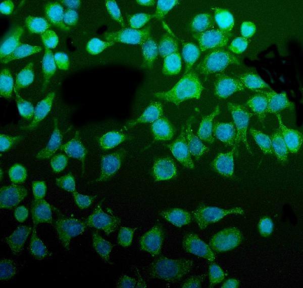
The catalog antibody has IHC images from human paraffin tissue sections and an IF image from CACO-2 cells; its listed reactivity includes human, mouse and rat (catalog: A09898 applications and reactivity; image captions).

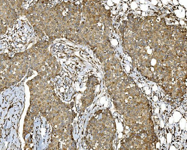
Real IHC data IHC analysis of PDIA5 using anti-PDIA5 antibody (A09898). PDIA5 was detected in paraffin-embedded section of human breast cancer tissue. Heat mediated antigen retrieval was performed in EDTA buffer (pH8.0, epitope retrieval solution). The tissue section was blocked with 10% goat serum. The tissue section was then incubated with 2μg/ml rabbit anti-PDIA5 Antibody (A09898) overnight at 4°C. Biotinylated goat anti-rabbit IgG was used as secondary antibody and incubated for 30 minutes at 37°C. The tissue section was developed using Strepavidin-Biotin-Complex (SABC) (Catalog # SA1022) with DAB as the chromogen.
Anti-PDIA5 Antibody ®
Cat # A09898

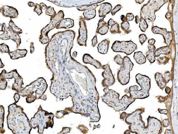
A09898 has IHC images from human breast cancer, placenta, gallbladder adenocarcinoma and rectal cancer paraffin sections (A09898 IHC image captions). It also has a CACO-2 cell IF image and is listed for IHC, ICC and IF (A09898 IF image caption; catalog: applications).

Which to pick: Choose A09898 for paraffin-section IHC: its tissue captions use EDTA retrieval at pH 8.0 and 2 μg/ml antibody; the fixative is unreported (A09898 IHC image captions). Choose the same SKU for IF/ICC based on its CACO-2 cell IF image at 5 μg/ml (A09898 IF image caption). For mouse or rat samples, the catalog lists reactivity and IHC use, while the supplied IHC images show human tissue only; clonality is unreported (catalog: A09898 reactivity and applications; A09898 IHC image captions).

Each figure is that product's own IHC / IF validation image from its datasheet.