PDK3 / [Pyruvate dehydrogenase (acetyl-transferring)] kinase isozyme 3, mitochondrial · IHC design guide

Design Immunohistochemistry for PDK3

This guide covers PDK3 staining in paraffin sections using the catalog antibody's IHC conditions (datasheet A05658-1). Compare stained hepatocytes or lung macrophages with adipocytes reported as not detected, and interpret results cautiously because staining and RNA data show low consistency (HPA tissue IHC).

Evidence assembled Oct 2026 · For research use; verify linked source records and product datasheet before use
Immunohistochemistry protocol sheet for PDK3 (IHC for PDK3): expected localisation Cytoplasmic tissue staining (HPA tissue IHC); mitochondrial matrix annotation (UniProt), antibody A05658-1, validated IHC image, and IHC protocol steps
Printable PDK3 IHC protocol sheet — expected localisation Cytoplasmic tissue staining (HPA tissue IHC); mitochondrial matrix annotation (UniProt), antibody A05658-1, controls and protocol steps. Open the full PDK3 IHC guide →

PDK3 Immunohistochemistry Experimental Design Guide

Expected localisation, validated protocols, controls and antibodies — the at-a-glance facts below, then the full design guide.

Must know before staining
Expected localisation Cytoplasmic tissue staining (HPA tissue IHC); mitochondrial matrix annotation (UniProt)
Staining pattern Cytoplasmic staining in lung macrophages (HPA tissue IHC)
Antigen retrieval EDTA pH 8.0 HIER, heat-mediated (datasheet A05658-1)
Positive control ⓘ Cerebral cortex+4 more · see all
Negative control ⓘ Adipose tissue+4 more · see all
Important caveats
Reasons your staining may differ from the expected pattern.
Fixation Keep fixation consistent across sections (standard IHC practice; not target-specific)
Caveat Staining shows low consistency with RNA data (HPA tissue IHC)
Regulation Expression regulation not specified (UniProt)
Isoform / epitope 2 isoforms; epitope coverage unspecified (UniProt)
Section 1

Recommended PDK3 IHC & IF Protocols

The catalog antibody has a datasheet IHC-P protocol (datasheet: A05658-1); the published options below report PDK3 staining in urothelial and prostate cancer samples (PMC8473833; PMC9174590).

Recommended immunohistochemistry (IHC-P) protocol parameters
SampleParaffin-embedded human meningioma tissue; fixative not specified (datasheet A05658-1)
FixationImage fixative and duration unreported (datasheet A05658-1); verify before use.
Sectioning4–5 µm sections on charged slides (standard)
DeparaffinisationXylene, graded ethanol series to water (standard)
Antigen retrievalHeat retrieval: EDTA pH 8.0 (datasheet A05658-1); 20 min, 95–100 °C (standard)
Peroxidase block3% H2O2, 10 min, room temperature (standard)
Blocking10% goat serum (datasheet A05658-1)
Primary antibodyRabbit anti-PDK3, 2-5 μg/ml (datasheet A05658-1)
Primary incubationOvernight at 4 °C (datasheet A05658-1)
DetectionHRP-conjugated secondary, DAB chromogen (datasheet A05658-1)
CounterstainHematoxylin, blue, dehydrate and mount (standard)
Expected resultPDK3-positive staining in neuronal cells of cerebral cortex (HPA tissue IHC: Medium). HPA tissue profile: Cytoplasmic expression in several tissues. No signal in the no-primary control.
💡Decision noteStart with heat-mediated EDTA retrieval at pH 8.0 for the catalog antibody (datasheet: A05658-1). The prostate study used pH 6 and pH 9 retrieval without assigning either to PDK3 (PMC9174590).
Section 2

What Is the Expected PDK3 Staining Pattern?

PDK3 is a mitochondrial matrix protein with no transmembrane segment (UniProt Q15120 topology). In paraffin IHC, expect cytoplasmic staining in selected cells, including cortical and hippocampal neurons, hepatocytes, and lung macrophages (HPA tissue IHC: Medium). HPA rates tissue staining Approved but reports low agreement with RNA expression and pending external verification (HPA tissue IHC: reliability). Interpret individual tissues with that limitation in mind.

What am I looking at on my slide?
Cytoplasmic staining in cortical or hippocampal neurons, hepatocytes, or lung macrophages (HPA tissue IHC: Medium).This matches the reported cell distribution (HPA tissue IHC). A chromogenic cytoplasmic signal is compatible with mitochondrial PDK3, but ordinary IHC cannot by itself resolve the mitochondrial matrix (UniProt Q15120: matrix; general IHC practice).
Predominantly nuclear or membrane staining, with little cytoplasmic signal.Recheck specificity and detection controls: these are not the expected dominant IHC compartments (UniProt Q15120: mitochondrial matrix; HPA tissue IHC: cytoplasmic). HPA also reports uncertain nucleolar localization by ICC-IF, so isolated nucleolar signal is not proof of either specificity or artefact (HPA subcellular).
Strong signal in a cell population reported as undetected, such as adipocytes or cardiomyocytes (HPA tissue IHC: Not detected).Consider cross-reactivity or endogenous detection activity and compare controls (general IHC practice). HPA's low staining–RNA agreement limits how confidently an apparent mismatch can be called false (HPA tissue IHC: reliability); UniProt reports expression in heart (UniProt Q15120: tissue specificity).
Diffuse staining across cells and tissue spaces, with poor separation between cells.Treat this as background until controls clarify it; excessive antibody or incomplete blocking can produce nonspecific staining (general IHC practice). A broad haze does not establish the cell-specific cytoplasmic pattern reported by HPA (HPA tissue IHC: profile).
No signal in a matched liver or cerebral cortex section, including the expected cell population (HPA tissue IHC: Medium).Check whether the assay detects its positive control before interpreting an experimental negative (general IHC practice). HPA reports medium staining in hepatocytes or cortical neurons, but its Approved rating has low agreement with RNA data (HPA tissue IHC: reliability).
💡Expected PDK3 appearanceA convincing positive shows discernible cytoplasmic staining in the relevant cells at the reported Medium level—for example, hepatocytes or cortical neurons—while dominant nuclear, membrane, or tissue-wide haze warrants control review (HPA tissue IHC: Medium, cytoplasmic; UniProt Q15120: mitochondrial matrix; general IHC practice).
How each factor affects the staining
Compartment and topology (UniProt Q15120: mitochondrial matrix; no transmembrane segment).A cytoplasmic IHC pattern is biologically plausible; matrix-level localization requires finer-resolution evidence than routine chromogenic sections provide (HPA tissue IHC: cytoplasmic; general IHC practice). Do not score membrane staining as the expected pattern (UniProt Q15120 topology).
Tissue choice and evidence strength (HPA tissue IHC: reliability Approved).HPA reports Medium staining in neuronal cells, hepatocytes, macrophages, and several glandular populations, but low staining–RNA agreement and pending external verification limit extrapolation (HPA tissue IHC). Its Not detected entries describe observed IHC results, not proven protein absence (HPA tissue IHC).
Antibody evidence (HPA antibodies: HPA046583 IHC Approved; HPA072492 ICC Supported).The supplied validation applies to different antibodies and applications (HPA antibodies). It does not establish that an ICC-IF pattern will be reproduced by the IHC antibody, or that either antibody has IHC Enhanced validation (HPA antibodies).
IF/ICC Q: Where should PDK3 appear? A: Mainly in mitochondria (HPA subcellular: supported).HPA additionally lists nucleoli with uncertain support and images from A-431, MCF-7, and RT-4 (HPA subcellular). Treat that nucleolar observation cautiously; this IHC section supplies no IF/ICC protocol option (HPA subcellular).
Target-specific fixation effects are not established by the supplied assay evidence. Verify with a matched IHC source before attributing a result to fixation.No target-specific fixation effect is reported in the supplied HPA or UniProt evidence. Choose and document retrieval conditions empirically for the IHC assay (general IHC practice); do not infer a PDK3-specific retrieval requirement from staining level or topology (HPA tissue IHC; UniProt Q15120).
Why is my staining missing, weak or wrong?
SituationLikely causeNext action
Expected hepatocyte or cortical neuronal signal is absent (HPA tissue IHC: Medium).Assay detection, primary-antibody concentration, or retrieval conditions may need optimization (general IHC practice); the supplied sources give no PDK3-specific cause.Run a matched reported-positive section with detection controls; titrate the IHC-validated antibody and compare documented retrieval conditions (HPA tissue IHC: Medium; general IHC practice).
Signal remains in a no-primary control.Endogenous detection activity or nonspecific secondary/detection chemistry can produce signal (general IHC practice).Address the detection system's endogenous activity and blocking steps, then repeat the control beside the test section (general IHC practice).
Haze obscures cytoplasmic cell boundaries.Excess primary reagent, inadequate blocking, or incomplete washing can raise background (general IHC practice).Titrate the IHC-validated antibody and optimize blocking and washes; retain the reported-positive tissue for comparison (HPA tissue IHC: Medium; general IHC practice).
Dominant membrane or nuclear staining appears.A compartment mismatch raises concern about nonspecific staining; nucleolar localization remains uncertain by ICC-IF (UniProt Q15120 topology; HPA subcellular).Compare no-primary and reported-positive sections before scoring; require a discernible cytoplasmic component consistent with tissue IHC (HPA tissue IHC: cytoplasmic; general IHC practice).
Cardiomyocytes stain despite HPA's Not detected IHC result (HPA tissue IHC).Cross-reactivity is possible, but UniProt reports heart expression and HPA reports low staining–RNA agreement (general IHC practice; UniProt Q15120; HPA tissue IHC).Check controls and repeat with independent specificity evidence before calling either the staining or HPA observation definitive (general IHC practice; HPA tissue IHC: reliability).
Cells outside the expected population stain strongly within a reported-positive tissue (HPA tissue IHC: cell-specific entries).Cross-reactivity or endogenous detection activity may mimic a new cell distribution (general IHC practice).Compare morphology and counterstain, review no-primary controls, and report the departure from HPA's specified cell pattern rather than assigning it to PDK3 alone (HPA tissue IHC; general IHC practice).

Sample controls for PDK3 IHC & IF

🧪Run cerebral cortex first: neuronal cells should show PDK3 staining at the HPA-reported Medium level (HPA: cerebral cortex, neuronal cells, Medium). Use cerebellum as a negative tissue, assessing its granular-layer cells, which HPA lists as Not detected; on the positive slide, use morphologically non-neuronal cells without signal above background as internal negative cells, while avoiding an assumption that all non-neuronal cells lack PDK3 (HPA: cerebellum, granular layer, Not detected; HPA: cerebral cortex, neuronal cells, Medium).
Positive control tissue: Cerebral cortex (Neuronal cells, HPA Medium)
Negative control tissue: Adipose tissue (HPA Not detected)
ICC-IF cell lines (HPA subcellular resource): HPA ICC-IF images show PDK3 in A-431, MCF-7, RT-4, with annotated localisation: Mitochondria (supported) (HPA subcellular).
Technical controls: Include a no-primary, secondary-only slide and a concentration-matched rabbit IgG isotype control for the rabbit primary (selected-SKU caption: rabbit anti-PDK3); use PDK3 knockout material, if available, as a biological specificity control (standard IHC practice). Block endogenous peroxidase for chromogenic detection and assess neuronal lipofuscin autofluorescence if imaging cerebral cortex by IF (selected-SKU caption: peroxidase/DAB detection; standard IHC/IF practice).
⚠️Feasibility: A target-specific fixation window or fixation effect is unreported in the supplied evidence, and the selected-SKU paraffin-section caption does not state a fixative (selected-SKU caption: fixative unreported). The illustrated IHC procedure uses heat retrieval in EDTA at pH 8.0, followed by 2 μg/ml primary antibody overnight at 4°C; this establishes a demonstrated retrieval condition, not a requirement for every specimen (selected-SKU caption). The supplied evidence does not establish whether frozen sections or IF are easier than paraffin IHC; neuronal lipofuscin autofluorescence can complicate cerebral-cortex IF interpretation (selected-SKU caption: paraffin IHC; HPA: cerebral cortex, neuronal cells; standard IF practice).

HPA tissue IHC evidence for PDK3

Comprehensive Human Protein Atlas IHC scoring per tissue (reliability: Approved — Low consistency between antibody staining and RNA expression data. Pending external verification.). Rows are taken directly from the HPA tissue chart — click any row's HPA link to view the source.

Positive expression · recommended positive controls

TissueCell typeLevelEvidenceSource
Cerebral cortex Neuronal cells Medium Protein (IHC) HPA →
Duodenum Glandular cells Medium Protein (IHC) HPA →
Endometrium Glandular cells Medium Protein (IHC) HPA →
Hippocampus Neuronal cells Medium Protein (IHC) HPA →
Liver Hepatocytes Medium Protein (IHC) HPA →

Undetected expression · recommended negative controls

TissueCell typeLevelEvidenceSource
Adipose tissue Adipocytes Not detected Protein (IHC) HPA →
Adrenal gland Glandular cells Not detected Protein (IHC) HPA →
Bronchus Basal cells Not detected Protein (IHC) HPA →
Cerebellum Cells in granular layer Not detected Protein (IHC) HPA →
Colon Endothelial cells Not detected Protein (IHC) HPA →
Section 3

Advanced PDK3 IHC Tips

Troubleshoot chromogenic PDK3 staining by checking retrieval, cellular context, and mitochondrial localisation; the catalog antibody has a paraffin-section IHC example (datasheet A05658-1).

How should I retrieve PDK3 when paraffin sections stain weakly?
Start with heat-mediated antigen retrieval in EDTA at pH 8.0 (datasheet A05658-1). The selected paraffin-section example used this retrieval before 10% goat serum blocking and 2 μg/ml primary antibody overnight at 4°C (datasheet A05658-1). If staining remains weak, compare a second retrieval condition on matched sections while keeping antibody concentration, detection, and development constant; assess tissue preservation alongside signal (standard IHC practice). Include a known positive tissue and a no-primary control, since weak staining alone cannot distinguish masked antigen from low expression or detection failure (standard IHC practice).
Could fixation explain weak or uneven PDK3 staining?
Target-specific fixation sensitivity is unknown: the selected paraffin-section caption does not state its fixative (datasheet A05658-1). Record each specimen’s fixative and fixation duration, then compare similarly processed sections with the same pH 8.0 EDTA retrieval and 2 μg/ml primary antibody conditions used in the example (datasheet A05658-1; standard IHC practice). Examine morphology and staining together, because uneven processing can produce regional variation that confounds interpretation (standard IHC practice). Do not attribute a weak result to a particular fixation effect without a controlled comparison; include a positive tissue and no-primary section in the same run (standard IHC practice).
What staining pattern is plausible for PDK3 in tissue sections?
PDK3 is assigned to the mitochondrial matrix and has no transmembrane segment (UniProt Q15120 topology). Expect signal within cell bodies in a cytoplasmic distribution compatible with mitochondria; HPA reports cytoplasmic expression across several tissues and a supported mitochondrial location in cell imaging (HPA tissue IHC; HPA subcellular). Evaluate cellular distribution at high magnification rather than treating diffuse background or extracellular deposits as evidence of PDK3 (standard IHC practice). Nucleolar localisation is listed as uncertain, so isolated nuclear or nucleolar chromogenic staining needs independent confirmation before it is scored as target signal (HPA subcellular; standard IHC practice).
Can the antibody distinguish PDK3 isoforms or a modified epitope?
PDK3 has 2 listed isoforms, and the supplied record identifies a histidine kinase domain at residues 131–362 and an N6-succinyllysine example at residue 278 (UniProt Q15120). The selected staining caption does not define the antibody’s epitope or establish isoform selectivity (datasheet A05658-1). Therefore, interpret positive chromogenic staining as antibody-reactive PDK3 signal without assigning it to one isoform or modification state (standard IHC interpretation). If that distinction matters, obtain epitope mapping and validate specificity with an independent perturbation or orthogonal assay under the same tissue-processing conditions (standard IHC practice).
How should I design an IF check alongside chromogenic PDK3 IHC?
Treat IF as a separate validation experiment: the selected antibody example establishes paraffin-section chromogenic IHC, while HPA reports mitochondrial localisation from cell imaging (datasheet A05658-1; HPA subcellular). For lung, multiplex PDK3 with a macrophage marker to assess the cell population reported as medium positive, and add a mitochondrial marker to inspect compartmental overlap (HPA tissue IHC; HPA subcellular). Choose fluorophores after examining unstained tissue autofluorescence, reserving a cleaner channel for the weaker signal (standard IF practice). Because PDK3 is assigned to the mitochondrial matrix, titrate permeabilisation to permit access across mitochondrial membranes while preserving morphology; include single-stain and no-primary controls (UniProt Q15120 topology; standard IF practice).
How can I reduce diffuse or punctate DAB background?
First compare the stained section with a no-primary control and inspect whether precipitate follows tissue edges, folds, or damaged areas (standard IHC practice). The selected example used 10% goat serum blocking, 2 μg/ml primary antibody overnight at 4°C, and a peroxidase-linked secondary for 30 minutes at 37°C (datasheet A05658-1). Add an endogenous peroxidase block before DAB development, then adjust washing, antibody concentration, or development time one variable at a time (standard chromogenic IHC practice). Reassess whether remaining cellular signal fits a cytoplasmic, mitochondria-compatible pattern rather than using overall brown intensity alone (HPA tissue IHC; HPA subcellular; standard IHC practice).
How should I score PDK3 staining across specimens? ⚠ ANSWER MARKED FOR VERIFICATION
Define the cell population and tissue compartment before scoring, then report the percentage of positive cells and an H-score calculated as 0–300 from intensity categories weighted by their positive-cell percentages (standard IHC practice). Record the number of evaluable cells or tissue area, and normalise counts or positive-cell density to evaluable cells or mm², respectively (standard IHC practice). Exclude necrosis, folds, and edge artefacts using the same rules across sections, and keep retrieval and DAB development consistent within a comparison (standard IHC practice). Because HPA notes low consistency between staining and RNA expression, interpret differences alongside controls and replicate sections (HPA tissue IHC; standard IHC practice).
When is a positive PDK3 IHC result convincing?
A convincing result has cellular staining compatible with cytoplasmic mitochondrial PDK3, appropriate negative-control behaviour, and reproducibility across comparable sections (UniProt Q15120 topology; HPA subcellular; standard IHC practice). Medium staining in lung macrophages or cerebral-cortex neurons is consistent with reported HPA observations, whereas a claim based solely on heart cardiomyocytes warrants caution because that HPA entry is not detected (HPA tissue IHC). Question isolated extracellular or strongly nuclear deposits, tissue-edge enrichment, necrotic staining, and DAB signal persisting without primary antibody (HPA subcellular; standard IHC practice). HPA describes low consistency between antibody staining and RNA expression and pending external verification, so resolve consequential findings with an independent antibody or orthogonal assay (HPA tissue IHC; standard IHC practice).
Boster reagents

Best PDK3 / [Pyruvate dehydrogenase (acetyl-transferring)] kinase isozyme 3, mitochondrial IHC Antibodies

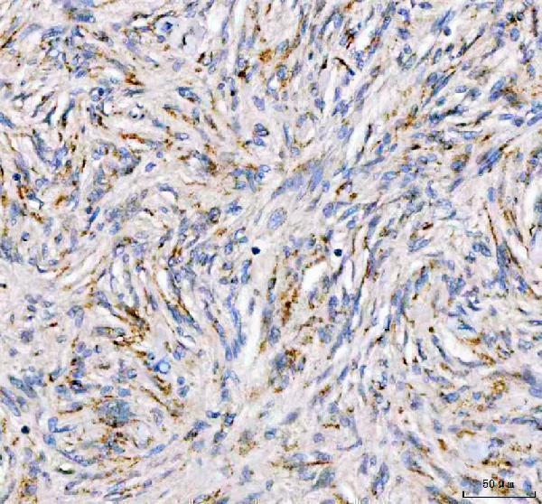
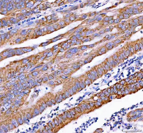
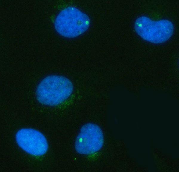
The IHC-validated anti-PDK3 antibody has paraffin-section images from human meningioma and rectum adenocarcinoma, plus mouse and rat testis; IF data come from HeLa cells (catalog image captions).

Real IHC data IHC analysis of PDK3 using anti-PDK3 antibody (A05658-1). PDK3 was detected in a paraffin-embedded section of human meningioma tissue. Heat mediated antigen retrieval was performed in EDTA buffer (pH 8.0, epitope retrieval solution). The tissue section was blocked with 10% goat serum. The tissue section was then incubated with 2 μg/ml rabbit anti-PDK3 Antibody (A05658-1) overnight at 4°C. Peroxidase Conjugated Goat Anti-rabbit IgG was used as secondary antibody and incubated for 30 minutes at 37°C. The tissue section was developed using HRP Conjugated Rabbit IgG Super Vision Assay Kit (Catalog # SV0002) with DAB as the chromogen.
Anti-PDK3 Antibody ®
Cat # A05658-1

A05658-1 is listed for IHC and shows PDK3 staining in paraffin sections of human meningioma and rectum adenocarcinoma, mouse testis, and rat testis (catalog applications; A05658-1 IHC captions). A05658-1 is also listed for IF/ICC, with an IF image from HeLa cells (catalog applications; A05658-1 IF caption).

Which to pick: Choose A05658-1 for paraffin-section tissue IHC: its own captions document EDTA retrieval at pH 8.0, primary antibody at 2 μg/ml, and HRP/DAB detection (A05658-1 IHC captions). For IF/ICC, its HeLa image documents 5 μg/ml; for cross-species tissue IHC, its images cover human, mouse, and rat (A05658-1 IF caption; A05658-1 IHC captions). Clonality and tissue fixative are unreported.

Each figure is that product's own IHC / IF validation image from its datasheet.

References

  1. UniProt Consortium. UniProt entry Q15120 (PDK3_HUMAN, [Pyruvate dehydrogenase (acetyl-transferring)] kinase isozyme 3, mitochondrial).
  2. Human Protein Atlas. PDK3 tissue IHC expression (reliability: Approved).
  3. Human Protein Atlas. PDK3 subcellular location (ICC-IF): Mainly localized to the mitochondria. In addition localized to the nucleoli..
  4. Human Protein Atlas. PDK3 antibody validation summary (2 antibodies).
  5. Overexpression of Pyruvate Dehydrogenase Kinase-3 Predicts Poor Prognosis in Urothelial Carcinoma. Frontiers in oncology 2021 — PMC8473833.
  6. Unveiling the oncogenic role of PDK3 in head and neck squamous cell carcinoma, an integrative in silico and an in vitro approach. Journal, genetic engineering & biotechnology 2026 — PMC13382302.
  7. Dual activation of cuproptosis and excessive autophagy by copper-bismuth metal-organic frameworks-loaded detachable microneedles in oral leukoplakia. Materials today. Bio 2026 — PMC13267564.
  8. Heterogeneous Expression and Subcellular Localization of Pyruvate Dehydrogenase Complex in Prostate Cancer. Frontiers in oncology 2022 — PMC9174590.
  9. PubMed PMID:7499431 — UniProt-cited evidence.
  10. PubMed PMID:14702039 — UniProt-cited evidence.
  11. PubMed PMID:15489334 — UniProt-cited evidence.