PDK4 / [Pyruvate dehydrogenase (acetyl-transferring)] kinase isozyme 4, mitochondrial · IHC design guide

Design Immunohistochemistry for PDK4

Plan PDK4 staining in human paraffin sections using the catalog antibody at 2 μg/mL (datasheet A02132-1). Compare cytoplasmic staining in high staining glandular cells with smooth muscle cells, where staining was not detected (HPA tissue IHC).

Evidence assembled Oct 2026 · For research use; verify linked source records and product datasheet before use
Immunohistochemistry protocol sheet for PDK4 (IHC for PDK4): expected localisation Cytoplasmic staining (HPA tissue IHC); mitochondrial matrix protein (UniProt), antibody A02132-1, validated IHC image, and IHC protocol steps
Printable PDK4 IHC protocol sheet — expected localisation Cytoplasmic staining (HPA tissue IHC); mitochondrial matrix protein (UniProt), antibody A02132-1, controls and protocol steps. Open the full PDK4 IHC guide →

PDK4 Immunohistochemistry Experimental Design Guide

Expected localisation, validated protocols, controls and antibodies — the at-a-glance facts below, then the full design guide.

Must know before staining
Expected localisation Cytoplasmic staining (HPA tissue IHC); mitochondrial matrix protein (UniProt)
Staining pattern General cytoplasmic staining, including glandular cells (HPA tissue IHC)
Antigen retrieval EDTA pH 8.0 HIER, heat-mediated (datasheet A02132-1)
Positive control ⓘ Adrenal gland+4 more · see all
Negative control ⓘ Smooth muscle
Important caveats
Reasons your staining may differ from the expected pattern.
Fixation Keep fixation consistent across sections. (standard IHC practice; not target-specific)
Caveat Staining and RNA show medium consistency (HPA tissue IHC)
Regulation Expression regulation not specified (UniProt)
Isoform / epitope No listed isoforms; epitope variation unreported (UniProt)
Section 1

Recommended PDK4 IHC & IF Protocols

The catalog antibody’s IHC-P protocol is accompanied by 2 published PDK4 chromogenic IHC protocols for comparison (PMC11335245; PMC6461828).

Recommended immunohistochemistry (IHC-P) protocol parameters
SampleParaffin-embedded human lung cancer tissue; fixative not specified (datasheet A02132-1)
FixationImage fixative and duration unreported (datasheet A02132-1); verify before use.
Sectioning4–5 µm sections on charged slides (standard)
DeparaffinisationXylene, graded ethanol series to water (standard)
Antigen retrievalHeat retrieval: EDTA pH 8.0 (datasheet A02132-1); 20 min, 95–100 °C (standard)
Peroxidase block3% H2O2, 10 min, room temperature (standard)
Blocking10% goat serum (datasheet A02132-1)
Primary antibodyRabbit anti-PDK4, 2-5 μg/ml (datasheet A02132-1)
Primary incubationOvernight at 4 °C (datasheet A02132-1)
DetectionHRP-conjugated secondary, DAB chromogen (datasheet A02132-1)
CounterstainHematoxylin, blue, dehydrate and mount (standard)
Expected resultPDK4-positive staining in glandular cells of adrenal gland (HPA tissue IHC: High). HPA tissue profile: General cytoplasmic expression. No signal in the no-primary control.
💡Decision noteStart with heat-mediated EDTA, pH 8.0, retrieval for the catalog antibody (datasheet: A02132-1); adjust for the tissue and staining result.
Section 2

What Is the Expected PDK4 Staining Pattern?

PDK4 localises to the mitochondrial matrix and has no transmembrane segment (UniProt Q16654). In paraffin-section IHC, expect cytoplasmic staining in cell populations reported as High, including adrenal glandular cells and bronchial respiratory epithelial cells (HPA tissue IHC). HPA describes the overall pattern as general cytoplasmic expression; its IHC evidence is Approved with medium consistency between staining and RNA data (HPA tissue IHC).

What am I looking at on my slide?
Cytoplasmic chromogen in adrenal glandular cells or bronchial respiratory epithelial cells (HPA tissue IHC).This fits populations scored High by HPA (HPA tissue IHC). A mitochondrial-matrix protein can appear cytoplasmic in routine chromogenic IHC when individual mitochondria are unresolved (UniProt Q16654; standard IHC practice). Judge the result against cellular distribution and controls, not an expectation of visibly outlined mitochondria.
Predominantly nuclear or sharply membranous staining, with little cytoplasmic signal.That distribution conflicts with the mitochondrial-matrix annotation and lack of a transmembrane segment (UniProt Q16654). Consider nonspecific antibody binding or detection artefact (standard IHC practice). Recheck the control slide and staining conditions before assigning the unexpected compartment to PDK4.
Strong staining in smooth muscle cells, while expected positive cells stain weakly.HPA lists smooth muscle cells as Not detected and several glandular or epithelial populations as High (HPA tissue IHC). The reversal raises concern for cross-reactivity or endogenous chromogenic activity (standard IHC practice); it does not by itself establish either mechanism. Compare the same run with a reagent-omission control.
Chromogen covers multiple compartments or the section background, obscuring cell boundaries.A broad haze cannot reliably be scored as PDK4 because HPA describes cellular cytoplasmic expression (HPA tissue IHC). Excess primary or detection reagent, incomplete blocking, or inadequate washing can cause diffuse background (standard IHC practice). Assess clean areas and controls before calling a cell population positive.
No cytoplasmic signal in a section containing HPA High cells.Absence in adrenal glandular cells or bronchial respiratory epithelial cells is a failed expected-pattern check for that run (HPA tissue IHC). Verify tissue identity, section quality, reagent performance and the catalog antibody's IHC-P instructions (standard IHC practice). HPA's Approved rating has medium consistency and cannot guarantee positivity in every specimen (HPA tissue IHC).
💡Expected PDK4 appearanceCall the slide positive when identifiable HPA High cells show clear cytoplasmic chromogen above local background (HPA tissue IHC), consistent with mitochondrial-matrix PDK4 (UniProt Q16654); dominant nuclear, membranous or uniform section-wide colour warrants an artefact check (standard IHC practice).
How each factor affects the staining
Cell population and reference level (HPA tissue IHC).Adrenal glandular and bronchial respiratory epithelial cells are High; smooth muscle cells are Not detected, while hepatocytes are Low (HPA tissue IHC). Score each identified population against its own reference level. Tissue-level RNA enrichment in skeletal muscle and tongue does not supply an IHC intensity call for those cells (HPA tissue IHC).
Compartment and optical resolution (UniProt Q16654; HPA tissue IHC).PDK4 is annotated in the mitochondrial matrix, whereas HPA reports general cytoplasmic IHC expression (UniProt Q16654; HPA tissue IHC). Routine chromogenic sections may not resolve individual mitochondria (standard IHC practice). Use a cytoplasmic call without claiming that a particular punctum marks a mitochondrion.
Strength of antibody evidence (HPA antibodies; HPA tissue IHC).HPA056731 is listed as IHC Approved, and the tissue profile reports medium consistency between staining and RNA data (HPA antibodies; HPA tissue IHC). The supplied record does not report IHC Enhanced validation. Treat an unusual cell type or compartment as a finding to verify, not an established PDK4 pattern.
Retrieval and detection conditions (standard IHC practice).Target-specific fixation effects are not established by the supplied assay evidence. Verify with a matched IHC source before attributing a result to fixation.
IF/ICC evidence boundary (HPA subcellular; HPA antibodies).Q: Can these IHC observations define an IF/ICC pattern? A: No; HPA provides no main ICC-IF location or cell-line images, and the listed antibody has no ICC status (HPA subcellular; HPA antibodies). UniProt's mitochondrial-matrix annotation offers a localisation hypothesis, but this record supplies no observed IF/ICC pattern (UniProt Q16654).
Why is my staining missing, weak or wrong?
SituationLikely causeNext action
No signal in an HPA High cell population (HPA tissue IHC).Tissue identification, section condition or a reagent step may have failed (standard IHC practice); HPA does not identify the cause for this run.Confirm the cell population, inspect the section and run a known-positive section alongside it; check the catalog IHC-P instructions and reagent controls before changing conditions (HPA tissue IHC; standard IHC practice).
Signal appears nuclear or confined to the cell edge.The location conflicts with matrix-localised PDK4 and its lack of a transmembrane segment (UniProt Q16654); nonspecific binding is possible (standard IHC practice).Compare with the expected cytoplasmic pattern in HPA High cells and inspect an appropriate omission control; withhold a PDK4 call until the discrepancy is resolved (HPA tissue IHC; standard IHC practice).
Smooth muscle cells stain strongly (HPA tissue IHC).HPA records these cells as Not detected; cross-reactivity or detection background is possible, but neither is proven by this slide (HPA tissue IHC; standard IHC practice).Check cell identity and compare an omission control and an HPA High population on the same run; reassess antibody and detection conditions if the mismatch persists (HPA tissue IHC; standard IHC practice).
Diffuse brown background makes cell-level scoring uncertain.Incomplete blocking or washing, or excess reagent, can raise chromogenic background (standard IHC practice).Inspect the reagent-omission control, review blocking and washes, and adjust reagents within the catalog IHC-P instructions; score only cells distinguishable from local background (standard IHC practice).
Hepatocytes show faint staining beside stronger positive cells.HPA lists hepatocytes as Low, so weak signal can fit the supplied profile (HPA tissue IHC). Background remains a separate possibility (standard IHC practice).Compare hepatocytes with local background and controls; report their intensity separately from HPA High populations rather than applying one threshold to every tissue (HPA tissue IHC; standard IHC practice).
An IF/ICC image lacks obvious mitochondrial puncta.The supplied HPA record has no ICC-IF images or main location, and HPA056731 has no ICC status (HPA subcellular; HPA antibodies).Use the separate IF/ICC guide for assay design; treat mitochondrial localisation as a UniProt-based expectation requiring independent IF/ICC validation, not as an HPA-confirmed image pattern (UniProt Q16654; HPA subcellular).

Sample controls for PDK4 IHC & IF

🧪Run appendix first: glandular cells should stain strongly (HPA: appendix glandular cells, High). Use smooth muscle as the negative tissue (HPA: smooth muscle cells, Not detected); smooth muscle in the appendix wall can also serve as an internal negative reference, though background should be judged against the dedicated negative section (HPA: smooth muscle cells, Not detected).
Positive control tissue: Adrenal gland (Glandular cells, HPA High)
Negative control tissue: Smooth muscle (HPA Not detected)
ICC-IF cell lines (HPA subcellular resource): HPA carries no ICC-IF cell line for PDK4; derive a cell-line control from the positive tissue's cell type (Glandular cells) and confirm it by RNA or western blot first.
Technical controls: Include a no-primary secondary-only section, a rabbit IgG control matched to the primary antibody’s clonality and concentration, and a validated PDK4 knockout specimen as a biological negative (caption: rabbit anti-PDK4, 2 μg/ml; standard IHC control practice). Block endogenous peroxidase and inspect appendiceal inflammatory cells for misleading DAB signal (caption: HRP/DAB detection; standard IHC practice).
⚠️Feasibility: The selected A02132-1 paraffin-section caption does not report a fixative, and no target-specific fixation window or fixation effect is reported in the supplied evidence (selected-SKU caption). Heat retrieval in EDTA at pH 8.0 is documented, but retrieval dependence has not been established (selected-SKU caption). Whether frozen sections or IF/ICC are easier is unreported; HPA supplies no ICC-IF image-bearing cell lines, and endogenous peroxidase in appendiceal inflammatory cells may complicate chromogenic interpretation (HPA: subcellular record; standard IHC practice).

HPA tissue IHC evidence for PDK4

Comprehensive Human Protein Atlas IHC scoring per tissue (reliability: Approved — Medium consistency between antibody staining and RNA expression data.). Rows are taken directly from the HPA tissue chart — click any row's HPA link to view the source.

Positive expression · recommended positive controls

TissueCell typeLevelEvidenceSource
Adrenal gland Glandular cells High Protein (IHC) HPA →
Appendix Glandular cells High Protein (IHC) HPA →
Bone marrow Hematopoietic cells High Protein (IHC) HPA →
Breast Glandular cells High Protein (IHC) HPA →
Bronchus Respiratory epithelial cells High Protein (IHC) HPA →

Undetected expression · recommended negative controls

TissueCell typeLevelEvidenceSource
Smooth muscle Smooth muscle cells Not detected Protein (IHC) HPA →
Section 3

Advanced PDK4 IHC Tips

Use the catalog antibody’s paraffin-section IHC conditions as the starting point, then judge staining against PDK4 localisation and tissue context (datasheet A02132-1; UniProt Q16654; HPA tissue IHC).

Which retrieval conditions should I try first for PDK4 paraffin-section IHC?
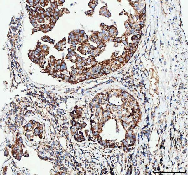
Start with heat-mediated antigen retrieval in EDTA at pH 8.0 for paraffin sections (datasheet A02132-1). The selected image used this retrieval before 2 μg/ml primary antibody overnight at 4°C, so keep those conditions comparable while optimizing retrieval (datasheet A02132-1). If staining is weak, vary heating duration in small increments on adjacent sections, with matched controls and checks of tissue integrity (standard IHC practice). Excessive heating can impair morphology or increase diffuse staining, so compare each condition with a no-primary control (standard IHC practice). Evaluate the expected cytoplasmic signal alongside background (HPA: general cytoplasmic expression; standard IHC practice).
How should I investigate weak PDK4 staining after tissue fixation?
The selected paraffin-section caption does not state a fixative, so target-specific PDK4 sensitivity to fixation remains unknown (datasheet A02132-1). Record the actual fixative, fixation duration and processing history for each specimen before comparing staining (standard IHC practice). When material permits, compare similarly processed sections using the documented EDTA pH 8.0 retrieval and 2 μg/ml primary antibody conditions (datasheet A02132-1; standard IHC practice). Include a no-primary control to separate weak signal from detection background (standard IHC practice). Interpret differences cautiously if specimens have different fixation histories, since processing can affect antigen accessibility in paraffin IHC (standard IHC practice).
Where should convincing PDK4 staining appear in a tissue section?
Expect PDK4 in the mitochondrial matrix, which can appear as cytoplasmic staining at chromogenic IHC resolution (UniProt Q16654 localisation; HPA: general cytoplasmic expression). Assess the cellular distribution and staining pattern against adjacent morphology rather than requiring individually resolved mitochondria (standard IHC practice). PDK4 has no transmembrane segment, so a sharp cell-surface rim alone is difficult to reconcile with its annotated location (UniProt Q16654 topology and localisation). If staining seems predominantly nuclear, compare serial sections, the no-primary control and a separately validated mitochondrial marker where available (standard IHC practice). Report cytoplasmic staining as compatible with PDK4, while treating compartment agreement alone as insufficient proof of specificity (UniProt Q16654 localisation; standard IHC practice).
Could an isoform or hidden epitope explain inconsistent PDK4 staining?
The supplied record lists 0 isoforms and no annotated glycosylation or modified-residue sites, so these annotations do not explain a staining discrepancy (UniProt Q16654 isoforms and PTMs). The record also does not map this antibody’s epitope, leaving its accessibility in processed sections uncertain (UniProt Q16654 record; datasheet A02132-1). PDK4 is annotated in the mitochondrial matrix and has no transmembrane segment; use that location to assess plausibility, without inferring epitope position (UniProt Q16654 topology and localisation). Compare adjacent sections under the documented EDTA pH 8.0 retrieval while changing only one staining variable at a time (datasheet A02132-1; standard IHC practice). Avoid assigning an unexpected pattern to an unlisted isoform without independent evidence (UniProt Q16654 isoforms; standard IHC practice).
How can I use IF to investigate an ambiguous PDK4 IHC pattern?
Use IF as an orthogonal localisation check for the mitochondrial-matrix assignment, while retaining chromogenic paraffin IHC as the primary readout (UniProt Q16654 localisation; datasheet A02132-1). Multiplex PDK4 with a validated marker for the expected cell type in the specimen and, if useful, a mitochondrial marker (standard IF practice; UniProt Q16654 localisation). Choose well-separated fluorophores and inspect unstained tissue for autofluorescence before assigning faint signal (standard IF practice). Because the expected antigen is inside mitochondria, test permeabilisation that permits antibody access to the matrix, then verify morphology and background (UniProt Q16654 localisation; standard IF practice). The supplied material contains no PDK4 IF/ICC image or protocol for this antibody, so optimize and validate IF separately (HPA subcellular record; datasheet A02132-1).
How do I reduce diffuse brown staining without losing PDK4 signal?
First compare stained and no-primary sections to identify signal arising from the detection system (standard IHC practice). The selected image used 10% goat serum blocking, a peroxidase-conjugated secondary antibody and DAB chromogen (datasheet A02132-1). Include a peroxidase-blocking step and check for endogenous pigment or enzyme activity before attributing brown deposits to PDK4 (standard chromogenic IHC practice). If background persists, titrate the primary around the documented 2 μg/ml condition and assess washing and secondary-antibody background on matched sections (datasheet A02132-1; standard IHC practice). Preserve a cytoplasmic signal that follows intact cells while rejecting staining concentrated at section edges or damaged areas (HPA: general cytoplasmic expression; standard IHC practice).
What is a defensible way to quantify PDK4 chromogenic IHC? ⚠ ANSWER MARKED FOR VERIFICATION
Define the cell population and region of interest before scoring, and exclude folds, edges and damaged tissue by a consistent rule (standard IHC quantification practice). Record the percentage of positive cells and staining intensity, then calculate an H-score from intensity categories if that scale fits the study design (standard IHC quantification practice). For spatial comparisons, report positive-cell density per mm² of evaluable tissue alongside the denominator used (standard IHC quantification practice). Normalize comparisons to the same cell population or evaluable tissue area, with matched staining batches and imaging settings (standard IHC quantification practice). State that an H-score measures staining under the chosen assay conditions, since HPA reports only medium consistency between antibody staining and RNA data (HPA: Approved, medium consistency; standard IHC practice).
When is an apparent PDK4-positive cell likely to be an artefact?
Favor signal in intact cytoplasm because PDK4 is annotated to the mitochondrial matrix and HPA describes general cytoplasmic staining (UniProt Q16654 localisation; HPA: tissue IHC profile). Treat isolated nuclear or cell-surface staining as suspect until an independent assay supports it (UniProt Q16654 topology and localisation; standard IHC practice). Compare the assigned cell type with tissue morphology: HPA reports high staining in bronchial respiratory epithelial cells but no detection in smooth muscle cells (HPA: bronchus respiratory epithelial cells high; smooth muscle cells not detected). Reject edge effects, necrotic regions and staining that persists in no-primary controls; assess endogenous peroxidase when interpreting DAB (standard chromogenic IHC practice). HPA’s medium staining–RNA consistency warrants caution when interpreting a surprising cell-specific result (HPA: Approved, medium consistency).
Boster reagents

Best PDK4 / [Pyruvate dehydrogenase (acetyl-transferring)] kinase isozyme 4, mitochondrial IHC Antibodies

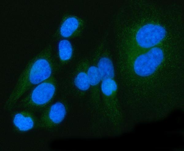
The IHC-validated antibody has paraffin-section IHC data from human lung cancer tissue and IF data from human squamous cell carcinoma tissue and HeLa cells (A02132-1 image captions).

Real IHC data IHC analysis of PDK4 using anti-PDK4 antibody (A02132-1). PDK4 was detected in a paraffin-embedded section of human lung cancer tissue. Heat mediated antigen retrieval was performed in EDTA buffer (pH 8.0, epitope retrieval solution). The tissue section was blocked with 10% goat serum. The tissue section was then incubated with 2 μg/ml rabbit anti-PDK4 Antibody (A02132-1) overnight at 4°C. Peroxidase Conjugated Goat Anti-rabbit IgG was used as secondary antibody and incubated for 30 minutes at 37°C. The tissue section was developed using HRP Conjugated Rabbit IgG Super Vision Assay Kit (Catalog # SV0002) with DAB as the chromogen.
Anti-PDK4 Antibody ®
Cat # A02132-1

A02132-1 will render with IHC data from a paraffin-embedded human lung cancer section (A02132-1 IHC image caption). Its IF image captions show human squamous cell carcinoma sections and HeLa cells; the catalog lists human, mouse and rat reactivity (A02132-1 IF image captions; catalog applications and reactivity).

Which to pick: Choose A02132-1 for paraffin-section tissue IHC because its own IHC caption documents that preparation; the fixative is unreported (A02132-1 IHC image caption). The same SKU is listed for IF and ICC and has IF images from tissue and cells (A02132-1 catalog applications; IF image captions). For mouse or rat samples, A02132-1 lists reactivity with both species, while its supplied IHC image documents a human sample (A02132-1 catalog reactivity; IHC image caption).

Each figure is that product's own IHC / IF validation image from its datasheet.

References

  1. UniProt Consortium. UniProt entry Q16654 (PDK4_HUMAN, [Pyruvate dehydrogenase (acetyl-transferring)] kinase isozyme 4, mitochondrial).
  2. Human Protein Atlas. PDK4 tissue IHC expression (reliability: Approved).
  3. Human Protein Atlas. PDK4 subcellular location (ICC-IF): Highest expression in HeLa: 65.4 nTPM.
  4. Human Protein Atlas. PDK4 antibody validation summary (1 antibodies).
  5. Decreased AMPK/SIRT1/PDK4 induced by androgen excess inhibits human endometrial stromal cell decidualization in PCOS. Cellular and molecular life sciences : CMLS 2024 — PMC11335245.
  6. Multi-dimensional analysis reveals the potential of PDK4 as a tumor biomarker and target for immunotherapy. Discover oncology 2026 — PMC13035999.
  7. High hepatic expression of PDK4 improves survival upon multimodal treatment of colorectal liver metastases. British journal of cancer 2019 — PMC6461828.
  8. Deficiency of TOP1MT enhances glycolysis through the stimulation of PDK4 expression in gastric cancer. Cancer & metabolism 2024 — PMC10777619.
  9. PubMed PMID:8798399 — UniProt-cited evidence.
  10. PubMed PMID:12853948 — UniProt-cited evidence.
  11. PubMed PMID:15489334 — UniProt-cited evidence.