This website uses cookies to ensure you get the best experience on our website.


- Table of Contents
Plan chromogenic PDLIM7 IHC in paraffin sections using cytoplasmic muscle staining as a reference (HPA tissue IHC). The guide covers fixation, controls and scoring, with 2–5 μg/ml as the catalog antibody’s starting concentration (datasheet A04377-1).

Expected localisation, validated protocols, controls and antibodies — the at-a-glance facts below, then the full design guide.
| Expected localisation | Cytoplasm (HPA tissue IHC); cytoskeleton (UniProt) | |
| Staining pattern | Cytoplasmic staining in myocytes and cardiomyocytes (HPA tissue IHC) | |
| Antigen retrieval | EDTA pH 8.0 HIER, heat-mediated (datasheet A04377-1) | |
| Positive control | Heart muscle+1 more · see all | |
| Negative control | Adipose tissue+4 more · see all |
| Fixation | Keep fixation consistent across sections. (standard IHC practice; not target-specific) | |
| Caveat | Low consistency between staining and RNA expression (HPA tissue IHC) | |
| Regulation | Most abundant in skeletal muscle (HPA tissue IHC) | |
| Isoform / epitope | Six isoforms; epitope coverage is unknown (UniProt) |
The catalog antibody’s IHC-P protocol uses EDTA pH 8.0 heat retrieval (datasheet A04377-1). One published PDLIM7 IHC protocol provides additional staining parameters (PMC12274469).
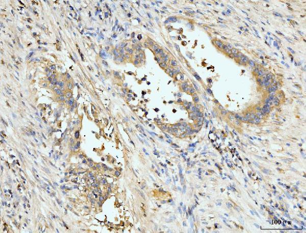
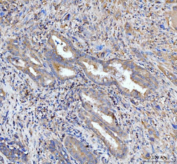
| Sample | Paraffin-embedded human appendiceal adenocarcinoma tissue; fixative not specified (datasheet A04377-1) |
| Fixation | Image fixative and duration unreported (datasheet A04377-1); verify before use. |
| Sectioning | 4–5 µm sections on charged slides (standard) |
| Deparaffinisation | Xylene, graded ethanol series to water (standard) |
| Antigen retrieval | Heat retrieval: EDTA pH 8.0 (datasheet A04377-1); 20 min, 95–100 °C (standard) |
| Peroxidase block | 3% H2O2, 10 min, room temperature (standard) |
| Blocking | 10% goat serum (datasheet A04377-1) |
| Primary antibody | Rabbit anti-PDLIM7, 2-5 μg/ml (datasheet A04377-1) |
| Primary incubation | Overnight at 4 °C (datasheet A04377-1) |
| Detection | Streptavidin-biotin complex (SABC), DAB chromogen (datasheet A04377-1) |
| Counterstain | Hematoxylin, blue, dehydrate and mount (standard) |
| Expected result | PDLIM7-positive staining in cardiomyocytes of heart muscle (HPA tissue IHC: Medium). HPA tissue profile: Cytoplasmic expression in several tissues, most abundant in skeletal muscle. No signal in the no-primary control. |
PDLIM7 is a cytoplasmic, cytoskeleton-associated protein with no transmembrane segment (UniProt Q9NR12). In paraffin-section IHC, expect cytoplasmic staining in skeletal myocytes and cardiomyocytes, both scored Medium by HPA (HPA tissue IHC). HPA describes cytoplasmic expression across several tissues, most abundant in skeletal muscle, but rates its tissue IHC evidence Approved with low consistency between staining and RNA expression (HPA tissue IHC).
| Cytoplasmic chromogen in skeletal myocytes or cardiomyocytes at roughly Medium intensity (HPA tissue IHC). | This matches HPA's observed positive cells and UniProt's cytoplasmic, cytoskeletal localisation (HPA tissue IHC; UniProt Q9NR12). Assess staining in the identified cells, since a tissue section contains more than one cell type. |
| Predominantly nuclear staining or a continuous cell-surface outline, without convincing cytoplasmic staining. | Treat this as a localisation mismatch: UniProt places PDLIM7 in the cytoplasm and cytoskeleton and lists no transmembrane segment (UniProt Q9NR12). Review antibody specificity and detection controls before scoring it as positive. |
| Strong staining in adipocytes or appendix glandular cells, where HPA reports Not detected (HPA tissue IHC). | This raises concern for cross-reactivity or endogenous detection activity; check a no-primary control and the detection reagents. HPA's negative calls describe those cell populations, not every cell in the section (HPA tissue IHC). |
| Widespread, poorly defined chromogen obscures cell boundaries and the expected cytoplasmic pattern. | A diffuse field cannot establish PDLIM7 localisation. General IHC practice is to inspect no-primary controls, wash quality, blocking and chromogen development before interpreting cell-specific staining. |
| No staining in skeletal myocytes or cardiomyocytes, the HPA Medium positive populations (HPA tissue IHC). | Consider an assay failure before calling the specimen negative. Check section integrity and the IHC-validated antibody's stated procedure, then compare a known-positive section processed in the same run (HPA tissue IHC). |
| Cell compartment and topology | UniProt places PDLIM7 in the cytoplasm and cytoskeleton and reports no transmembrane segment (UniProt Q9NR12). Judge paraffin IHC by cellular localisation; a membrane-only outline does not match that annotation. |
| Tissue and cell selection | Skeletal myocytes and cardiomyocytes are Medium IHC positives; adipocytes and appendix glandular cells are Not detected in HPA's observations (HPA tissue IHC). Select controls by cell type. |
| Isoforms and antibody coverage | UniProt lists 6 isoforms and reports isoform 2 predominating in skeletal muscle (UniProt Q9NR12). The supplied evidence gives no antibody epitope, so it cannot establish which isoforms an IHC stain detects. |
| Strength of IHC evidence | HPA rates tissue IHC Approved but notes low staining–RNA consistency; antibody HPA018794 is IHC Approved, not IHC Enhanced (HPA tissue IHC; HPA antibodies). Interpret unexpected positives cautiously. |
| IF/ICC Q: What localisation should I expect? | A: HPA ICC-IF places PDLIM7 mainly on actin filaments and focal adhesion sites; images are listed for A-431, U-251MG and U2OS (HPA subcellular). Those ICC-IF observations do not specify a paraffin IHC protocol. |
| Situation | Likely cause | Next action |
|---|---|---|
| The known-positive muscle section has no chromogen. | The run may have failed; HPA reports Medium staining in skeletal myocytes and cardiomyocytes (HPA tissue IHC). | Check antibody dilution and detection against the IHC-validated antibody's instructions; inspect section integrity and repeat with a positive control in the same run. |
| Signal is mainly nuclear or forms a crisp membrane ring. | That pattern conflicts with cytoplasmic and cytoskeletal localisation and the lack of a transmembrane segment (UniProt Q9NR12). | Compare the no-primary control, examine cell boundaries with the counterstain and reassess specificity before assigning a positive score. |
| Adipocytes or appendix glandular cells stain strongly. | Those cells are Not detected in HPA tissue IHC; cross-reactivity or endogenous detection activity is possible (HPA tissue IHC). | Run a no-primary control and check endogenous enzyme blocking where applicable; score the named cell population separately from neighboring cells. |
| The section has diffuse brown haze. | General IHC practice: background can arise from detection reagents, insufficient washing or overdevelopment. | Inspect a no-primary control, review blocking and washing, and shorten chromogen development if the control shows background. |
| A low or negative cell population differs from the HPA image. | HPA's tissue IHC has low consistency with RNA data, and its staining levels describe observed samples (HPA tissue IHC). | Confirm cell identity, compare same-run positive and negative controls, and describe the observed pattern without treating HPA levels as absolute thresholds. |
| Staining varies markedly across one section. | General IHC practice: uneven reagent coverage or section quality can create spatial variation. | Inspect tissue preservation and reagent coverage; repeat the section with uniform incubation and use the same-run muscle control to assess the run. |
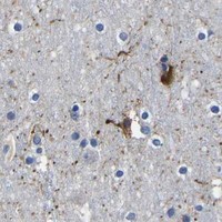


Comprehensive Human Protein Atlas IHC scoring per tissue (reliability: Approved — Low consistency between antibody staining and RNA expression data.). Rows are taken directly from the HPA tissue chart — click any row's HPA link to view the source.
| Tissue | Cell type | Level | Evidence | Source |
|---|---|---|---|---|
| Heart muscle | Cardiomyocytes | Medium | Protein (IHC) | HPA → |
| Skeletal muscle | Myocytes | Medium | Protein (IHC) | HPA → |
| Tissue | Cell type | Level | Evidence | Source |
|---|---|---|---|---|
| Adipose tissue | Adipocytes | Not detected | Protein (IHC) | HPA → |
| Appendix | Glandular cells | Not detected | Protein (IHC) | HPA → |
| Bone marrow | Hematopoietic cells | Not detected | Protein (IHC) | HPA → |
| Breast | Adipocytes | Not detected | Protein (IHC) | HPA → |
| Bronchus | Respiratory epithelial cells | Not detected | Protein (IHC) | HPA → |
Troubleshoot PDLIM7 chromogenic IHC in paraffin sections with attention to retrieval, cytoplasmic localisation, and cell specific controls (caption A04377-1; HPA tissue IHC).
The IHC-validated antibody A04377-1 has real staining data from human paraffin-embedded carcinoma sections (catalog image captions). Mouse and Rat reactivity is listed, while IF/ICC validation is unreported (catalog applications and reactivity).
A04377-1 has IHC images from human paraffin-embedded appendiceal adenocarcinoma, gall bladder adenosquamous carcinoma, and gastric adenocarcinoma sections (catalog image captions). Its listed applications include IHC, and its listed reactivity is Human, Mouse, and Rat; the images document human IHC only (catalog applications, reactivity, and image captions).
Which to pick: Choose A04377-1 for human paraffin-section IHC: its caption uses 2 μg/ml after EDTA retrieval at pH 8.0, and its listed IHC range is 2–5 μg/ml (A04377-1 image caption; catalog dilution). The caption does not report the fixative, and IF/ICC is absent from the application list, so there is no documented IF/ICC option here (A04377-1 image caption; catalog applications). A04377-1 lists Mouse and Rat reactivity, but the supplied IHC images show human tissue only; cross-species tissue IHC would require validation (catalog reactivity and image captions).