This website uses cookies to ensure you get the best experience on our website.
- Table of Contents
Facts about 3-phosphoinositide-dependent protein kinase 1.
Plays a central role in the transduction of signals from insulin by supplying the activating phosphorylation to PKB/AKT1, thus propagating the signal to downstream targets controlling cell proliferation and survival, in addition to glucose and amino acid uptake and storage. Negatively regulates the TGF-beta-induced signaling by: modulating the institution of SMAD3 and SMAD7 using TGF-beta receptor, phosphorylating SMAD2, SMAD3, SMAD4 and SMAD7, preventing the nuclear translocation of SMAD3 and SMAD4 and the translocation of SMAD7 from the nucleus to the cytoplasm in response to TGF-beta.
Human | |
---|---|
Gene Name: | PDPK1 |
Uniprot: | O15530 |
Entrez: | 5170 |
Belongs to: |
---|
protein kinase superfamily |
3-phosphoinositide-dependent protein kinase 1; 3-phosphoinositide-dependent protein kinase 2 pseudogene; PDK1; PDK-1; PDPK1; PDPK2; PDPK2P; PkB kinase like gene 1; PKB kinase
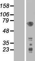
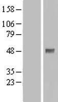
Mass (kDA):
63.152 kDA
Human | |
---|---|
Location: | 16p13.3 |
Sequence: | 16; NC_000016.10 (2538014..2603190) |
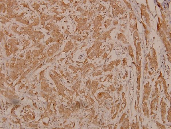
Appears to be expressed ubiquitously. The Tyr- 9 phosphorylated form is markedly increased in diseased tissue compared with normal tissue from lung, liver, colon and breast.
Cytoplasm. Nucleus. Cell membrane; Peripheral membrane protein. Cell junction, focal adhesion. Tyrosine phosphorylation seems to occur only at the cell membrane. Translocates to the cell membrane following insulin stimulation by a mechanism that involves binding to GRB14 and INSR. SRC and HSP90 promote its localization to the cell membrane. Its nuclear localization is dependent on its association with PTPN6 and its phosphorylation at Ser-396. Restricted to the nucleus in neuronal cells while in non-neuronal cells it is found in the cytoplasm. The Ser-241 phosphorylated form is distributed alon
Read on if interested in the PDPK1 marker. This article provides an overview of this PI3K modulator and its many applications. You are likely to be able to utilize this tool to aid your research. If you're uncertain, here are some things to keep in mind. Keep reading! Read on!
Fibrosis can be linked to the process of fibrosis and PDPK1. PDK1 activates PDC by phosphorylating PDH-E1a on the Ser232 and Ser300 sites. TGF-b1 significantly decreased PDC activity while TGF b1 increased expression the PGK1. The relative abundance of mRNA transcripts was compared to the protein levels, and flow cytometry used to determine levels of protein.
TGF-b1 inhibits PDC activity, which leads to a worldwide decrease of the acetylation. TGF-b1 has therefore an immediate impact on the levels of PDK1 proteins. UUOF had higher levels of PDK1 protein than NRKF and the molecule caused PDK1 be expressed more frequently in fibrosis cells that in normal cells.
This gene is expressed in cardiomyocytes. The Ang II receptor stimulates its expression in cardiomyocytes. Cardiomyocytes infected with PDPK1 deficiencies were unable to differentiate to M2 phenotypes, which can cause the development of cardiac fibrosis. The gene is essential to cause cardiac fibrosis. This study provides new insight into cardiac fibrosis. Thus, PDPK1 can be a useful fibrosis marker to monitor the progress of the disease.
Chronic kidney disease (CKD) is described by the non-recoverable process of remodeling and tubulointerstitial fibrosis. The interstitial fibrillary cell is the principal cell that is the effector. It is a heterogeneous group. The de novo expression of the protein asmooth muscle actin (ASM) differentiates the different subsets of fibroblasts. Myofibroblasts are a source for collagen and are involved in the development of fibrosis.
PDPK1 is an important regulator of the AGC family. It has been found to phosphorylate 23 closely related protein kinases. It performs multiple functions, including initiating tumorigenesis and activating serum-induced protein-kinase. It is an effective inhibitor of Akt Kinase signaling pathways. This pathway is blocked and cells undergo apoptosis or cell death.
In an FP-based SPR assay, Rettenmaier et al. identified novel noncovalent allosteric activators through site-directed chemical screen. To identify compounds that disrupt PIF-pocket interaction, Molecular mimicry (FP) and an assay for competitive binding (FP) were used. After screening 1280 molecules the top ten hits were further assessed in a dose-response manner. The most powerful of the hit compounds was determined by iterative synthesizing.
To enhance PDK1's catalytic activity, the small molecules mimic the phosphorylation process and alteration of conformation in PI3K. Small molecules, such as 1 have been proven to enhance PI3K activity by targeting the allosteric PIF pocket of PDK189. It was also determined that 1 is the smallest of molecules that increase the catalytic capacity of the PDK1.
The PDPK1 protein phosphorylates substrate kinases via docking into the PIF-pocket. The PIF-pocket's carboxyl group is wrapped around it. Variations in the Arg131 residue can cause loss or retention the positive charge. The carboxyl group on the aB helix is transformed into an ester. These modifications have a profound impact on PDK1 activity.
In the realm of drug discovery and development, non-competitive inhibitors for PDK1 are becoming more attractive. PIF-competitive inhibitors have higher sensitivity and have less adverse side effects than ATP. They also inhibit or activate PI3K. Allosteric modulators and ATP-competitive inhibits are also able to inhibit the activity PDK1.
Another inhibitor of PI3K activity is PEICT. PEICT blocks the expression of PI3K and Akt, thereby inhibiting their function. PEICT could also be associated with genes that regulate the expression of mTOR or Akt. When these pathways are activated, they lead to angiogenesis and cell growth. Further research is required to establish the role of PDPK1 in cellular progression.
PDPK1 is a key inhibitor of PI3K signaling in breast cancer cells. It has been found to inhibit survivin expression, which is a different downstream target of PI3K/Akt. These inhibitors have proven to be effective in the treatment of leukemia. They also inhibit the activity of several other proteins, including Akt. In addition, they hinder the process of apoptosis as well as the growth of breast cancer cells.
PDPK1 blocks PI3K at Tyr199, and p38 MAPK at Thr180/Tyr182, which increases autophagic cell death. It also inhibits the transcription factors forkhead and mTOR. PLB also triggers autophagy in prostate cancer cells. These results suggest that PLB may directly influence autophagic cell death.
One of the main features of fibrosis is an overexpression the PDPK1. This enzyme is involved in regulating the synthesis of proteins. Fibrosis develops when the fibrosis causing proteinkinase is over-exposed in the liver. Other indicators of fibrosis include the expression of PDGFB and TGFb mRNAs.
The role of PDPK1 in fibrosis is unclear. Studies have suggested that fibroblasts fight apoptosis and stop apoptosis. The inhibition of PDK1 as well as the knockdown of the DDR2 gene decreased the level of pThr308-Akt. However, PDPK1 expression was not affected in fibroblasts lacking DDR2.
Fibrosis is a progressive degenerative disease that has a variable course. Fibrosis can be caused by a variety factors, such as lung injury chronic inflammation, secondary and primary illnesses that are not properly treated. Lack of effective treatment options increases the chance of disability and death. PDPK1 can help predict progression in fibrosis. The PDPK1 gene has been linked to increased cellularity in fibrotic tissue.
In this study, PDPK1 is also a fibrosis marker and it is linked to profibrotic responses to collagen. Researchers discovered that mice lacking DDR2 showed less fibrosis following Bleomycin injury. They did discover that mice that did not have DDR2 had reduced myeloid cell recruitment and expressions of profibrotic cytokines. It is possible that DDR2 is involved in collagen-induced activation of fibroblasts.
The loss of Neu1 causes changes in fibroblasts that form the connective tissue in muscles. These changes result in the activation of fibroblasts as well as their transdifferentiation into myofibroblasts which are the dominant cell type that causes fibrosis. This mechanism could also be involved in human idiopathic fibrosis. Although the study only includes mice, it does prove that it is a prognostic indicator.
While the role of PDPK1 in inflammatory fibrosis is unknown however, it has been shown to play a role in innate immune responses and differential activation of macrophages. This gene could be involved in the development and maintaining inflammatory responses to common bacteria as well as infection-related organ injuries like acute respiratory distress syndrome (ARDS).
DDR2 regulates fibroblast survival through Akt. Lung fibroblasts with a deficiency in PDPK1 were immunoprecipitated in order to obtain phospho-tyrosine. The levels of phospho-Tyr9-PDK1 in cells that were not null were comparable to those observed in WT fibroblasts. Their PDK1 total content was identical. Therefore, lung fibroblasts with DDR2-deficient genes are profibrotic in a broad sense.
PDK1 is involved in the inflammatory response and the activation of M1 macrophages. NO production that is induced by LPS was impaired through the knockdown or inhibition of PDK1. These findings are not clinically relevant, but they have been shown to possess anti-fibrotic properties. This enzyme blocks a variety of genes that are involved in the inflammatory response and is a contributor to fibrosis.
PMID: 9094314 by Alessi D.R., et al. Characterization of a 3-phosphoinositide-dependent protein kinase which phosphorylates and activates protein kinase B alpha.
PMID: 9368760 by Alessi D.R., et al. 3-phosphoinositide-dependent protein kinase-1 (PDK1): structural and functional homology with the Drosophila DSTPK61 kinase.