This website uses cookies to ensure you get the best experience on our website.


- Table of Contents
Use lung type I alveolar cells and breast myoepithelial cells as positive controls for PDPN IHC (HPA tissue IHC). Assess staining with its expected membrane location (UniProt) and account for positive lymphatic vessels (HPA tissue IHC).

Expected localisation, validated protocols, controls and antibodies — the at-a-glance facts below, then the full design guide.
| Expected localisation | Plasma membrane, including apical and basolateral surfaces (UniProt) | |
| Staining pattern | Alveoli, lymphatics and myoepithelium (HPA tissue IHC); membrane expected (UniProt) | |
| Antigen retrieval | Citrate pH 6.0 HIER, 95–98 °C, 20 min (rule: cytoplasmic / membrane antigen) | |
| Positive control | Breast+2 more · see all | |
| Negative control | Adipose tissue+4 more · see all |
| Fixation | Matched tissue-IHC evidence does not establish the fixation claim. Validate the specimen-specific method before use. (selected-SKU IHC image M01124-4) | |
| Caveat | Lymphatic vessels may complicate lesional scoring (HPA tissue IHC) | |
| Regulation | Up-regulated in colorectal tumors (UniProt) | |
| Isoform / epitope | 6 isoforms; check extracellular vs cytoplasmic epitope (UniProt) |
The catalog antibody’s IHC-P protocol is accompanied by four published PDPN IHC protocols covering thoracic tumors, placenta, canine testicular tumors, and pancreatic cancer (PMC12109644; PMC11202497; PMC12771865; PMC5711987).
| Sample | Tissue sections; selected-image fixative not specified (standard IHC workflow) |
| Fixation | Image fixative and duration unreported (datasheet M01124-4); verify before use. |
| Sectioning | 4–5 µm sections on charged slides (standard) |
| Deparaffinisation | Xylene, graded ethanol series to water (standard) |
| Antigen retrieval | Heat-induced epitope retrieval in citrate buffer, pH 6.0, 20 min at 95–98 °C (standard rule: cytoplasmic / membrane antigen) |
| Peroxidase block | 3% H2O2, 10 min, room temperature (standard) |
| Blocking | 10% normal serum of the secondary host, 30 min, room temperature (standard) |
| Primary antibody | Rabbit anti-PDPN, 1:200-1:1000 (datasheet M01124-4) |
| Primary incubation | Overnight at 4 °C (standard) |
| Detection | HRP-polymer secondary, DAB chromogen 5–10 min (standard) |
| Counterstain | Hematoxylin, blue, dehydrate and mount (standard) |
| Expected result | PDPN-positive staining in myoepithelial cells of breast (HPA tissue IHC: High). HPA tissue profile: Distinct expression in alveoli, lymphatic vessels, placental and ovarian stroma, myoepithelium and basal cells of squamous epithelia. No signal in the no-primary control. |
PDPN is a transmembrane protein whose main observed location is the plasma membrane (UniProt Q86YL7 topology; HPA subcellular ICC-IF). In paraffin-section IHC, expect staining in breast myoepithelial cells and lung alveolar type I cells, both rated High by HPA (HPA tissue IHC). HPA rates tissue IHC reliability Enhanced, with medium consistency between staining and RNA expression (HPA tissue IHC).
| Crisp staining outlines breast myoepithelial cells or lung alveolar type I cells; nearby cells remain distinguishable. | This fits the main plasma-membrane location and HPA's High staining in those cell populations (HPA subcellular ICC-IF; HPA tissue IHC). Judge the named cells within their tissue architecture rather than treating every cell in the section as a positive control (general IHC practice). |
| Signal is chiefly nuclear or uniformly fills cells, with no convincing membrane outline. | That distribution does not match PDPN's main observed plasma-membrane location (HPA subcellular ICC-IF). UniProt also lists cytosol, so cytoplasmic signal alone is not proof of an artifact (UniProt Q86YL7 subcellular location). Recheck localization and controls before scoring it as specific (general IHC practice). |
| Strong staining appears in adipocytes or adrenal glandular cells while the expected cell populations are hard to identify. | HPA reports PDPN as Not detected in those named cell populations (HPA tissue IHC). Consider cross-reactivity or endogenous chromogenic activity, and compare with a control lacking primary antibody (general IHC practice). A negative HPA call applies to the specified cells, not every structure in that tissue. |
| Brown color spreads across the section or collects over many unrelated cells without clear borders. | Diffuse deposition cannot establish PDPN's expected membrane pattern (HPA subcellular ICC-IF; general IHC practice). Check the control lacking primary antibody, reagent background, blocking and washes before interpreting intensity (general IHC practice). |
| No staining is visible in breast myoepithelial cells or lung alveolar type I cells. | These are High-staining HPA cell populations, so their absence raises a technical or sampling question (HPA tissue IHC). Confirm that the relevant cells are present, then review the IHC-validated antibody's documented procedure, detection reagents and control performance (general IHC practice). |
| Membrane topology (UniProt Q86YL7 topology) | PDPN has an extracellular region at 23–131, a transmembrane segment at 132–152 and a short cytoplasmic tail at 153–162 (UniProt Q86YL7 topology). These regions support a membrane-centered interpretation; they do not identify the catalog antibody's epitope. |
| Cell-specific tissue pattern (HPA tissue IHC) | HPA reports High staining in breast myoepithelial and lung alveolar type I cells, but Not detected in adipocytes and several named glandular cell populations (HPA tissue IHC). Select controls and score results by cell type. |
| Subcellular evidence (HPA subcellular ICC-IF) | Plasma membrane and cell-junction localization are supported in ICC-IF; mitochondrial localization is uncertain (HPA subcellular ICC-IF). ICC-IF localization helps interpret compartment, but does not establish a separate IF/ICC protocol. |
| IHC evidence strength (HPA tissue IHC) | The tissue IHC profile is rated Enhanced, although HPA reports medium consistency between antibody staining and RNA expression (HPA tissue IHC). Use the pattern as a reference, then evaluate the actual slide and controls. |
| Sequence variation and glycosylation (UniProt Q86YL7) | UniProt lists 6 isoforms and multiple glycosylation sites in the extracellular region (UniProt Q86YL7 isoforms; glycosylation; topology). Without an antibody epitope or isoform-specific validation, these annotations cannot predict which variants this stain detects. |
| Antigen retrieval choice (general IHC practice) | Use the IHC-validated antibody's documented paraffin-section conditions and assess retrieval with controls (general IHC practice). Neither the supplied HPA pattern nor UniProt topology establishes a PDPN-specific retrieval requirement or fixation sensitivity. |
| Situation | Likely cause | Next action |
|---|---|---|
| Expected positive cells are present, but the slide has no signal. | A failed detection step, unsuitable antibody conditions or ineffective retrieval can cause a negative IHC run (general IHC practice); the supplied sources do not identify a PDPN-specific failure mechanism. | Verify the run control and detection reagents, then follow the IHC-validated antibody's documented paraffin-section procedure (general IHC practice). Use HPA's High-staining breast myoepithelial or lung alveolar type I cells as a cell-specific reference (HPA tissue IHC). |
| Color is widespread, including over tissue spaces or unrelated cells. | Non-specific reagent deposition, incomplete blocking or insufficient washing may obscure cell boundaries (general IHC practice). This is inconsistent with a readable membrane-centered pattern (HPA subcellular ICC-IF). | Inspect the control lacking primary antibody; review blocking, washes and chromogen development, then reassess whether positive cell borders remain distinct (general IHC practice). |
| Adipocytes or adrenal glandular cells look strongly positive. | Those named cells are Not detected in HPA tissue IHC, so cross-reactivity or endogenous chromogenic activity is plausible (HPA tissue IHC; general IHC practice). | Compare the same cell type with a control lacking primary antibody and check endogenous enzyme blocking appropriate to the detection system (general IHC practice). Do not extend HPA's cell-specific negative call to the whole tissue. |
| Signal is mainly nuclear or diffuse cytoplasmic. | The result departs from HPA's supported main plasma-membrane location, although UniProt also annotates cytosol (HPA subcellular ICC-IF; UniProt Q86YL7 subcellular location). | Check the positive-control compartment, counterstain and control lacking primary antibody; score membrane staining separately from ambiguous intracellular color (general IHC practice). |
| A suspected positive field looks weak or entirely negative. | The sampled field may lack the specific cells HPA identifies as High, even if their tissue is present elsewhere on the section (HPA tissue IHC; general IHC practice). | Locate and assess breast myoepithelial cells or lung alveolar type I cells before calling the specimen negative; record the cell population scored (HPA tissue IHC; general IHC practice). |
| IF/ICC shows extra intracellular signal that seems to conflict with IHC. | HPA supports plasma-membrane and cell-junction ICC-IF localization, but labels mitochondrial localization uncertain; modalities and images require their own controls (HPA subcellular ICC-IF; general IHC practice). | Use the supported locations to interpret the image and treat mitochondrial assignment cautiously (HPA subcellular ICC-IF). Consult the separate IF/ICC guide for that application's procedure. |
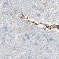


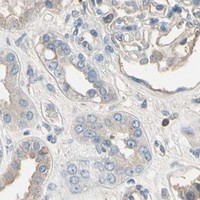


Comprehensive Human Protein Atlas IHC scoring per tissue (reliability: Enhanced — Medium consistency between antibody staining and RNA expression data.). Rows are taken directly from the HPA tissue chart — click any row's HPA link to view the source.
| Tissue | Cell type | Level | Evidence | Source |
|---|---|---|---|---|
| Breast | Myoepithelial cells | High | Protein (IHC) | HPA → |
| Lung | Alveolar cells type I | High | Protein (IHC) | HPA → |
| Testis | Round or early spermatids | Medium | Protein (IHC) | HPA → |
| Tissue | Cell type | Level | Evidence | Source |
|---|---|---|---|---|
| Adipose tissue | Adipocytes | Not detected | Protein (IHC) | HPA → |
| Adrenal gland | Glandular cells | Not detected | Protein (IHC) | HPA → |
| Appendix | Glandular cells | Not detected | Protein (IHC) | HPA → |
| Bone marrow | Hematopoietic cells | Not detected | Protein (IHC) | HPA → |
| Caudate | Glial cells | Not detected | Protein (IHC) | HPA → |
These questions address PDPN staining in paraffin sections using chromogenic IHC, with one entry on related IF experiments.
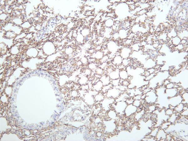
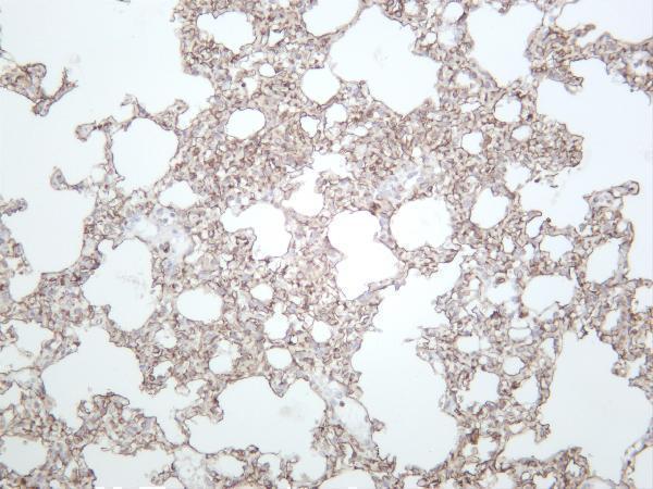
M01124-4 has IHC images from mouse lung, rat lung and human lung squamous carcinoma; it is listed for IHC and IF in human, mouse and rat (catalog image captions, applications and reactivity).
M01124-4 is the SKU that will render, with IHC and IF listed for human, mouse and rat (catalog applications and reactivity). Its IHC captions identify mouse lung, rat lung and human lung squamous carcinoma; no IF image is supplied (catalog image captions).
Which to pick: For tissue IHC, choose M01124-4 when a pictured tissue example is useful: it is a rabbit monoclonal with an IHC figure captioned mouse lung (catalog host, clonality and IHC caption). For IF planning and cross-species work, M01124-4 is listed for IF and for human, mouse and rat reactivity; ICC validation and an IF image are unreported (catalog applications, reactivity and IF image alts). M01124-2 is a human-reactive monoclonal IHC alternative without an IHC image (catalog clone, applications, reactivity and image alts); M01124-4’s IHC captions do not report paraffin processing or fixative (catalog IHC captions).