This website uses cookies to ensure you get the best experience on our website.


- Table of Contents
Plan PDRG1 staining in paraffin sections using the reported cytoplasmic pattern, especially in Leydig cells (HPA tissue IHC). This guide covers fixation consistency, controls and chromogenic scoring.

Expected localisation, validated protocols, controls and antibodies — the at-a-glance facts below, then the full design guide.
| Expected localisation | Cytoplasmic staining in several tissues (HPA tissue IHC) | |
| Staining pattern | Cytoplasmic staining, strongest in Leydig cells (HPA tissue IHC) | |
| Antigen retrieval | Tris-EDTA pH 8.0 HIER, heat-mediated (datasheet A13826) | |
| Positive control | Testis+4 more · see all | |
| Negative control | Adipose tissue+4 more · see all |
| Fixation | Keep fixation consistent across sections (standard IHC practice; not target-specific) | |
| Caveat | Staining has medium RNA concordance; verify the pattern (HPA tissue IHC) | |
| Regulation | Expression predominates in normal testis (UniProt) | |
| Isoform / epitope | One 1–133 chain; no annotated isoforms (UniProt) |
Compare the catalog antibody’s IHC-P protocol (datasheet A13826) with a published PDRG1 IHC protocol (PMC13050440 methods).
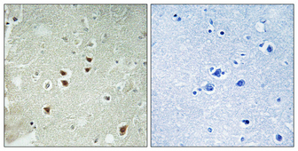
| Sample | Paraffin-embedded Human brain tissue; fixative not specified (datasheet A13826) |
| Fixation | Image fixative and duration unreported (datasheet A13826); verify before use. |
| Sectioning | 4–5 µm sections on charged slides (standard) |
| Deparaffinisation | Xylene, graded ethanol series to water (standard) |
| Antigen retrieval | Heat retrieval: Tris-EDTA pH 8.0 (datasheet A13826); 20 min, 95–100 °C (standard) |
| Peroxidase block | 3% H2O2, 10 min, room temperature (standard) |
| Blocking | 10% normal serum of the secondary host, 30 min, room temperature (standard) |
| Primary antibody | Rabbit anti-PDRG1, 1:100-1:300 (datasheet A13826) |
| Primary incubation | Overnight at 4 °C (standard) |
| Detection | HRP-polymer secondary, DAB chromogen 5–10 min (standard) |
| Counterstain | Hematoxylin, blue, dehydrate and mount (standard) |
| Expected result | PDRG1-positive staining in leydig cells of testis (HPA tissue IHC: High). HPA tissue profile: Cytoplasmic expression in several tissues, mainly in Leydig cells. No signal in the no-primary control. |
PDRG1 is cytoplasmic and has no transmembrane segment (UniProt Q9NUG6). In tissue IHC, expect cytoplasmic staining in several tissues, strongest in testicular Leydig cells (HPA tissue IHC: High in Leydig cells). The HPA tissue profile is Approved, with medium consistency between staining and RNA data, and awaits external verification (HPA tissue IHC: reliability).
| Distinct cytoplasmic staining in testicular Leydig cells, with weaker staining in some other cell populations. | This fits the strongest reported tissue result: High in Leydig cells (HPA tissue IHC). Medium staining also occurs in adrenal glandular cells, bronchial respiratory epithelial cells, and cerebellar Purkinje cells (HPA tissue IHC). Compare the named cell population, rather than treating an entire organ as uniformly positive. |
| Signal appears predominantly nuclear, membranous, or extracellular, without a convincing cytoplasmic component. | That distribution conflicts with the cytoplasmic tissue profile (HPA tissue IHC) and UniProt localisation and topology (UniProt Q9NUG6). Check morphology, counterstain, detection background, and antibody specificity before interpreting it as PDRG1. The ICC-IF actin-filament report is a separate observation (HPA subcellular ICC-IF). |
| Strong staining appears in a cell population reported as Not detected, such as adipocytes or liver cholangiocytes. | That result warrants scrutiny, especially beside a correctly stained positive section (HPA tissue IHC: Not detected in these populations). Cross-reactivity or endogenous detection activity may explain it (standard IHC practice). An HPA Not detected result is a sampled staining observation, not proof of universal absence. |
| Diffuse colour covers stroma, blank regions, and multiple cell types without clear cellular boundaries. | An unstructured pattern cannot establish the reported cytoplasmic localisation (HPA tissue IHC). Review the no-primary control, blocking, washes, and chromogen development for nonspecific or endogenous signal (standard IHC practice). Judge any remaining staining against intact cells and the expected positive population. |
| No convincing staining appears in testicular Leydig cells. | This conflicts with the reported High Leydig-cell signal (HPA tissue IHC). Confirm that Leydig cells are present and identifiable, then review the IHC-validated antibody, antigen retrieval, detection reagents, and a positive control (standard IHC practice). HPA's Approved rating still has medium consistency and pending external verification (HPA tissue IHC). |
| Tissue and cell selection | Testicular Leydig cells provide the clearest reported positive comparison (HPA tissue IHC: High). Adipocytes and bone-marrow hematopoietic cells were Not detected in the cited profile (HPA tissue IHC); use their results as context, not absolute negative standards. |
| Compartment and molecular topology | UniProt places PDRG1 in the cytoplasm and reports no transmembrane segment (UniProt Q9NUG6). Interpret chromogenic signal within cell boundaries; dominant membrane staining lacks support from the supplied localisation evidence. |
| Strength of tissue evidence | The tissue profile is Approved, but staining and RNA show medium consistency, with external verification pending (HPA tissue IHC). HPA047084 has Approved IHC status; the supplied record does not report IHC Enhanced validation (HPA antibodies). |
| IF/ICC pattern? | For IF/ICC interpretation, HPA reports actin-filament localisation using an ICC-Approved antibody, HPA063542 (HPA subcellular ICC-IF; HPA antibodies). This differs in resolution and assay from cytoplasmic tissue IHC; it does not establish an IHC-P protocol or require filament resolution on a chromogenic section. |
| Situation | Likely cause | Next action |
|---|---|---|
| The Leydig-cell positive control is blank. | A failed staining run or missing target cell population is possible (standard IHC practice); High Leydig-cell staining is reported (HPA tissue IHC). | Verify cell identity on the section, then check antibody application, retrieval conditions, detection reagents, and run controls (standard IHC practice). Record the result as inconclusive until the positive control works. |
| Signal is widespread in cells reported as Not detected. | Nonspecific antibody binding or endogenous detection activity may contribute (standard IHC practice); HPA lists selected populations as Not detected (HPA tissue IHC). | Compare a no-primary control and a known positive section; inspect whether the same signal appears without primary antibody (standard IHC practice). Reassess specificity before calling those cells PDRG1-positive. |
| Only nuclei or cell borders stain strongly. | The dominant compartment disagrees with cytoplasmic PDRG1 localisation (UniProt Q9NUG6; HPA tissue IHC). | Check counterstain and cell boundaries, examine no-primary staining, and repeat with the IHC-validated antibody if needed (standard IHC practice). Score only a supported, cellular pattern. |
| The whole section has weak, diffuse colour. | Incomplete blocking or washing, or excess detection development, can obscure specific staining (standard IHC practice). | Review controls and reagent timing, then adjust the general IHC workflow as indicated by those controls (standard IHC practice). Reassess Leydig-cell cytoplasm against nearby background (HPA tissue IHC). |
| A medium-staining tissue gives little or no signal. | Its reported level is below the High Leydig-cell reference; for example, adrenal glandular cells are Medium (HPA tissue IHC). | Check a Leydig-cell positive section in the same run before inferring assay failure (HPA tissue IHC; standard IHC practice). Document the specific cell population and staining threshold. |
| Tissue IHC lacks visible actin filaments seen in an ICC-IF reference. | HPA reports actin-filament localisation in ICC-IF and broader cytoplasmic staining in tissue IHC (HPA subcellular ICC-IF; HPA tissue IHC). | Judge the chromogenic section by its cytoplasmic cell-level pattern (HPA tissue IHC). Consult the separate IF/ICC guide for filament-level interpretation. |


Comprehensive Human Protein Atlas IHC scoring per tissue (reliability: Approved — Medium consistency between antibody staining and RNA expression data. Pending external verification.). Rows are taken directly from the HPA tissue chart — click any row's HPA link to view the source.
| Tissue | Cell type | Level | Evidence | Source |
|---|---|---|---|---|
| Testis | Leydig cells | High | Protein (IHC) | HPA → |
| Adrenal gland | Glandular cells | Medium | Protein (IHC) | HPA → |
| Appendix | Glandular cells | Medium | Protein (IHC) | HPA → |
| Breast | Glandular cells | Medium | Protein (IHC) | HPA → |
| Bronchus | Respiratory epithelial cells | Medium | Protein (IHC) | HPA → |
| Tissue | Cell type | Level | Evidence | Source |
|---|---|---|---|---|
| Adipose tissue | Adipocytes | Not detected | Protein (IHC) | HPA → |
| Bone marrow | Hematopoietic cells | Not detected | Protein (IHC) | HPA → |
| Caudate | Glial cells | Not detected | Protein (IHC) | HPA → |
| Cervix | Glandular cells | Not detected | Protein (IHC) | HPA → |
| Liver | Cholangiocytes | Not detected | Protein (IHC) | HPA → |
Troubleshoot PDRG1 staining in paraffin sections by checking retrieval, cellular localisation and controls before comparing signal across tissues.
A13826 has a human paraffin-section IHC image (A13826 IHC caption) and is listed for IF and for human, mouse, and rat reactivity (catalog: applications/reactivity).
A13826 will render with an IHC image of human paraffin-embedded brain, stained at 1:100 after Tris-EDTA retrieval at pH 8.0 (A13826 IHC caption). IF is listed as an application, but no IF image is supplied (catalog: applications; A13826 IF image alts).
Which to pick: For tissue IHC, choose A13826 based on its own human paraffin-section image; the fixative is unreported (A13826 IHC caption). For IF, A13826 is a listed rabbit polyclonal antibody with a catalog dilution of 1:50; ICC-specific validation is unreported (catalog: host/applications/if_dilution). For cross-species work, A13826 lists human, mouse, and rat reactivity, while its displayed IHC evidence is limited to human tissue (catalog: reactivity; A13826 IHC caption).