PDX1 / Pancreas/duodenum homeobox protein 1 · IHC design guide

Design Immunohistochemistry for PDX1

Plan chromogenic IHC for PDX1 in paraffin sections using the IHC-validated antibody (datasheet M00491). Compare nuclear staining in pancreatic islets and intercalated ducts with intestinal epithelium, and score by cell type (HPA tissue IHC).

Evidence assembled Oct 2026 · For research use; verify linked source records and product datasheet before use
Immunohistochemistry protocol sheet for PDX1 (IHC for PDX1): expected localisation Nuclear staining in pancreatic islets and ducts (HPA tissue IHC), antibody M00491, validated IHC image, and IHC protocol steps
Printable PDX1 IHC protocol sheet — expected localisation Nuclear staining in pancreatic islets and ducts (HPA tissue IHC), antibody M00491, controls and protocol steps. Open the full PDX1 IHC guide →

PDX1 Immunohistochemistry Experimental Design Guide

Expected localisation, validated protocols, controls and antibodies — the at-a-glance facts below, then the full design guide.

Must know before staining
Expected localisation Nuclear staining in pancreatic islets and ducts (HPA tissue IHC)
Staining pattern Islet, intercalated-duct and intestinal epithelial nuclei (HPA tissue IHC)
Antigen retrieval EDTA pH 8.0 HIER, heat-mediated (datasheet M00491)
Positive control ⓘ Duodenum+4 more · see all
Negative control ⓘ Adipose tissue+4 more · see all
Important caveats
Reasons your staining may differ from the expected pattern.
Fixation Keep fixation consistent across sections. (standard IHC practice; not target-specific)
Caveat Enterocyte staining calls for cell-type-aware scoring (HPA tissue IHC)
Regulation No staining-intensity regulator specified (UniProt)
Isoform / epitope One 1–283 chain; no annotated isoforms or cleavage (UniProt)
Section 1

Recommended PDX1 IHC & IF Protocols

The catalog antibody’s IHC-P protocol (datasheet: M00491) is accompanied by four published PDX1 IHC protocols (PMC3123421; PMC7667445; PMC5608307; PMC9710085).

Recommended immunohistochemistry (IHC-P) protocol parameters
SampleParaffin-embedded human stomach cancer tissue; fixative not specified (datasheet M00491)
FixationImage fixative and duration unreported (datasheet M00491); verify before use.
Sectioning4–5 µm sections on charged slides (standard)
DeparaffinisationXylene, graded ethanol series to water (standard)
Antigen retrievalHeat retrieval: EDTA pH 8.0 (datasheet M00491); 20 min, 95–100 °C (standard)
Peroxidase block3% H2O2, 10 min, room temperature (standard)
Blocking10% goat serum (datasheet M00491)
Primary antibodyRabbit monoclonal (clone AEC-16) anti-PDX1, 1:50 (datasheet M00491)
Primary incubationOvernight at 4 °C (datasheet M00491)
DetectionHRP-conjugated secondary, DAB chromogen (datasheet M00491)
CounterstainHematoxylin, blue, dehydrate and mount (standard)
Expected resultPDX1-positive staining in enterocytes of duodenum (HPA tissue IHC: High). HPA tissue profile: Selective nuclear expression in islets of Langerhans and intercalated ducts in pancreas, as well as in duodenum and small intestine. No signal in the no-primary control.
💡Decision noteStart with EDTA pH 8.0 heat retrieval for the catalog antibody (datasheet: M00491); use each paper’s reported retrieval when reproducing its protocol.
Section 2

What Is the Expected PDX1 Staining Pattern?

PDX1 is predominantly a nuclear IHC target: HPA reports selective nuclear staining in pancreatic islets and intercalated ducts, duodenum, and small intestine, with Enhanced tissue reliability (HPA tissue IHC). Expect the clearest signal in pancreatic endocrine cells and duodenal enterocytes (HPA: High). UniProt also lists cytosol, but PDX1 has no transmembrane segment, so membrane staining is not an expected pattern (UniProt P52945 topology and subcellular location).

What am I looking at on my slide?
Distinct nuclear chromogen in pancreatic endocrine cells or duodenal enterocytes, with recognizable tissue structure (HPA: High in both cell groups).This matches the principal positive pattern (HPA tissue IHC). Score the fraction and strength of stained nuclei within the identified cell population; a dark deposit outside cells should not be counted as nuclear signal (general IHC practice).
Cytoplasmic-only or membrane-like staining, without convincing stained nuclei in expected positive cells (HPA tissue IHC; UniProt P52945 topology).HPA tissue IHC supports a nuclear pattern, although UniProt also lists cytosol (HPA tissue IHC; UniProt P52945 subcellular location). Treat isolated cytoplasmic signal as uncertain and membrane-like signal as discordant; check localisation and controls before calling either specific (general IHC practice).
Strong staining in a cell population listed as undetected, such as adipocytes in adipose tissue or adrenal glandular cells (HPA: Not detected).That cell-level result conflicts with the supplied HPA observations; it does not establish that every cell in those tissues is negative (HPA tissue IHC). Consider cross-reactivity or endogenous chromogenic activity, and compare reagent controls (general IHC practice).
Diffuse chromogen across nuclei, cytoplasm, and spaces between cells, with little separation from the expected positive cells (general IHC practice).This is background rather than a scorable PDX1 distribution when it obscures the selective nuclear pattern (HPA tissue IHC; general IHC practice). Review blocking, washes, and detection controls before interpreting weakly stained nuclei (general IHC practice).
No nuclear signal in pancreatic endocrine cells or duodenal enterocytes on a slide intended as a positive control (HPA: High in both cell groups).The result fails to reproduce the reported tissue pattern, but alone cannot distinguish an assay problem from an unsuitable specimen (HPA tissue IHC; general IHC practice). Check tissue identity, antibody use, and detection performance before scoring test sections negative (general IHC practice).
💡Expected PDX1 appearanceCall PDX1 positive when distinct nuclear staining appears in pancreatic endocrine cells or duodenal enterocytes at the reported High level; diffuse, membrane-like, or isolated staining in listed undetected cell types warrants scrutiny (HPA tissue IHC; UniProt P52945 topology).
How each factor affects the staining
Tissue and cell selectionPancreatic endocrine cells and duodenal enterocytes are High; enterocytes in appendix, colon, rectum, and small intestine are Medium (HPA tissue IHC). Compare the named cells, not whole-organ averages, when choosing and reading controls (general IHC practice).
Antibody validationTwo listed antibodies, HPA059146 and CAB025873, have Enhanced IHC validation (HPA antibodies). HPA describes its tissue result as consistent with RNA expression; this supports the reported pattern but does not validate every antibody or staining condition (HPA tissue IHC).
Chromogenic detectionEndogenous enzyme activity can resemble specific color deposition in chromogenic IHC (general IHC practice). A detection-reagent control helps distinguish that activity from antibody-dependent staining; assess deposits against nuclear cell outlines and the expected tissue distribution (general IHC practice; HPA tissue IHC).
IF/ICC Q: what localisation is supported?A: HPA reports supported nucleoplasmic localisation and lists HeLa among cell lines with ICC-IF images (HPA subcellular ICC-IF). Use that as a compartment check; it does not establish an IF/ICC protocol or change the tissue IHC interpretation (HPA subcellular ICC-IF; HPA tissue IHC).
Why is my staining missing, weak or wrong?
SituationLikely causeNext action
Known-positive pancreatic or duodenal control has no nuclear stain (HPA: High in pancreatic endocrine cells and duodenal enterocytes).Specimen identity or assay performance may be wrong; this result alone does not identify which step failed (general IHC practice).Confirm the expected cells are present, then check antibody application and detection reagents against the catalog antibody's IHC-P instructions (general IHC practice).
Color is mainly cytoplasmic or membrane-like in an otherwise positive tissue (HPA tissue IHC).Cytosol is listed by UniProt, but a cytoplasmic-only result lacks the HPA nuclear pattern; membrane staining conflicts with the lack of a transmembrane segment (UniProt P52945; HPA tissue IHC).Inspect nuclei and compare a positive tissue and reagent control before assigning specificity to the nonnuclear signal (general IHC practice).
Adipocytes in adipose tissue or adrenal glandular cells stain strongly (HPA: Not detected in those cell groups).The result is discordant with the listed HPA cell observations; cross-reactivity or endogenous detection activity is possible (HPA tissue IHC; general IHC practice).Use an appropriate reagent control and reassess staining by cell type; avoid calling the entire tissue positive from an unidentified deposit (general IHC practice).
Diffuse background obscures stained nuclei across the section (general IHC practice).Nonspecific reagent binding or incomplete washing can reduce contrast in chromogenic IHC (general IHC practice).Review blocking, washes, and detection controls, then rescore only nuclei distinguishable from background (general IHC practice).
Small-intestine enterocytes stain less strongly than duodenal enterocytes (HPA: Medium versus High).That relative difference matches the supplied HPA levels and does not, by itself, show an assay failure (HPA tissue IHC).Compare each sample with its own expected cell population and record nuclear intensity and distribution separately (HPA tissue IHC; general IHC practice).
A reagent control shows chromogen deposits in the same places as the test section (general IHC practice).Detection chemistry or tissue-associated activity may be contributing signal independently of the primary antibody (general IHC practice).Resolve the control signal before interpreting weak test staining as PDX1; if using peroxidase detection, review the enzyme-blocking step (general IHC practice).

Sample controls for PDX1 IHC & IF

🧪Run pancreas first: pancreatic endocrine cells should show strong PDX1 staining (HPA: High in pancreatic endocrine cells). Use adipose tissue as a negative, with no detectable signal expected in adipocytes (HPA: Not detected in adipocytes); on the pancreatic slide, acinar cells should stain less strongly than endocrine cells, though low acinar expression means they are not an absolute negative (UniProt P52945 tissue specificity).
Positive control tissue: Duodenum (Enterocytes, HPA High)
Negative control tissue: Adipose tissue (HPA Not detected)
ICC-IF cell lines (HPA subcellular resource): HPA ICC-IF images show PDX1 in HeLa, with annotated localisation: Nucleoplasm (supported) (HPA subcellular).
Technical controls: Include no-primary (secondary-only) and host-species- and isotype-matched control immunoglobulin controls, plus PDX1 knockout material or a validated peptide-block control. For pancreatic HRP/DAB IHC, quench endogenous peroxidase; for IF, check tissue autofluorescence with an unstained section (standard IHC/IF practice).
⚠️Feasibility: The selected M00491 paraffin-section IHC caption uses heat retrieval in EDTA at pH 8.0, but reports no fixative; no target-specific fixation window or fixation effect is supplied (M00491 tissue-IHC caption). Frozen-section performance and whether IF is easier than paraffin IHC are unreported in the supplied application evidence. Low PDX1 expression in pancreatic acinar cells can complicate interpretation of faint acinar staining as background (UniProt P52945 tissue specificity).

HPA tissue IHC evidence for PDX1

Comprehensive Human Protein Atlas IHC scoring per tissue (reliability: Enhanced — High consistency between antibody staining and RNA expression data.). Rows are taken directly from the HPA tissue chart — click any row's HPA link to view the source.

Positive expression · recommended positive controls

TissueCell typeLevelEvidenceSource
Duodenum Enterocytes High Protein (IHC) HPA →
Pancreas Pancreatic endocrine cells High Protein (IHC) HPA →
Appendix Enterocytes Medium Protein (IHC) HPA →
Colon Enterocytes Medium Protein (IHC) HPA →
Rectum Enterocytes Medium Protein (IHC) HPA →

Undetected expression · recommended negative controls

TissueCell typeLevelEvidenceSource
Adipose tissue Adipocytes Not detected Protein (IHC) HPA →
Adrenal gland Glandular cells Not detected Protein (IHC) HPA →
Bone marrow Hematopoietic cells Not detected Protein (IHC) HPA →
Breast Adipocytes Not detected Protein (IHC) HPA →
Bronchus Respiratory epithelial cells Not detected Protein (IHC) HPA →
Section 3

Advanced PDX1 IHC Tips

Troubleshoot PDX1 staining in paraffin sections by checking retrieval, nuclear localisation, tissue controls and scoring before interpreting chromogenic signal.

What retrieval conditions should I try when nuclear PDX1 staining is weak?
Start with heat-mediated retrieval in EDTA at pH 8.0 for paraffin sections (datasheet M00491). The documented staining workflow then used 10% goat serum, antibody at 1:50 overnight at 4°C, and a peroxidase-conjugated secondary for 30 minutes at 37°C (datasheet M00491). If nuclei remain faint, compare a longer retrieval cycle on adjacent sections while holding antibody dilution and DAB development constant (standard IHC practice). Judge the result against pancreatic endocrine cells or duodenal enterocytes, where PDX1 staining is reported as high, and inspect tissue integrity before intensifying retrieval further (HPA tissue IHC; standard IHC practice).
How should I assess whether fixation is limiting PDX1 IHC?
Target-specific sensitivity to fixation is unknown from the supplied evidence; the M00491 caption identifies a paraffin section but does not state its fixative (datasheet M00491). Record the actual fixative, fixation duration and processing history for each specimen, then compare matched sections processed under documented conditions (standard IHC practice). Apply the documented EDTA retrieval at pH 8.0 and 1:50 primary dilution consistently before attributing weak staining to fixation (datasheet M00491; standard IHC practice). Check whether nuclear staining is retained in an expected positive cell population, such as pancreatic endocrine cells, alongside preserved morphology (HPA tissue IHC; standard IHC practice).
Should PDX1 stain nuclei, cytoplasm or both in tissue sections?
Prioritise a crisp nuclear signal: PDX1 is reported in the nucleus and cytosol, while tissue IHC shows selective nuclear expression in pancreatic islets, intercalated ducts, duodenum and small intestine (UniProt P52945 subcellular location; HPA tissue IHC). Nucleoplasmic localisation is independently supported by subcellular imaging (HPA subcellular). If DAB appears mainly cytoplasmic, compare its distribution with the nuclear counterstain and assess whether the same pattern persists in a no-primary control (standard IHC practice). Score nuclear and cytoplasmic signal separately, and interpret diffuse cytoplasmic staining cautiously until its specificity is supported by appropriate controls (standard IHC practice).
Could an isoform or epitope change explain inconsistent PDX1 staining?
No alternative isoforms are annotated for the 283-residue PDX1 record, so an isoform switch is not established by the supplied evidence (UniProt P52945 isoforms and processing). PDX1 has no annotated transmembrane segment and includes reported phosphorylation at Thr151 and Ser268, but the M00491 epitope is unspecified here (UniProt P52945 topology and modified residues; datasheet M00491). Do not assign loss of staining to either modification without epitope information and direct experimental evidence (standard IHC practice). Instead, compare adjacent sections with identical retrieval, antibody incubation and detection, then evaluate nuclear signal in an expected positive cell population (datasheet M00491; HPA tissue IHC; standard IHC practice).
How can I adapt PDX1 localisation checks to a multiplex IF experiment?
Treat IF as a separate application from this chromogenic paraffin-section workflow; the supplied M00491 evidence documents tissue IHC, while subcellular imaging supports nucleoplasmic PDX1 localisation (datasheet M00491; HPA subcellular). Pair PDX1 with a validated marker of the expected cell population, such as an endocrine-cell marker when examining pancreatic islets, and include a nuclear counterstain (HPA tissue IHC; standard IF practice). Select spectrally separated fluorophores after checking tissue autofluorescence and single-stain controls, especially if native fluorescence overlaps the chosen emission channel (standard IF practice). Because PDX1 has no transmembrane segment and is nuclear, optimise permeabilisation for nuclear access without assuming an extracellular epitope (UniProt P52945 topology and subcellular location; standard IF practice).
What should I change when DAB background obscures PDX1-positive nuclei?
Separate widespread DAB deposition from discrete nuclear staining in expected cells before changing the primary antibody concentration (HPA tissue IHC; standard IHC practice). The documented workflow used 10% goat serum, M00491 at 1:50, a peroxidase-conjugated secondary and DAB development (datasheet M00491). Include no-primary and detection-only controls, block endogenous peroxidase as a general chromogenic IHC step, and compare shorter DAB development or stronger washing on adjacent sections (standard IHC practice). If background remains high, assess tissue folds, section edges and necrotic areas separately, and retain a positive tissue control while adjusting one condition at a time (standard IHC practice).
How should I quantify PDX1 staining across paraffin sections? ⚠ ANSWER MARKED FOR VERIFICATION
Define the cell population and scoring region before measuring nuclear PDX1, because reported expression differs across pancreatic and intestinal cell types (HPA tissue IHC). Record the percentage of positive nuclei and an intensity-based H-score, or count positive nuclei per mm² when cell density varies (standard IHC practice). Normalise positive counts to the total number of intact nuclei in the same annotated cell population, and report tissue area when using density (standard IHC practice). Keep retrieval, 1:50 primary dilution, imaging and DAB development consistent across compared sections, and exclude folds or necrosis by prespecified rules (datasheet M00491; standard IHC practice).
How do I distinguish true PDX1 staining from chromogenic artefacts?
A convincing result is predominantly nuclear in an expected population, such as pancreatic endocrine cells or duodenal enterocytes, with the counterstain confirming cellular boundaries (HPA tissue IHC; standard IHC practice). Diffuse staining in an unexpected compartment or cell population warrants comparison with a no-primary control and review of morphology (UniProt P52945 subcellular location; standard IHC practice). Discount signal confined to section edges, folds or necrotic regions, and check whether endogenous peroxidase contributes to DAB colour (standard IHC practice). The reported stomach-cancer section demonstrates staining under the M00491 workflow but does not, by itself, establish the specificity of every stained cell in a new specimen (datasheet M00491; standard IHC practice).
Boster reagents

Best PDX1 / Pancreas/duodenum homeobox protein 1 IHC Antibodies

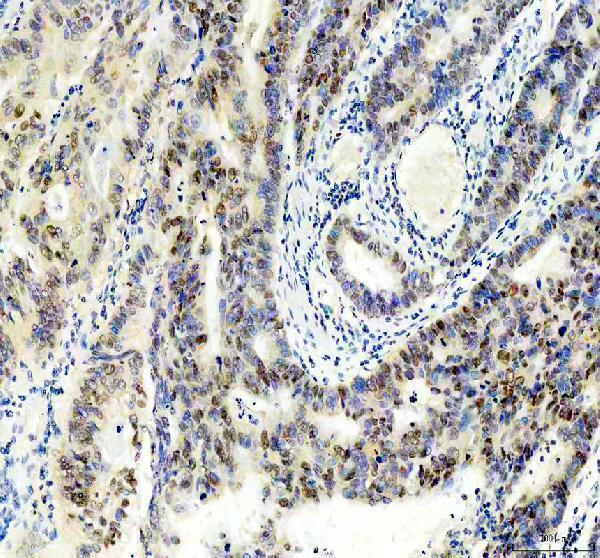
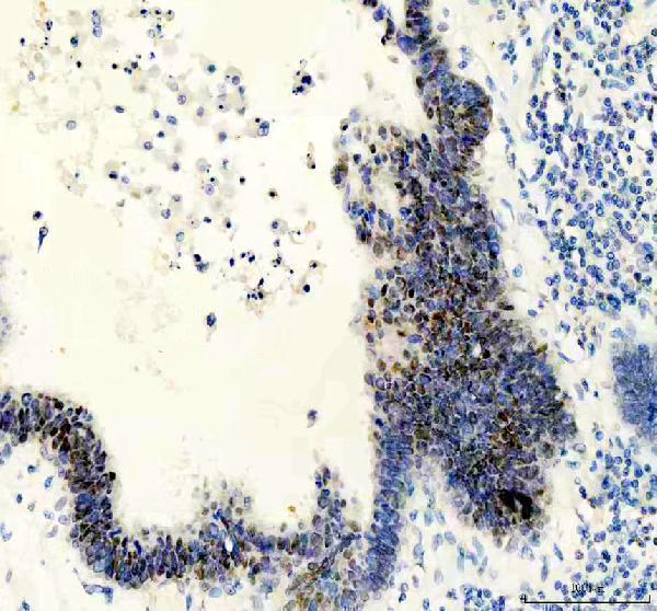
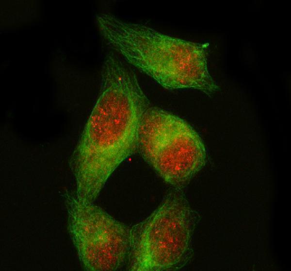
Anti-PDX1 antibodies have IHC data from human stomach cancer paraffin sections (M00491 image caption) and IF data from HELA cells (A00491-4 image caption).

Real IHC data IHC analysis of PDX1/Ipf1 using anti-PDX1/Ipf1 antibody (M00491). PDX1/Ipf1 was detected in a paraffin-embedded section of human stomach cancer tissue. Heat mediated antigen retrieval was performed in EDTA buffer (pH 8.0, epitope retrieval solution). The tissue section was blocked with 10% goat serum. The tissue section was then incubated with 1:50 rabbit anti-PDX1/Ipf1 Antibody (M00491) overnight at 4°C. Peroxidase Conjugated Goat Anti-rabbit IgG was used as secondary antibody and incubated for 30 minutes at 37°C. The tissue section was developed using HRP Conjugated Rabbit IgG Super Vision Assay Kit (Catalog # SV0002) with DAB as the chromogen.
Anti-PDX1/Ipf1 Rabbit Monoclonal Antibody
Cat # M00491
Real IF data IF analysis of PDX1 using anti-PDX1 antibody (A00491-4) and anti-Beta Tubulin antibody (M01857-3). PDX1 was detected in immunocytochemical section of HELA cell. Enzyme antigen retrieval was performed using IHC enzyme antigen retrieval reagent (AR0022) for 15 mins. The cells were blocked with 10% goat serum. And then incubated with 5 μg/mL rabbit anti-PDX1 Antibody (A00491-4) and mouse anti-Beta Tubulin antibody (M01857-3) overnight at 4°C. Cy3 Conjugated Goat Anti-Rabbit IgG (BA1032) and FITC Conjugated Goat Anti-Mouse IgG (BA1101) were used as secondary antibody at 1:500 dilution and incubated for 30 minutes at 37°C. Visualize using a fluorescence microscope and filter sets appropriate for the label used.
Anti-PDX1 Antibody ®
Cat # A00491-4

M00491 is listed for human IHC and has a chromogenic image from a paraffin section of human stomach cancer tissue (catalog applications and reactivity; M00491 image caption). A00491-4 is listed for human ICC/IF and has an IF image from HELA cells (catalog applications and reactivity; A00491-4 image caption).

Which to pick: Choose M00491 for tissue IHC: it is a rabbit monoclonal listed for IHC, and its image shows staining in a human paraffin section after EDTA pH 8.0 retrieval at 1:50; the fixative is unreported (catalog clone and applications; M00491 image caption). Choose A00491-4 for IF/ICC: its HELA cell image uses 5 μg/mL, while no IHC application is listed for it (A00491-4 image caption; catalog applications). Neither SKU has documented cross-species reactivity; both are listed as human-reactive only (catalog reactivity).

Each figure is that product's own IHC / IF validation image from its datasheet.

References

  1. UniProt Consortium. UniProt entry P52945 (PDX1_HUMAN, Pancreas/duodenum homeobox protein 1).
  2. Human Protein Atlas. PDX1 tissue IHC expression (reliability: Enhanced).
  3. Human Protein Atlas. PDX1 subcellular location (ICC-IF): Localized to the nucleoplasm..
  4. Human Protein Atlas. PDX1 antibody validation summary (2 antibodies).
  5. Pdx1 expression in pancreatic precursor lesions and neoplasms. Applied immunohistochemistry & molecular morphology : AIMM 2011 — PMC3123421.
  6. Mucins, trefoil factors and pancreatic duodenal homeobox 1 expression in spasmolytic polypeptide expressing metaplasia and intestinal metaplasia adjacent to gastric carcinomas. Archives of medical science : AMS 2020 — PMC7667445.
  7. Generation of a pancreatic cancer model using a Pdx1-Flp recombinase knock-in allele. PloS one 2017 — PMC5608307.
  8. 2D and 3D cultured human umbilical cord-derived mesenchymal stem cell-conditioned medium has a dual effect in type 1 diabetes model in rats: immunomodulation and beta-cell regeneration. Inflammation and regeneration 2022 — PMC9710085.
  9. PubMed PMID:7590740 — UniProt-cited evidence.
  10. PubMed PMID:8635654 — UniProt-cited evidence.
  11. PubMed PMID:9252422 — UniProt-cited evidence.