PDXK / Pyridoxal kinase · IHC design guide

Design Immunohistochemistry for PDXK

Plan chromogenic PDXK IHC in paraffin sections using the observed cytoplasmic tissue pattern (HPA tissue IHC). Compare staining in cerebral cortex glial cells, where it is high, with cardiomyocytes, where it is not detected (HPA tissue IHC).

Evidence assembled Oct 2026 · For research use; verify linked source records and product datasheet before use
Immunohistochemistry protocol sheet for PDXK (IHC for PDXK): expected localisation Mainly cytoplasmic in most cell types (HPA tissue IHC), antibody A06683-1, validated IHC image, and IHC protocol steps
Printable PDXK IHC protocol sheet — expected localisation Mainly cytoplasmic in most cell types (HPA tissue IHC), antibody A06683-1, controls and protocol steps. Open the full PDXK IHC guide →

PDXK Immunohistochemistry Experimental Design Guide

Expected localisation, validated protocols, controls and antibodies — the at-a-glance facts below, then the full design guide.

Must know before staining
Expected localisation Mainly cytoplasmic in most cell types (HPA tissue IHC)
Staining pattern Mainly cytoplasmic staining across most cell types (HPA tissue IHC)
Antigen retrieval EDTA pH 8.0 HIER, heat-mediated (datasheet A06683-1)
Positive control ⓘ Cerebral cortex+4 more · see all
Negative control ⓘ Endometrium+4 more · see all
Important caveats
Reasons your staining may differ from the expected pattern.
Fixation Keep fixation consistent across sections (standard IHC practice; not target-specific)
Caveat Staining has medium RNA concordance (HPA tissue IHC)
Regulation Brain-enhanced RNA expression (HPA tissue RNA)
Isoform / epitope 3 isoforms; epitope coverage needs validation (UniProt)
Section 1

Recommended PDXK IHC & IF Protocols

The catalog antibody’s IHC-P protocol uses EDTA pH 8.0 heat retrieval (datasheet A06683-1). The published protocols below describe PDXK staining in NSCLC and HCC samples (PMC8231002; PMC9903524).

Recommended immunohistochemistry (IHC-P) protocol parameters
SampleParaffin-embedded human colon adenocarcinoma tissue; fixative not specified (datasheet A06683-1)
FixationImage fixative and duration unreported (datasheet A06683-1); verify before use.
Sectioning4–5 µm sections on charged slides (standard)
DeparaffinisationXylene, graded ethanol series to water (standard)
Antigen retrievalHeat retrieval: EDTA pH 8.0 (datasheet A06683-1); 20 min, 95–100 °C (standard)
Peroxidase block3% H2O2, 10 min, room temperature (standard)
Blocking10% goat serum (datasheet A06683-1)
Primary antibodyRabbit anti-PDXK, 2-5 μg/ml (datasheet A06683-1)
Primary incubationOvernight at 4 °C (datasheet A06683-1)
DetectionHRP-conjugated secondary, DAB chromogen (datasheet A06683-1)
CounterstainHematoxylin, blue, dehydrate and mount (standard)
Expected resultPDXK-positive staining in glial cells of cerebral cortex (HPA tissue IHC: High). HPA tissue profile: Mainly cytoplasmic expression in most cell types. No signal in the no-primary control.
💡Decision noteStart with EDTA pH 8.0 heat retrieval for the catalog antibody (datasheet A06683-1); the NSCLC protocol used a different retrieval solution (PMC8231002).
Section 2

What Is the Expected PDXK Staining Pattern?

In paraffin IHC, expect mainly cytoplasmic PDXK staining, consistent with a cytosolic protein lacking a transmembrane segment (HPA: tissue IHC; UniProt O00764: location and topology). Cerebral cortex glial cells show high staining; adipocytes and several glandular cell populations show medium staining (HPA: tissue IHC). HPA rates the tissue profile Enhanced, with medium consistency against RNA data and external verification pending (HPA: tissue IHC).

What am I looking at on my slide?
Cerebral cortex glial cells show strong cytoplasmic staining, with discernible cell boundaries.This matches the reported high glial signal and mainly cytoplasmic tissue pattern (HPA: cerebral cortex; HPA: tissue IHC). Compare signal within the same section before judging other cell types.
Nuclei dominate the IHC signal while cytoplasm is weak or unstained.Treat this as a possible compartment artefact and investigate specificity: HPA reports mainly cytoplasmic tissue IHC, although its separate ICC-IF assessment reports nucleoplasmic localization (HPA: tissue IHC; HPA: subcellular ICC-IF).
Strong staining appears in cardiomyocytes or skeletal myocytes while expected positive cells are weak.Those cell types were not detected in HPA tissue IHC (HPA: heart muscle; HPA: skeletal muscle). Consider cross-reactivity or endogenous chromogenic detection activity, and review controls before assigning PDXK expression (general IHC practice).
Color spreads across tissue, extracellular spaces, or the whole section without clear cell-associated contrast.This is diffuse background rather than the mainly cytoplasmic cellular pattern (HPA: tissue IHC). Assess blocking, detection background, washing, and section integrity using the appropriate controls (general IHC practice).
Cerebral cortex glial cells show no convincing signal.They are a reported high-staining population, so an absent result warrants a technical check (HPA: cerebral cortex). Examine tissue preservation, retrieval, antibody handling, detection, and a positive-control section before interpreting absence (general IHC practice).
💡Expected PDXK appearanceCall a positive IHC result when cell-associated cytoplasmic staining is high in cerebral cortex glia or medium in reported adipocyte or glandular populations; dominant nuclear or diffuse noncellular color needs review (HPA: tissue IHC).
How each factor affects the staining
Which compartment guides paraffin IHC scoring?Use the mainly cytoplasmic tissue pattern, consistent with UniProt's cytosolic annotation and absence of a transmembrane segment (HPA: tissue IHC; UniProt O00764: location and topology).
Which cells provide useful comparisons?Cerebral cortex glia stain high; adipocytes in adipose tissue and glandular cells in adrenal gland, appendix, colon, and duodenum stain medium (HPA: listed tissue IHC observations). A negative cell population does not make its entire tissue a negative control.
What about IF/ICC localization?HPA's separate ICC-IF assessment reports mainly nucleoplasmic PDXK (HPA: subcellular ICC-IF). Keep that assay-specific observation distinct from the mainly cytoplasmic paraffin IHC pattern; IF/ICC has its own guide.
Do isoforms or processing establish a different IHC pattern?UniProt lists three isoforms, one chain spanning residues 1–312, and no signal peptide or propeptide (UniProt O00764: isoforms and processing). These annotations alone do not identify an antibody epitope or establish isoform-specific staining.
How strong is the pattern evidence?Four listed antibodies have Enhanced IHC status, while the tissue profile has medium consistency with RNA expression and awaits external verification (HPA: antibodies; HPA: tissue reliability). Validate the catalog antibody in the intended tissue and detection workflow (general IHC practice).
Why is my staining missing, weak or wrong?
SituationLikely causeNext action
Known-positive cortex glia are unstained.A weak assay or failed detection is possible; these cells are reported high in HPA tissue IHC (HPA: cerebral cortex).Check a positive-control section, antibody preparation, retrieval, and detection reagents in sequence; avoid calling the tissue negative until the controls work (general IHC practice).
Nuclei dominate staining in paraffin sections.The result differs from mainly cytoplasmic tissue IHC; HPA's nucleoplasmic finding comes from ICC-IF (HPA: tissue IHC; HPA: subcellular ICC-IF).Compare antibody-omission and tissue controls, review the nuclear counterstain and detection channel, and score the paraffin result independently of ICC-IF (general IHC practice).
Cardiomyocytes or skeletal myocytes stain strongly.HPA reports PDXK as not detected in those respective cell types; antibody cross-reactivity or endogenous detection activity is possible (HPA: heart muscle; HPA: skeletal muscle; general IHC practice).Run an antibody-omission control and assess endogenous enzyme blocking for chromogenic detection; compare with a reported positive cell population (general IHC practice; HPA: cerebral cortex).
Background covers cells and extracellular areas.Poor contrast can arise from detection background, incomplete blocking, or insufficient washing (general IHC practice).Inspect the antibody-omission control, then adjust blocking, washes, or detection conditions according to the assay controls; retain recognizable cytoplasmic cell-associated staining as the readout (general IHC practice; HPA: tissue IHC).
A reported negative cell population has faint staining.HPA's not-detected calls are cell-specific observations, and its tissue profile still awaits external verification (HPA: negative tissue entries; HPA: tissue reliability).Identify the stained cell type, compare it with adjacent reported positive cells and controls, and record weak ambiguous signal separately from a confident positive call (general IHC practice).
Staining varies across the section.Different cell populations can have different reported levels; HPA lists high cortex glia, medium populations, and several low or not-detected populations (HPA: tissue IHC). Technical gradients are also possible (general IHC practice).Score named cell populations separately, check whether controls and section quality vary across the slide, and repeat staining if a technical gradient prevents interpretation (general IHC practice).

Sample controls for PDXK IHC & IF

🧪Run cerebral cortex first and require staining in glial cells (HPA: High in cerebral cortex glial cells). Use skeletal muscle myocytes as the negative tissue (HPA: Not detected in skeletal muscle myocytes); within the cortex slide, any cells used as internal negatives should be intact and show only background chromogen, but the supplied HPA row does not identify a validated negative cortical cell type (HPA: cerebral cortex glial cells High).
Positive control tissue: Cerebral cortex (Glial cells, HPA High)
Negative control tissue: Endometrium (HPA Not detected)
ICC-IF cell lines (HPA subcellular resource): HPA ICC-IF images show PDXK in A-431, U-251MG, U2OS, with annotated localisation: Nucleoplasm (enhanced) (HPA subcellular).
Technical controls: Include a no-primary, secondary-only control and a rabbit isotype control matched to the primary antibody’s clonality where known (selected-SKU caption: rabbit anti-PDXK); a PDXK-knockout specimen, if available, provides a biological negative. Quench endogenous peroxidase and inspect vascular regions for residual signal in the cortical section (standard chromogenic IHC practice).
⚠️Feasibility: A target-specific fixation window or fixation effect is unreported, and the selected-SKU paraffin-section caption does not state the fixative (selected-SKU caption: fixative not stated). Heat retrieval in EDTA at pH 8.0 is documented for the catalog antibody, but its necessity has not been established by a retrieval comparison (selected-SKU caption: heat-mediated EDTA retrieval). The supplied evidence does not establish whether frozen sections or IF/ICC would be easier; assess cortical pigment and autofluorescence if using IF, and validate localization because UniProt reports cytosol while HPA ICC-IF reports mainly nucleoplasm (UniProt O00764: cytosol; HPA subcellular: nucleoplasm enhanced; standard IF practice).

HPA tissue IHC evidence for PDXK

Comprehensive Human Protein Atlas IHC scoring per tissue (reliability: Enhanced — Medium consistency between antibody staining and RNA expression data. Pending external verification.). Rows are taken directly from the HPA tissue chart — click any row's HPA link to view the source.

Positive expression · recommended positive controls

TissueCell typeLevelEvidenceSource
Cerebral cortex Glial cells High Protein (IHC) HPA →
Adipose tissue Adipocytes Medium Protein (IHC) HPA →
Adrenal gland Glandular cells Medium Protein (IHC) HPA →
Appendix Glandular cells Medium Protein (IHC) HPA →
Breast Adipocytes Medium Protein (IHC) HPA →

Undetected expression · recommended negative controls

TissueCell typeLevelEvidenceSource
Endometrium Cells in endometrial stroma Not detected Protein (IHC) HPA →
Heart muscle Cardiomyocytes Not detected Protein (IHC) HPA →
Parathyroid gland Glandular cells Not detected Protein (IHC) HPA →
Skeletal muscle Myocytes Not detected Protein (IHC) HPA →
Skin Fibroblasts Not detected Protein (IHC) HPA →
Section 3

Advanced PDXK IHC Tips

Use the catalog antibody’s paraffin-section IHC example as a starting point, then assess staining by cell type and compartment (datasheet A06683-1; HPA tissue IHC).

Which retrieval conditions should I start with for paraffin-section PDXK IHC?
Start with heat-mediated retrieval in EDTA at pH 8.0 for paraffin-section PDXK IHC (datasheet A06683-1). In the documented example, the section was then blocked with 10% goat serum and incubated with 2 μg/ml primary antibody overnight at 4°C (datasheet A06683-1). If staining is weak, compare a carefully controlled alternative retrieval condition with the documented EDTA condition on adjacent sections, while keeping detection and imaging settings consistent (standard IHC practice). Judge improvement by coherent cellular staining rather than stronger staining everywhere: tissue IHC is mainly cytoplasmic, while the subcellular IF record reports nucleoplasm (HPA tissue IHC; HPA subcellular).
How should I troubleshoot fixation-related variation in PDXK staining?
The catalog image documents a paraffin-embedded colon adenocarcinoma section, but does not state its fixative; target-specific fixation sensitivity is therefore unknown (datasheet A06683-1). Record the fixative, fixation interval, tissue thickness and processing history for every comparison, and evaluate matched sections under identical retrieval and detection conditions (standard IHC practice). If a batch stains poorly, examine tissue preservation and compare a known staining control before attributing the result to altered PDXK abundance (standard IHC practice). Do not infer fixation tolerance from the cytoplasmic tissue pattern, nucleoplasmic IF pattern, or annotated phosphorylation sites; none directly tests fixation effects (HPA tissue IHC; HPA subcellular; UniProt O00764).
Should PDXK staining appear in cytoplasm or nuclei?
Expect predominantly cytoplasmic staining when assessing paraffin-section tissue IHC: the tissue profile describes mainly cytoplasmic expression in most cell types (HPA tissue IHC). The protein record places PDXK in the cytosol and annotates no transmembrane segment, supporting a soluble intracellular pattern rather than a membrane outline (UniProt O00764). A separate subcellular IF assessment calls nucleoplasm the main location, so nuclear staining deserves examination rather than automatic rejection (HPA subcellular). Compare cellular morphology, a matched control and staining across compartments; isolated nuclear signal that tracks section edges or background should be treated cautiously until independently verified (standard IHC practice).
Can this antibody distinguish PDXK isoforms or modified epitopes in tissue?
PDXK has 3 annotated isoforms, but the supplied tissue-IHC caption does not identify the antibody’s epitope or establish isoform selectivity (UniProt O00764; datasheet A06683-1). The record also lists phosphorylation at serines 59, 164, 213 and 285; those annotations alone do not show whether modification changes tissue staining (UniProt O00764). If staining varies between specimens, first compare fixation, retrieval and cell composition using matched sections (standard IHC practice). Interpret a positive DAB signal as antibody-reactive PDXK staining, without assigning an isoform or phosphorylation state unless epitope mapping or an orthogonal assay establishes that specificity (standard IHC practice).
How can IF help resolve ambiguous PDXK localisation seen by IHC?
Use IF as a complementary localisation check: tissue IHC is mainly cytoplasmic, whereas the subcellular IF record reports enhanced nucleoplasmic localisation in A-431, U-251MG and U2OS cells (HPA tissue IHC; HPA subcellular). Multiplex PDXK with a validated marker for the cell population being assessed, such as glial cells when investigating the high cerebral-cortex signal (HPA tissue IHC; standard IF practice). Choose a fluorophore channel with low tissue autofluorescence, and inspect single-channel images before interpreting overlap (standard IF practice). Because PDXK has no transmembrane segment, permeabilise sufficiently for intracellular access; check both cytosol and nucleus, since the antibody epitope is unspecified (UniProt O00764; HPA subcellular; datasheet A06683-1).
What should I check when PDXK DAB staining looks diffuse?
The documented paraffin-section example used 10% goat-serum block, 2 μg/ml primary antibody, a peroxidase-conjugated secondary and DAB development (datasheet A06683-1). For diffuse signal, compare a primary-omission control, verify peroxidase blocking and assess whether DAB development or insufficient washing is raising background (standard IHC practice). Keep the documented primary concentration as a reference while adjusting detection conditions systematically, and inspect unstained areas and section edges for nonspecific deposits (datasheet A06683-1; standard IHC practice). Accept staining only when it resolves into plausible cells and compartments: the tissue profile is mainly cytoplasmic and includes cell types with no detected signal (HPA tissue IHC).
How should I score PDXK IHC across heterogeneous tissue regions? ⚠ ANSWER MARKED FOR VERIFICATION
Define the tissue region, cell population and cytoplasmic scoring compartment before measuring, because PDXK tissue IHC is mainly cytoplasmic and varies by cell type (HPA tissue IHC). Report either the percentage of positive cells with intensity, an H-score from 0–300, or positive-cell density per mm², using the same threshold and imaging conditions across specimens (standard IHC practice). Normalise cell counts to the number of evaluable cells in the defined population, or report density against evaluable tissue area while excluding folds and necrosis (standard IHC practice). Record nuclear staining separately so the discrepant subcellular IF localisation does not silently alter the tissue-IHC score (HPA subcellular; standard IHC practice).
How do I separate credible PDXK positivity from tissue artefact?
Look for reproducible intracellular staining in anatomically appropriate cells: HPA reports high signal in cerebral-cortex glia and medium signal in colon glandular cells, with mainly cytoplasmic tissue staining (HPA tissue IHC). Apparent positivity confined to necrosis, section edges or broadly stained cell-free areas should prompt review of morphology and controls before scoring (standard IHC practice). Check a primary-omission control and peroxidase block when DAB deposits suggest endogenous enzyme activity or detection background (standard IHC practice). Treat an exclusively membrane-shaped pattern cautiously because PDXK is annotated as cytosolic without a transmembrane segment; evaluate nuclear staining in light of the separate nucleoplasmic IF record (UniProt O00764; HPA subcellular).
Boster reagents

Best PDXK / Pyridoxal kinase IHC Antibodies

Two anti-PDXK antibodies have real paraffin-section IHC images: human and mouse samples are shown; rat reactivity is listed for A06683-1 (catalog and image captions). Neither has IF data (catalog).

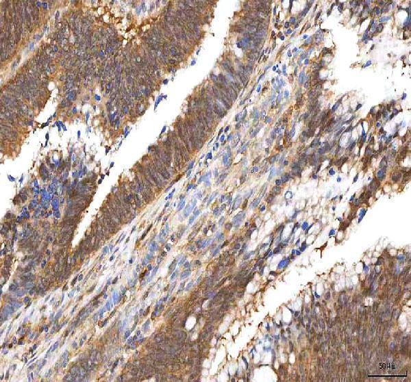
Real IHC data IHC analysis of PDXK using anti-PDXK antibody (A06683-1). PDXK was detected in a paraffin-embedded section of human colon adenocarcinoma tissue. Heat mediated antigen retrieval was performed in EDTA buffer (pH 8.0, epitope retrieval solution). The tissue section was blocked with 10% goat serum. The tissue section was then incubated with 2 μg/ml rabbit anti-PDXK Antibody (A06683-1) overnight at 4°C. Peroxidase Conjugated Goat Anti-rabbit IgG was used as secondary antibody and incubated for 30 minutes at 37°C. The tissue section was developed using HRP Conjugated Rabbit IgG Super Vision Assay Kit (Catalog # SV0002) with DAB as the chromogen.
Anti-PDXK Antibody ®
Cat # A06683-1
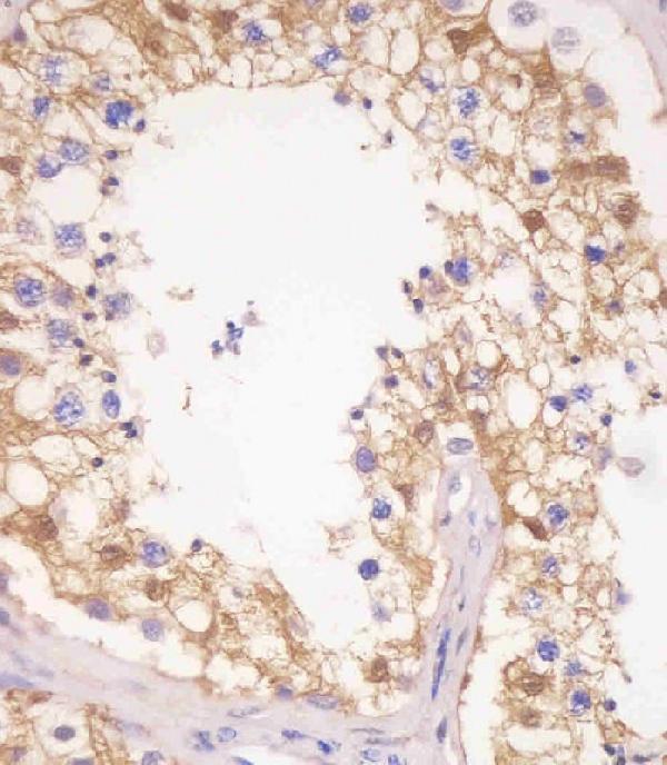
Real IHC data M06683 staining PDXK in human testis tissue sections by Immunohistochemistry (IHC-P -paraformaldehyde-fixed, paraffin-embedded sections). Tissue was fixed with formaldehyde and blocked with 3% BSA for 0. 5 hour at room temperature; antigen retrieval was by heat mediation with a citrate buffer (pH6). Samples were incubated with primary antibody (1/25) for 1 hours at 37°C. A undiluted biotinylated goat polyvalent antibody was used as the secondary antibody.
Anti-PDXK Antibody (N-term)
Cat # M06683

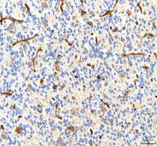
A06683-1 is listed for human, mouse and rat IHC, with images from human colon adenocarcinoma, glioblastoma and thyroid papillary carcinoma, plus mouse kidney (catalog and A06683-1 image captions). M06683 is listed for human IHC-P and shown on human testis sections (catalog and M06683 image caption).

Which to pick: For paraffin-section IHC, choose A06683-1 for human or mouse samples: its captions show EDTA retrieval at pH 8.0 and 2 μg/ml primary antibody; the fixative is unreported (A06683-1 image captions). Choose the mouse monoclonal M06683 for human testis sections if its fixation, citrate retrieval at pH 6 and 1:25 dilution fit your workflow (catalog and M06683 image caption). For rat IHC, only A06683-1 lists reactivity, without a rat IHC image; neither SKU lists IF/ICC validation, so there is no supported IF/ICC pick here (catalog and image captions). The selected A06683-1 tissue-IHC caption documents paraffin sections, but does not specify the fixative (selected-SKU IHC image A06683-1).

Each figure is that product's own IHC / IF validation image from its datasheet.

References

  1. UniProt Consortium. UniProt entry O00764 (PDXK_HUMAN, Pyridoxal kinase).
  2. Human Protein Atlas. PDXK tissue IHC expression (reliability: Enhanced).
  3. Human Protein Atlas. PDXK subcellular location (ICC-IF): Mainly localized to the nucleoplasm..
  4. Human Protein Atlas. PDXK antibody validation summary (4 antibodies).
  5. Metabolic features of cancer cells impact immunosurveillance. Journal for immunotherapy of cancer 2021 — PMC8231002.
  6. Lentivirus-mediated microRNA-124 gene-modified bone marrow mesenchymal stem cell transplantation promotes the repair of spinal cord injury in rats. Experimental & molecular medicine 2017 — PMC5454445.
  7. Identification of a prognostic cuproptosis-related signature in hepatocellular carcinoma. Biology direct 2023 — PMC9903524.
  8. Pyridoxal 5'-phosphate alleviates prenatal pyridaben exposure-induced anxiety-like behaviors in offspring. Environmental science and ecotechnology 2023 — PMC9691908.
  9. PubMed PMID:9099727 — UniProt-cited evidence.
  10. PubMed PMID:15154080 — UniProt-cited evidence.
  11. PubMed PMID:10830953 — UniProt-cited evidence.