This website uses cookies to ensure you get the best experience on our website.


- Table of Contents
Plan PDZD2 paraffin-section IHC around mainly cytoplasmic staining in connective tissue (HPA tissue IHC). Tonsil squamous epithelial cells show medium staining, but tissue results need cautious interpretation because antibody staining has low concordance with RNA expression (HPA tissue IHC).

Expected localisation, validated protocols, controls and antibodies — the at-a-glance facts below, then the full design guide.
| Expected localisation | Mainly cytoplasmic staining in connective tissue (HPA tissue IHC) | |
| Staining pattern | Connective tissue: mainly cytoplasmic staining (HPA tissue IHC) | |
| Antigen retrieval | Tris-EDTA pH 9.0 HIER, 95–98 °C, 20 min (rule: nuclear antigen) | |
| Positive control | Tonsil | |
| Negative control | Adipose tissue+4 more · see all |
| Fixation | Keep fixation consistent across sections (standard IHC practice; not target-specific) | |
| Caveat | Antibody staining has low concordance with RNA expression (HPA tissue IHC) | |
| Regulation | No specific induction cue annotated (UniProt) | |
| Isoform / epitope | 2 isoforms; processed chain spans 2493–2839. Check epitope location (UniProt) |
Compare the catalog antibody’s IHC-P protocol with a published PDZD2 tumor-tissue IHC procedure (PMC6448123).
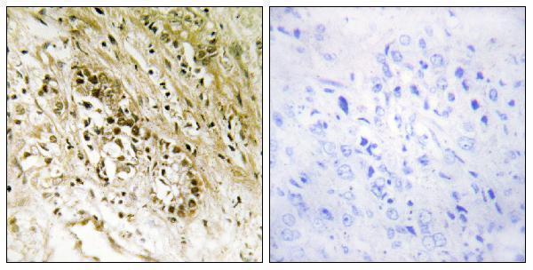
| Sample | Paraffin-embedded human heart tissue; fixative not specified (datasheet A10309-1) |
| Fixation | Image fixative and duration unreported (datasheet A10309-1); verify before use. |
| Sectioning | 4–5 µm sections on charged slides (standard) |
| Deparaffinisation | Xylene, graded ethanol series to water (standard) |
| Antigen retrieval | Heat-induced epitope retrieval in Tris-EDTA buffer, pH 9.0, 20 min at 95–98 °C (standard rule: nuclear antigen) |
| Peroxidase block | 3% H2O2, 10 min, room temperature (standard) |
| Blocking | 10% normal serum of the secondary host, 30 min, room temperature (standard) |
| Primary antibody | Rabbit anti-PDZD2, 1:100 - 1:300 (datasheet A10309-1) |
| Primary incubation | Overnight at 4 °C (standard) |
| Detection | HRP-polymer secondary, DAB chromogen 5–10 min (standard) |
| Counterstain | Hematoxylin, blue, dehydrate and mount (standard) |
| Expected result | PDZD2-positive staining in squamous epithelial cells of tonsil (HPA tissue IHC: Medium). HPA tissue profile: Mainly expressed in connective tissue and cytoplasm. No signal in the no-primary control. |
PDZD2 is reported in cytoplasm and connective tissue by tissue IHC, with medium staining in tonsil squamous epithelial cells (HPA tissue IHC). Plasma membrane and cytosol localisation is supported by ICC-IF; UniProt also lists nucleus, endoplasmic reticulum and secretion, with no transmembrane segment (HPA subcellular; UniProt O15018 topology). Treat the tissue pattern as provisional: HPA rates its IHC reliability Uncertain because antibody staining and RNA expression have low consistency (HPA tissue IHC).
| Medium chromogenic signal in tonsil squamous epithelial cells, chiefly cytoplasmic, with cell boundaries sometimes apparent (HPA tissue IHC; HPA subcellular). | This fits the reported tonsil level and cytoplasmic tissue pattern (HPA tissue IHC). Boundary staining is plausible given supported plasma membrane localisation in ICC-IF, but that evidence does not establish an IHC membrane pattern (HPA subcellular). Score the named cells and compartments separately (standard IHC practice). |
| Strong staining confined to an unexpected compartment, without the reported cytoplasmic pattern (HPA tissue IHC). | Consider artefact or off-target staining and inspect morphology and controls (standard IHC practice). Nuclear or endoplasmic reticulum signal alone cannot rule out PDZD2 because UniProt lists both locations; HPA tissue IHC reliability is Uncertain (UniProt O15018; HPA tissue IHC). |
| Prominent signal in adipocytes or appendix glandular cells, or broadly across unrelated cell types (HPA tissue IHC). | HPA reports those named cells as Not detected, so the result warrants review (HPA tissue IHC). Cross-reactivity or endogenous chromogenic activity are possible explanations, especially if signal also appears in a no-primary control (standard IHC practice). |
| Diffuse colour across tissue, stroma and blank areas obscures cell boundaries (standard IHC practice). | This does not support a cell-specific localisation call (standard IHC practice). Examine the no-primary control and detection chemistry, then optimise blocking, washing or primary concentration as indicated by the control result (standard IHC practice). |
| No signal in tonsil squamous epithelial cells reported as Medium by HPA (HPA tissue IHC). | Check tissue identity, morphology, retrieval conditions, antibody dilution and detection controls before interpreting a negative result (standard IHC practice). The HPA positive is provisional because its tissue IHC reliability is Uncertain; absence alone cannot establish biological loss (HPA tissue IHC). |
| IHC evidence strength (HPA tissue IHC; HPA antibodies). | The tissue profile is Uncertain, and each listed antibody with an IHC rating is Uncertain (HPA tissue IHC; HPA antibodies). Use the reported pattern as a comparison point, not an independently verified specificity standard (HPA tissue IHC). |
| Compartment and topology (HPA subcellular; UniProt O15018 topology). | ICC-IF supports plasma membrane and cytosol localisation, while UniProt also lists nucleus, endoplasmic reticulum and secretion (HPA subcellular; UniProt O15018). With no transmembrane segment annotated, membrane staining should not be interpreted as evidence that PDZD2 spans the membrane (UniProt O15018 topology). |
| Isoforms and tissue context (UniProt O15018; HPA tissue IHC). | UniProt records 2 isoforms and reports isoform 2 protein in prostate and many prostate tumors (UniProt O15018). That does not supply an IHC intensity or distinguish isoforms in a stained section; interpret the tonsil observation within HPA's Uncertain tissue profile (HPA tissue IHC). |
| Processing and epitope position (UniProt O15018). | UniProt lists a full-length chain at residues 1–2839 and a processed chain at 2493–2839 (UniProt O15018). Without an antibody epitope in the supplied evidence, neither chain's detection nor an effect of processing on staining can be predicted. |
| Antigen retrieval (standard IHC practice). | Document retrieval conditions and compare control sections when optimising paraffin IHC (standard IHC practice). The supplied HPA and UniProt records report no PDZD2-specific retrieval or fixation sensitivity; a changed signal cannot be assigned to PDZD2 epitope masking from these sources. |
| What does IF/ICC add? (HPA subcellular). | HPA supports plasma membrane and cytosol localisation in ICC-IF and lists images from HeLa, PC-3, U2OS, EFO-21, SiHa and NIH 3T3 (HPA subcellular). That is a localisation cross-check; this section makes no IF/ICC protocol recommendation. |
| Situation | Likely cause | Next action |
|---|---|---|
| Tonsil squamous epithelium is blank despite the reported Medium stain (HPA tissue IHC). | Tissue selection, retrieval, dilution or detection may be responsible; HPA's positive profile is itself Uncertain (standard IHC practice; HPA tissue IHC). | Confirm the cell type and section quality, then check the run's detection control and recorded retrieval and primary conditions before revising the biological interpretation (standard IHC practice). |
| Adipocytes or appendix glandular cells stain strongly although HPA reports Not detected (HPA tissue IHC). | Off-target binding or endogenous chromogenic activity is possible; HPA's negative observations are not definitive specificity tests (standard IHC practice; HPA tissue IHC). | Inspect a no-primary control and compare signal placement with the tissue morphology and the tonsil reference cells; report the discrepancy rather than calling it confirmed PDZD2 expression (standard IHC practice; HPA tissue IHC). |
| Colour is diffuse or appears in blank regions of the slide (standard IHC practice). | Background from detection chemistry, insufficient blocking or washing, or excessive primary concentration can obscure a cell-specific pattern (standard IHC practice). | Use a no-primary control to locate the background source, then adjust blocking, washes or primary concentration one condition at a time; rescore only interpretable cells (standard IHC practice). |
| Signal appears only in nuclei or another compartment, with little cytoplasmic staining (HPA tissue IHC). | This departs from HPA's chiefly cytoplasmic tissue description, although UniProt includes nuclear localisation; compartment alone cannot settle identity (HPA tissue IHC; UniProt O15018). | Check counterstain, cellular boundaries and controls, and record nuclear and cytoplasmic scores separately; treat the result as unresolved if the controls do not explain it (standard IHC practice). |
| Connective tissue staining makes epithelial scoring difficult (HPA tissue IHC). | HPA describes connective tissue expression as part of its tissue profile, so stromal colour can complicate assignment to nearby squamous epithelial cells (HPA tissue IHC). | Use morphology and the counterstain to mark epithelial and connective tissue regions, then score each separately; avoid assigning stromal colour to epithelial cells (standard IHC practice). |
| Two antibodies yield different tissue patterns (HPA antibodies). | HPA assigns Uncertain IHC status to every listed antibody with an IHC rating, and the tissue profile has low staining-to-RNA consistency (HPA antibodies; HPA tissue IHC). | Compare the same cell types, compartments and controls across sections, document the disagreement, and avoid treating agreement with one antibody as proof of specificity (standard IHC practice; HPA antibodies). |


Comprehensive Human Protein Atlas IHC scoring per tissue (reliability: Uncertain — Low consistency between antibody staining and RNA expression data. Pending external verification.). Rows are taken directly from the HPA tissue chart — click any row's HPA link to view the source.
| Tissue | Cell type | Level | Evidence | Source |
|---|---|---|---|---|
| Tonsil | Squamous epithelial cells | Medium | Protein (IHC) | HPA → |
| Tissue | Cell type | Level | Evidence | Source |
|---|---|---|---|---|
| Adipose tissue | Adipocytes | Not detected | Protein (IHC) | HPA → |
| Appendix | Glandular cells | Not detected | Protein (IHC) | HPA → |
| Bone marrow | Hematopoietic cells | Not detected | Protein (IHC) | HPA → |
| Breast | Adipocytes | Not detected | Protein (IHC) | HPA → |
| Caudate | Glial cells | Not detected | Protein (IHC) | HPA → |
Troubleshoot PDZD2 staining by keeping retrieval, detection, and scoring consistent while checking cellular location and the limits of the available tissue evidence.
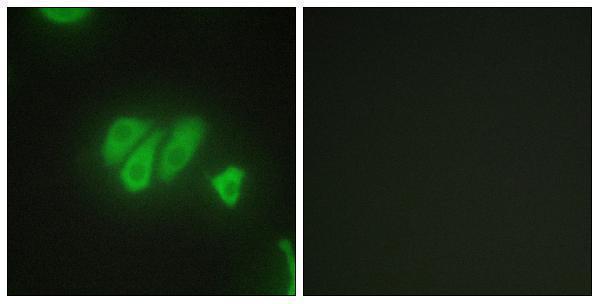
A10309-1 has IHC data from paraffin-embedded human heart and IF data from HepG2 cells (catalog: image captions); listed reactivity covers human, mouse, and rat (catalog: reactivity).
A10309-1 will render with its IHC figure from paraffin-embedded human heart, including a synthesized-peptide blocking image (catalog: IHC image caption). The same SKU lists IHC, IF, and ICC, with IF shown in HepG2 cells (catalog: applications and IF image caption).
Which to pick: For tissue IHC, choose A10309-1 at 1:100–1:300; its own figure shows paraffin-embedded human heart, but the fixative is unreported (catalog: IHC dilution and IHC image caption). For IF/ICC, choose A10309-1, a rabbit polyclonal listed for both applications at 1:200–1:1000; its IF example uses HepG2 cells (catalog: host, dilution_raw, applications, and IF image caption). For cross-species work, A10309-1 lists human, mouse, and rat reactivity; the pictured IHC example is human heart tissue (catalog: reactivity and IHC image caption).