PDZD8 / PDZ domain-containing protein 8 · IHC design guide

Design Immunohistochemistry for PDZD8

Plan chromogenic PDZD8 IHC in paraffin sections using the catalog antibody's IHC conditions (datasheet A10427-1). Compare high staining in adipocytes with undetected staining in skeletal muscle myocytes, while treating the reported tissue pattern as uncertain pending external verification (HPA tissue IHC).

Evidence assembled Oct 2026 · For research use; verify linked source records and product datasheet before use
Immunohistochemistry protocol sheet for PDZD8 (IHC for PDZD8): expected localisation Cytoplasmic tissue staining (HPA tissue IHC); ER membrane at organelle contacts (UniProt), antibody A10427-1, validated IHC image, and IHC protocol steps
Printable PDZD8 IHC protocol sheet — expected localisation Cytoplasmic tissue staining (HPA tissue IHC); ER membrane at organelle contacts (UniProt), antibody A10427-1, controls and protocol steps. Open the full PDZD8 IHC guide →

PDZD8 Immunohistochemistry Experimental Design Guide

Expected localisation, validated protocols, controls and antibodies — the at-a-glance facts below, then the full design guide.

Must know before staining
Expected localisation Cytoplasmic tissue staining (HPA tissue IHC); ER membrane at organelle contacts (UniProt)
Staining pattern General cytoplasmic staining; high in adipocytes and glandular cells (HPA tissue IHC)
Antigen retrieval EDTA pH 8.0 HIER, heat-mediated (datasheet A10427-1)
Positive control ⓘ Adipose tissue+4 more · see all
Negative control ⓘ Bone marrow+2 more · see all
Important caveats
Reasons your staining may differ from the expected pattern.
Fixation Keep fixation consistent across paraffin sections (standard IHC practice; not target-specific)
Caveat Tissue staining awaits external verification (HPA tissue IHC)
Regulation No expression regulator annotated (UniProt)
Isoform / epitope No isoforms listed; epitope orientation is unspecified (UniProt)
Section 1

Recommended PDZD8 IHC & IF Protocols

The catalog antibody protocol uses EDTA pH 8.0 retrieval (datasheet A10427-1). Two published PDZD8 IHC protocols provide additional tissue preparation, staining, and scoring details (PMC11621647; PMC8999764).

Recommended immunohistochemistry (IHC-P) protocol parameters
SampleParaffin-embedded human liver cancer tissue; fixative not specified (datasheet A10427-1)
FixationImage fixative and duration unreported (datasheet A10427-1); verify before use.
Sectioning4–5 µm sections on charged slides (standard)
DeparaffinisationXylene, graded ethanol series to water (standard)
Antigen retrievalHeat retrieval: EDTA pH 8.0 (datasheet A10427-1); 20 min, 95–100 °C (standard)
Peroxidase block3% H2O2, 10 min, room temperature (standard)
Blocking10% goat serum (datasheet A10427-1)
Primary antibodyRabbit anti-PDZD8, 2-5 μg/ml (datasheet A10427-1)
Primary incubationOvernight at 4 °C (datasheet A10427-1)
DetectionHRP-conjugated secondary, DAB chromogen (datasheet A10427-1)
CounterstainHematoxylin, blue, dehydrate and mount (standard)
Expected resultPDZD8-positive staining in adipocytes of adipose tissue (HPA tissue IHC: High). HPA tissue profile: General cytoplasmic expression. No signal in the no-primary control.
💡Decision noteStart with heat-mediated EDTA pH 8.0 retrieval for the catalog antibody (datasheet A10427-1); neither published excerpt specifies a retrieval method (PMC11621647; PMC8999764).
Section 2

What Is the Expected PDZD8 Staining Pattern?

PDZD8 is an endoplasmic reticulum membrane protein with one transmembrane segment at residues 2–24 and reported contacts with mitochondria, late endosomes and lysosomes (UniProt Q8NEN9 topology and subcellular location). In paraffin tissue sections, expect predominantly cytoplasmic staining in cells such as adipocytes, bronchial respiratory epithelium and cerebellar Purkinje cells (HPA tissue IHC: High). Treat this as a provisional pattern because HPA rates its tissue IHC reliability Uncertain, pending external verification (HPA tissue IHC).

What am I looking at on my slide?
Cytoplasmic staining is clear in adipocytes, bronchial respiratory epithelial cells or Purkinje cells (HPA tissue IHC: High).This agrees with HPA’s general cytoplasmic tissue profile and with an endoplasmic reticulum membrane protein (HPA tissue IHC; UniProt Q8NEN9 subcellular location). Record the stained cell population and intensity; HPA’s Uncertain reliability means agreement is supportive, not independent validation (HPA tissue IHC).
Staining is predominantly nuclear, nucleolar or confined to the cell surface in a paraffin section.That differs from HPA’s general cytoplasmic tissue IHC profile (HPA tissue IHC). Review morphology and controls before calling it PDZD8; nucleolar fibrillar center and additional plasma membrane staining are approved observations in ICC-IF, a different application (HPA subcellular ICC-IF).
Strong signal appears in bone marrow hematopoietic cells, lymph node germinal center cells or skeletal myocytes.These populations are listed as Not detected by HPA tissue IHC (HPA tissue IHC). Check whether the signal follows the intended cells, persists in a no-primary control, or reflects nonspecific binding; this discrepancy alone cannot establish cross-reactivity because HPA tissue IHC reliability is Uncertain (HPA tissue IHC; general IHC practice).
Weak, diffuse color covers multiple tissue compartments and obscures cell boundaries.This is harder to interpret than a cell-associated cytoplasmic pattern (HPA tissue IHC: general cytoplasmic expression). Diffuse color can reflect detection background, excess antibody or inadequate washing (general IHC practice). Compare a no-primary control and the expected positive cell population before scoring (general IHC practice).
No convincing signal appears in adipocytes, bronchial epithelium or Purkinje cells.A negative result conflicts with HPA’s High staining observations in those respective cells (HPA tissue IHC). First assess tissue preservation, retrieval, antibody and detection controls using general IHC practice; a failed run should not be interpreted as PDZD8 absence (general IHC practice).
💡Expected PDZD8 appearanceCall a result provisionally positive when distinct cytoplasmic staining is present in HPA High cell populations, such as adipocytes or bronchial respiratory epithelial cells; isolated nuclear signal, diffuse background or strong signal in HPA Not detected populations warrants control review (HPA tissue IHC: profile, levels and Uncertain reliability).
How each factor affects the staining
Membrane location and topology (UniProt Q8NEN9)PDZD8 has one transmembrane segment at residues 2–24 and is assigned to the endoplasmic reticulum membrane and organelle contact sites (UniProt Q8NEN9 topology and subcellular location). Its location supports a cytoplasmic compartment call; the supplied record does not identify the IHC antibody epitope (UniProt Q8NEN9; supplied evidence).
Tissue evidence and validation (HPA tissue IHC)HPA reports general cytoplasmic expression but rates the tissue IHC pattern Uncertain, pending external verification; antibody HPA015716 is also listed as IHC Uncertain (HPA tissue IHC; HPA antibodies). Use the listed cell patterns as comparison points, not definitive specificity proof (HPA tissue IHC).
RNA and protein readouts (HPA tissue IHC)HPA describes RNA as tissue enhanced in bone marrow while reporting hematopoietic cells there as protein Not detected by tissue IHC (HPA tissue IHC). Do not turn the RNA label into an expected positive IHC control; compare protein staining with the protein IHC observations (HPA tissue IHC).
Why is my staining missing, weak or wrong?
SituationLikely causeNext action
A HPA High cell population shows no chromogenic signal (HPA tissue IHC).The staining run may have failed, or the selected conditions may not yield detectable signal (general IHC practice). HPA’s observed intensity does not establish sensitivity in every preparation (HPA tissue IHC: Uncertain).Check tissue morphology, the run’s positive control, reagent activity and whether retrieval followed the selected IHC-P protocol; repeat with controlled changes if needed (general IHC practice).
Staining is weak across the section, including cells expected to be High (HPA tissue IHC).Antibody concentration, retrieval or detection conditions may be insufficient for this run (general IHC practice); the supplied sources give no PDZD8-specific fixation sensitivity (supplied evidence).Confirm the antibody and detection reagents, then optimize retrieval and dilution against a positive tissue control under the chosen IHC-P protocol (general IHC practice).
Diffuse color obscures the expected cytoplasmic pattern (HPA tissue IHC: general cytoplasmic expression).Excess primary or detection reagent, inadequate blocking or washing, or endogenous detection activity can increase background (general IHC practice).Compare no-primary and detection controls; adjust blocking, washing and reagent concentration according to the detection system (general IHC practice).
Strong color appears in Not detected hematopoietic cells, germinal center cells or myocytes (HPA tissue IHC).The result may reflect nonspecific binding or endogenous detection activity (general IHC practice), although HPA’s tissue IHC pattern remains Uncertain (HPA tissue IHC).Check a no-primary control and cell morphology, then compare staining with a HPA High population on the same run before assigning specificity (general IHC practice; HPA tissue IHC).
The apparent signal is chiefly nuclear or nucleolar in paraffin tissue IHC.It conflicts with HPA’s general cytoplasmic tissue IHC profile (HPA tissue IHC), though HPA reports approved nucleolar fibrillar center staining in ICC-IF (HPA subcellular ICC-IF).Inspect counterstain, localization and controls; report the discrepancy by application rather than treating the ICC-IF localization as validation of the paraffin IHC result (general IHC practice; HPA tissue IHC; HPA subcellular ICC-IF).
Q: Should nucleolar or plasma membrane ICC-IF images define the positive IHC-P pattern?HPA approves a main nucleolar fibrillar center location and an additional plasma membrane location in ICC-IF, while its tissue IHC profile is generally cytoplasmic (HPA subcellular ICC-IF; HPA tissue IHC).A: Interpret ICC-IF on its own guide page; score paraffin IHC against tissue morphology, controls and the provisional HPA tissue IHC profile (HPA tissue IHC: Uncertain; general IHC practice).

Sample controls for PDZD8 IHC & IF

🧪Run bronchus first: respiratory epithelial cells should stain (HPA: High in bronchus respiratory epithelial cells). Use skeletal muscle myocytes as the negative tissue (HPA: Not detected in skeletal muscle myocytes); on the bronchus slide, unstained non-epithelial cells provide a background reference, but the supplied HPA rows do not establish them as PDZD8-negative.
Positive control tissue: Adipose tissue (Adipocytes, HPA High)
Negative control tissue: Bone marrow (HPA Not detected)
ICC-IF cell lines (HPA subcellular resource): HPA ICC-IF images show PDZD8 in A-431, U-251MG, U2OS, NIH 3T3, with annotated localisation: Nucleoli fibrillar center (approved) (HPA subcellular).
Technical controls: Include a no-primary, secondary-only slide, a rabbit IgG isotype control matched to the primary, and a PDZD8 knockout biological control (selected-SKU caption: rabbit primary; standard IHC practice). For bronchus, quench endogenous peroxidase before DAB detection and distinguish any staining in airway mucus from cellular staining (selected-SKU caption: HRP/DAB; standard IHC practice).
⚠️Feasibility: The selected-SKU tissue-IHC caption reports heat retrieval in EDTA at pH 8.0, but does not establish that retrieval is required; its fixative and any target-specific fixation window or fixation effect are unreported (selected-SKU tissue-IHC caption). Neither frozen sections nor IF is shown to be easier for tissue detection; HPA reports ICC-IF images in cell lines, which do not establish frozen-section performance (HPA subcellular; selected-SKU tissue-IHC caption). Bronchial mucus can complicate interpretation of chromogenic signal, so score staining in respiratory epithelial cells separately from luminal material (standard IHC practice; HPA: High in bronchus respiratory epithelial cells).

HPA tissue IHC evidence for PDZD8

Comprehensive Human Protein Atlas IHC scoring per tissue (reliability: Uncertain — Pending external verification.). Rows are taken directly from the HPA tissue chart — click any row's HPA link to view the source.

Positive expression · recommended positive controls

TissueCell typeLevelEvidenceSource
Adipose tissue Adipocytes High Protein (IHC) HPA →
Adrenal gland Glandular cells High Protein (IHC) HPA →
Breast Glandular cells High Protein (IHC) HPA →
Bronchus Respiratory epithelial cells High Protein (IHC) HPA →
Caudate Glial cells High Protein (IHC) HPA →

Undetected expression · recommended negative controls

TissueCell typeLevelEvidenceSource
Bone marrow Hematopoietic cells Not detected Protein (IHC) HPA →
Lymph node Germinal center cells Not detected Protein (IHC) HPA →
Skeletal muscle Myocytes Not detected Protein (IHC) HPA →
Section 3

Advanced PDZD8 IHC Tips

Troubleshoot PDZD8 chromogenic IHC in paraffin sections by checking retrieval, cellular pattern, controls, and scoring before interpreting signal.

What retrieval conditions should I try first when PDZD8 staining is weak?
Start with heat-mediated retrieval in EDTA at pH 8.0 (datasheet A10427-1). The selected paraffin-section example used that retrieval before incubation with 2 μg/ml primary antibody overnight at 4°C (datasheet A10427-1). If staining is weak, compare retrieval times on adjacent sections while holding antibody concentration, detection, and development constant (standard IHC practice). Check tissue integrity and background alongside signal: excessive heating can damage morphology and make apparent gains difficult to interpret (standard IHC practice). The caption does not report a fixative, so its result cannot establish retrieval performance across fixation conditions (datasheet A10427-1).
Could fixation explain weak or uneven PDZD8 staining?
Target-specific fixation sensitivity is unknown from the supplied evidence (datasheet A10427-1: fixative not stated). The selected image shows PDZD8 detection in a paraffin section, but its caption gives no fixation reagent or duration (datasheet A10427-1). Record fixation conditions and compare sections processed together before changing retrieval or primary-antibody concentration (standard IHC practice). Inspect poorly stained areas for uneven preservation, tissue detachment, and lost morphology, then repeat with consistently processed sections if those problems are present (standard IHC practice). Do not assign a fixation effect to PDZD8 from its membrane topology or phosphorylation sites alone (UniProt Q8NEN9 topology and modified residues).
Which cellular pattern is plausible for PDZD8 in tissue IHC?
Look first for cell-associated cytoplasmic staining: the tissue IHC profile describes general cytoplasmic expression (HPA tissue IHC). PDZD8 is assigned to the endoplasmic-reticulum membrane and organelle contact sites, which gives a reason to examine intracellular signal closely (UniProt Q8NEN9 subcellular location). The HPA subcellular IF record instead reports a nucleolar fibrillar-center location and additional plasma-membrane signal (HPA subcellular). Compare any predominantly nuclear or surface pattern with controls before accepting it as the tissue IHC pattern (HPA tissue IHC; HPA subcellular; standard IHC practice). Because HPA rates its tissue IHC reliability Uncertain, localisation alone cannot validate a stain (HPA tissue IHC).
How should epitope uncertainty affect PDZD8 IHC troubleshooting?
Do not assume which side of the membrane the antibody recognizes: the supplied record identifies a transmembrane segment at residues 2–24 but gives no antibody epitope (UniProt Q8NEN9 topology; datasheet A10427-1). The record lists no isoforms, so an isoform-specific staining explanation is unsupported here (UniProt Q8NEN9 isoforms). Its SMP-LTD and PDZ domains occupy residues 91–294 and 366–449, respectively, but those positions do not identify the antibody binding site (UniProt Q8NEN9 domains; datasheet A10427-1). Compare retrieval conditions on adjacent sections and use an independent specificity control before attributing a changed pattern to epitope exposure (standard IHC practice).
How can IF help assess an ambiguous PDZD8 IHC pattern?
Use IF as a separate localisation check, since HPA reports nucleolar fibrillar-center and additional plasma-membrane signal in its subcellular IF record (HPA subcellular). Multiplex PDZD8 with a validated marker for the cell population being assessed, such as an endothelial-cell marker when examining colon endothelium (HPA tissue IHC: High in colon endothelial cells; standard IF practice). Choose a fluorophore in a channel with low tissue autofluorescence and include an unstained section to measure that background (standard IF practice). Because the antibody epitope and its membrane-facing side are unspecified, compare gentle permeabilisation with an unpermeabilised control before interpreting absent intracellular signal (UniProt Q8NEN9 topology; standard IF practice).
What should I check when PDZD8 DAB staining looks diffuse?
Separate chromogen background from cellular staining with a no-primary control and a matched section processed through the full detection sequence (standard IHC practice). Block endogenous peroxidase before HRP detection, and adjust DAB development if the no-primary section darkens (standard IHC practice). The selected example used 10% goat serum, a peroxidase-conjugated secondary antibody, and DAB; those conditions document one staining example, not a universal background threshold (datasheet A10427-1). If staining remains diffuse, compare primary-antibody concentration and wash conditions while keeping retrieval constant (standard IHC practice). Judge changes against preserved cell morphology and the expected cytoplasmic tissue profile (HPA tissue IHC; standard IHC practice).
How should I score PDZD8 staining across tissue sections? ⚠ ANSWER MARKED FOR VERIFICATION
Define the cell population and tissue area before scoring, then exclude folds, edges, and damaged regions consistently (standard IHC practice). Record the percentage of positive cells and staining intensity, or calculate an H-score from intensity categories for a 0–300 scale (standard IHC practice). For sparse populations, report positive-cell density per mm² and normalise it to the evaluated tissue area or the number of cells in that population (standard IHC practice). Keep retrieval, antibody incubation, DAB development, and imaging conditions matched across sections (standard IHC practice). Report cell-type scores separately because HPA lists both High and Not detected populations and rates its tissue IHC reliability Uncertain (HPA tissue IHC).
When is a PDZD8-positive IHC result convincing rather than artefactual?
A convincing result has reproducible cellular staining, appropriate control behaviour, and a pattern that can be assessed against the reported cytoplasmic tissue profile (HPA tissue IHC; standard IHC practice). For example, HPA lists colon endothelial cells as High and bone-marrow hematopoietic cells as Not detected, but calls the tissue IHC evidence Uncertain (HPA tissue IHC). Investigate predominantly nuclear staining, edge-only signal, and staining in necrotic areas before assigning them to PDZD8 (HPA tissue IHC; standard IHC practice). If a no-primary section develops DAB signal, check endogenous peroxidase activity and detection background (standard IHC practice). Treat disagreement with the reported intracellular membrane association as a reason for further validation, not proof of misidentification (UniProt Q8NEN9 subcellular location; standard IHC practice).
Boster reagents

Best PDZD8 / PDZ domain-containing protein 8 IHC Antibodies

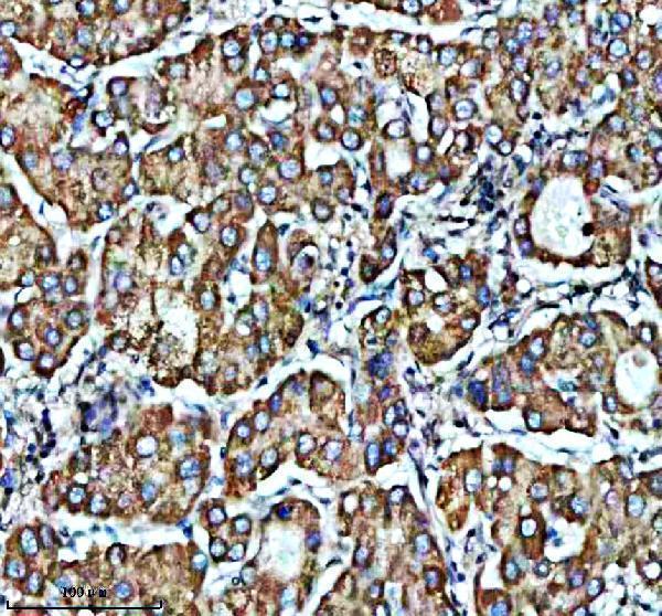
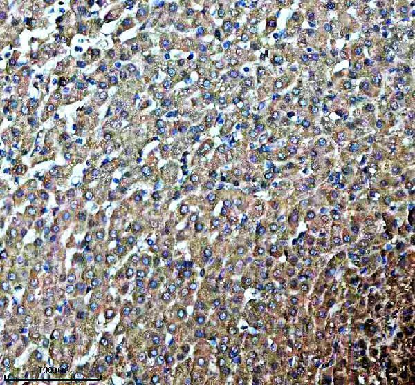
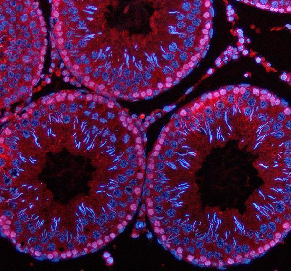
A10427-1 has IHC images from paraffin sections of human liver cancer, human liver and rat testis, plus an IF image from rat testis (catalog IHC/IF captions).

Real IHC data IHC analysis of PDZD8 using anti-PDZD8 antibody (A10427-1). PDZD8 was detected in a paraffin-embedded section of human liver cancer tissue. Heat mediated antigen retrieval was performed in EDTA buffer (pH 8.0, epitope retrieval solution). The tissue section was blocked with 10% goat serum. The tissue section was then incubated with 2 μg/ml rabbit anti-PDZD8 Antibody (A10427-1) overnight at 4°C. Peroxidase Conjugated Goat Anti-rabbit IgG was used as secondary antibody and incubated for 30 minutes at 37°C. The tissue section was developed using HRP Conjugated Rabbit IgG Super Vision Assay Kit (Catalog # SV0002) with DAB as the chromogen.
Anti-PDZD8 Antibody ®
Cat # A10427-1

A10427-1 is listed for IHC and has images from paraffin sections of human liver cancer, human liver and rat testis (datasheet applications; catalog IHC captions). It is also listed for IF, with an image from a rat testis paraffin section (datasheet applications; catalog IF caption).

Which to pick: For tissue IHC, choose A10427-1: its paraffin-section images use EDTA retrieval at pH 8.0 and 2 μg/ml primary antibody; the fixative is unreported (catalog IHC captions). For IF, A10427-1 has a rat testis paraffin-section image; ICC validation and clonality are unreported (catalog IF caption; datasheet). For cross-species IHC, A10427-1 lists Human, Mouse and Rat reactivity, with IHC images for Human and Rat only (datasheet reactivity; catalog IHC captions).

Each figure is that product's own IHC / IF validation image from its datasheet.

References

  1. UniProt Consortium. UniProt entry Q8NEN9 (PDZD8_HUMAN, PDZ domain-containing protein 8).
  2. Human Protein Atlas. PDZD8 tissue IHC expression (reliability: Uncertain).
  3. Human Protein Atlas. PDZD8 subcellular location (ICC-IF): Mainly localized to the nucleoli fibrillar center. In addition localized to the plasma membrane..
  4. Human Protein Atlas. PDZD8 antibody validation summary (2 antibodies).
  5. PDZD8 Augments Endoplasmic Reticulum-Mitochondria Contact and Regulates Ca2+ Dynamics and Cypd Expression to Induce Pancreatic β-Cell Death during Diabetes. Diabetes & metabolism journal 2024 — PMC11621647.
  6. Sunitinib and Pterostilbene Combination Treatment Exerts Antitumor Effects in Gastric Cancer via Suppression of PDZD8. International journal of molecular sciences 2022 — PMC8999764.
  7. Overexpression of miR-1283 inhibits cell proliferation and migration of colorectal cancer cells by targeting PDZD8. Translational cancer research 2025 — PMC12432640.
  8. PubMed PMID:15164054 — UniProt-cited evidence.
  9. PubMed PMID:15489334 — UniProt-cited evidence.
  10. PubMed PMID:12601173 — UniProt-cited evidence.