This website uses cookies to ensure you get the best experience on our website.


- Table of Contents
Plan chromogenic PEG10 IHC in paraffin sections using placental cytotrophoblasts as a strong-staining reference (HPA tissue IHC). Start within the catalog antibody's 1:50–1:200 IHC dilution range (datasheet A03240-2) and score cytoplasmic staining by cell type.

Expected localisation, validated protocols, controls and antibodies — the at-a-glance facts below, then the full design guide.
| Expected localisation | Cytoplasm in placental cytotrophoblasts (HPA tissue IHC) | |
| Staining pattern | Cytoplasmic staining in placental cytotrophoblasts (HPA tissue IHC) | |
| Antigen retrieval | Citrate pH 6.0 HIER, heat-mediated (datasheet A03240-2) | |
| Positive control | Placenta+3 more · see all | |
| Negative control | Adipose tissue+4 more · see all |
| Fixation | Keep fixation consistent across paraffin sections. (standard IHC practice; not target-specific) | |
| Caveat | Breast and prostate carcinomas may stain despite negative normal epithelium (UniProt) | |
| Regulation | Expressed in breast and prostate carcinomas (UniProt) | |
| Isoform / epitope | 5 isoforms; epitope coverage is unknown, with no transmembrane split between extracellular and cytoplasmic epitopes (UniProt) |
Compare the catalog antibody’s IHC-P protocol (datasheet A03240-2) with published PEG10 protocols for placenta, hepatocellular carcinoma, breast tumors, and gallbladder carcinoma (PMC10355026; PMC4614193; PMC10683152; PMC3961444).
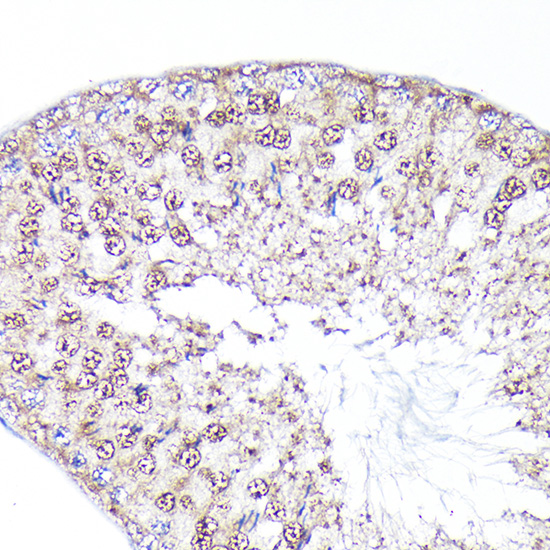
| Sample | Paraffin-embedded rat testis tissue; fixative not specified (datasheet A03240-2) |
| Fixation | Image fixative and duration unreported (datasheet A03240-2); verify before use. |
| Sectioning | 4–5 µm sections on charged slides (standard) |
| Deparaffinisation | Xylene, graded ethanol series to water (standard) |
| Antigen retrieval | Heat retrieval: Citrate pH 6.0 (datasheet A03240-2); 20 min, 95–100 °C (standard) |
| Peroxidase block | 3% H2O2, 10 min, room temperature (standard) |
| Blocking | 10% normal serum of the secondary host, 30 min, room temperature (standard) |
| Primary antibody | Rabbit anti-PEG10, 1:50-1:200 (datasheet A03240-2) |
| Primary incubation | Overnight at 4 °C (standard) |
| Detection | HRP-polymer secondary, DAB chromogen 5–10 min (standard) |
| Counterstain | Hematoxylin, blue, dehydrate and mount (standard) |
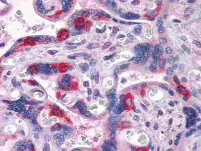
| Expected result | PEG10-positive staining in cytotrophoblasts of placenta (HPA tissue IHC: High). HPA tissue profile: Cytoplasmic expression in placental cytotrophoblasts and adrenal gland. No signal in the no-primary control. |
PEG10 staining should be predominantly cytoplasmic in placental cytotrophoblasts, with cytoplasmic expression also reported in adrenal glandular cells (HPA tissue IHC: Enhanced reliability). High staining is reported in placental cytotrophoblasts and late spermatids; adrenal glandular cells show medium staining (HPA tissue IHC). UniProt also lists cytoplasmic, nuclear and extracellular vesicle membrane localization, with no transmembrane segment (UniProt Q86TG7 topology and subcellular location).
| Placental cytotrophoblasts show strong cytoplasmic chromogen, while the overlying syncytiotrophoblast is unstained. | This is the clearest cell-level reference pattern: cytotrophoblast staining is High (HPA tissue IHC), and expression is reported in cytotrophoblasts but not overlying syncytiotrophoblast (UniProt Q86TG7 tissue specificity). Score the two layers separately rather than treating the whole placenta as uniformly positive. |
| Late spermatids stain strongly, or adrenal glandular cells stain at moderate intensity. | These are additional reported positive cell populations: High in elongated or late spermatids and Medium in adrenal glandular cells (HPA tissue IHC). The supplied HPA profile specifies cytoplasmic expression for placenta and adrenal gland; it does not specify the compartment within spermatids (HPA tissue IHC). |
| A section shows predominantly nuclear staining, with little cytoplasmic staining. | Reassess the IHC result against the predominantly cytoplasmic tissue pattern (HPA tissue IHC). Nuclear PEG10 cannot be ruled out categorically: UniProt lists nucleus, and HPA ICC-IF reports additional approved nucleoplasmic localization (UniProt Q86TG7 subcellular location; HPA subcellular ICC-IF). Nuclear-only IHC is therefore a discrepancy to investigate, not proof of specificity. |
| Strong chromogen appears in an HPA-listed negative cell population, or color spreads uniformly across the section. | Adipocytes and appendix glandular cells are reported Not detected (HPA tissue IHC). Unexpected cell staining warrants a cross-reactivity or endogenous detection-activity check (general IHC practice). Diffuse color without cell boundaries is background and cannot establish PEG10 localization (general IHC interpretation). |
| Placental cytotrophoblasts show no discernible signal. | A negative result conflicts with the reported High cytotrophoblast staining (HPA tissue IHC). Review the positive control and detection workflow before interpreting other tissues as negative (general IHC practice). The supplied sources do not establish a PEG10-specific fixation effect or an antigen retrieval requirement. |
| Cell population and tissue | PEG10 staining is cell-specific: cytotrophoblasts and late spermatids are High, while adrenal glandular cells and skin granular-layer cells are Medium (HPA tissue IHC). Adipocytes are Not detected in HPA tissue IHC, although UniProt lists expression in adipose tissue without assigning it to adipocytes (UniProt Q86TG7 tissue specificity). Compare like cell populations when scoring. |
| Subcellular location | The tissue IHC profile emphasizes cytoplasmic expression in placental cytotrophoblasts and adrenal gland (HPA tissue IHC). UniProt additionally lists nucleus and extracellular vesicle membrane and describes released virion-like vesicles (UniProt Q86TG7 subcellular location). Those annotations do not, by themselves, establish a visible membrane staining pattern in a paraffin section. |
| Isoforms and antibody epitope | UniProt lists five PEG10 isoforms (UniProt Q86TG7 isoforms). The supplied evidence does not identify the epitope recognized by a particular IHC antibody or establish equal recognition of all isoforms. Interpret staining as antibody-dependent PEG10 detection; do not assign an isoform from chromogen distribution alone. |
| Antibody validation | Three listed antibodies have Enhanced IHC status (HPA antibodies: HPA021324, HPA029915, HPA051038). The tissue profile also has Enhanced reliability, described as high consistency between staining and RNA expression (HPA tissue IHC). These support the reported pattern, while unexpected staining still needs controls on the actual section (general IHC practice). |
| Situation | Likely cause | Next action |
|---|---|---|
| Known-positive placenta is blank. | The observed result differs from High cytotrophoblast staining (HPA tissue IHC); the failed step cannot be identified from the image alone. | Check tissue preservation, retrieval, primary-antibody incubation and chromogenic detection with appropriate controls (general IHC practice). Treat retrieval as a workflow variable; no PEG10-specific retrieval or fixation sensitivity is supplied. |
| Color covers both tissue and empty slide areas. | A distribution without cell boundaries is consistent with nonspecific deposition or detection background (general IHC practice), rather than the cytoplasmic placental profile (HPA tissue IHC). | Inspect no-primary and detection controls, blocking, washes and chromogen development (general IHC practice). Re-score only bounded cellular staining after background is controlled. |
| Unexpected cells stain as strongly as cytotrophoblasts. | This may reflect cross-reactivity or endogenous detection activity (general IHC practice). HPA reports some populations, including adipocytes, as Not detected (HPA tissue IHC). | Identify the exact stained cell population, compare it with the HPA tissue entry, and review an appropriate negative control (general IHC practice). Do not label an entire tissue negative when its cell populations differ. |
| Staining is mainly nuclear in placenta. | It departs from the cytoplasmic placental IHC profile (HPA tissue IHC), although nuclear localization is listed by UniProt and nucleoplasmic localization is approved in HPA ICC-IF (UniProt Q86TG7; HPA subcellular ICC-IF). | Check whether cytoplasmic signal is also present, review controls, and compare another IHC-validated antibody if available (HPA antibodies: three Enhanced IHC entries; general IHC practice). Report the compartment observed rather than forcing a cytoplasmic score. |
| Adipocytes are negative despite a general expression listing for adipose tissue. | The annotations have different resolution: HPA reports adipocytes Not detected, while UniProt lists expression in adipose tissue without specifying adipocytes (HPA tissue IHC; UniProt Q86TG7 tissue specificity). | Score the identified adipocytes against the cell-level HPA result. Keep the tissue-level UniProt listing as context rather than treating it as a predicted positive adipocyte control. |
| Can an IF/ICC image settle an ambiguous paraffin IHC result? | The assays provide related but different observations: HPA ICC-IF approves mainly cytosolic and additional nucleoplasmic localization, whereas its tissue IHC profile describes cytoplasmic placental and adrenal expression (HPA subcellular ICC-IF; HPA tissue IHC). | Use the IF/ICC localization as context, then resolve the paraffin-section result with its own cell-level pattern and IHC controls (general IHC practice). Follow the separate IF/ICC guide for that application. |


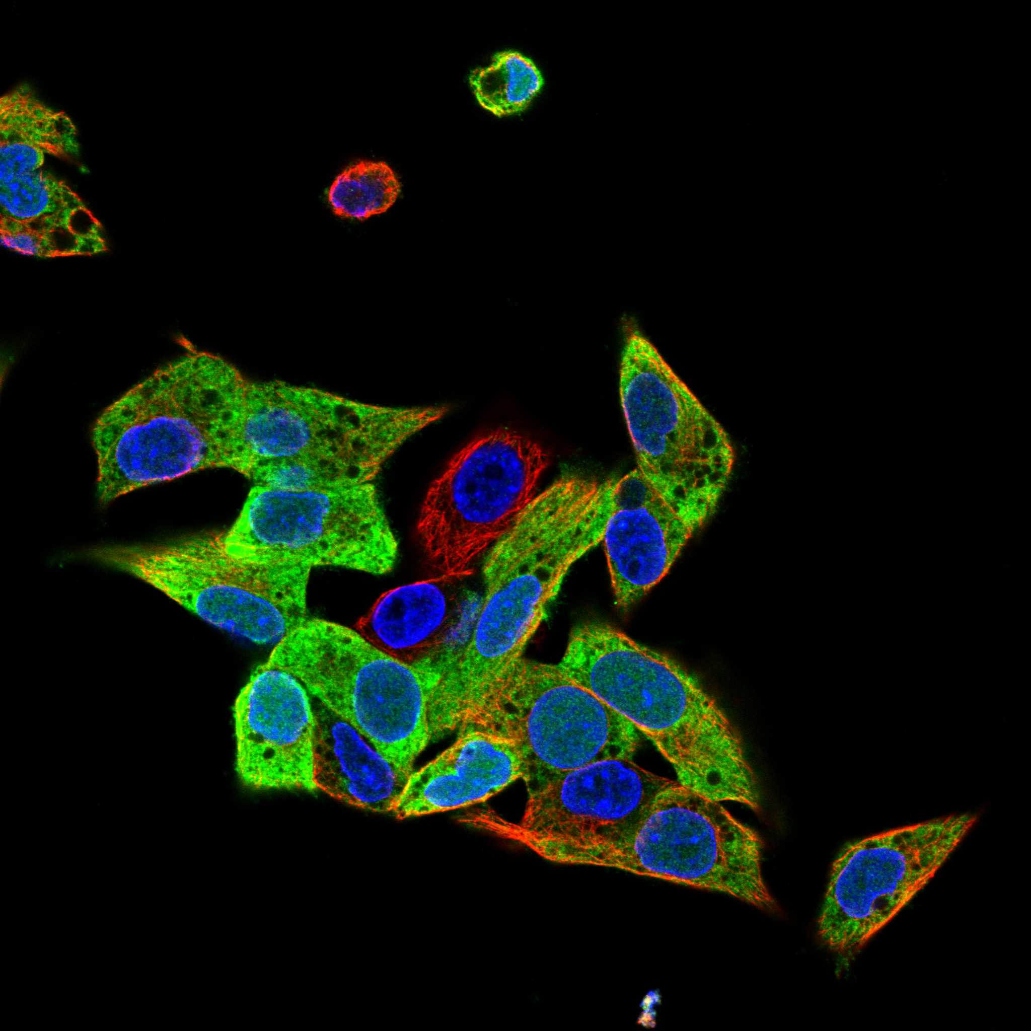
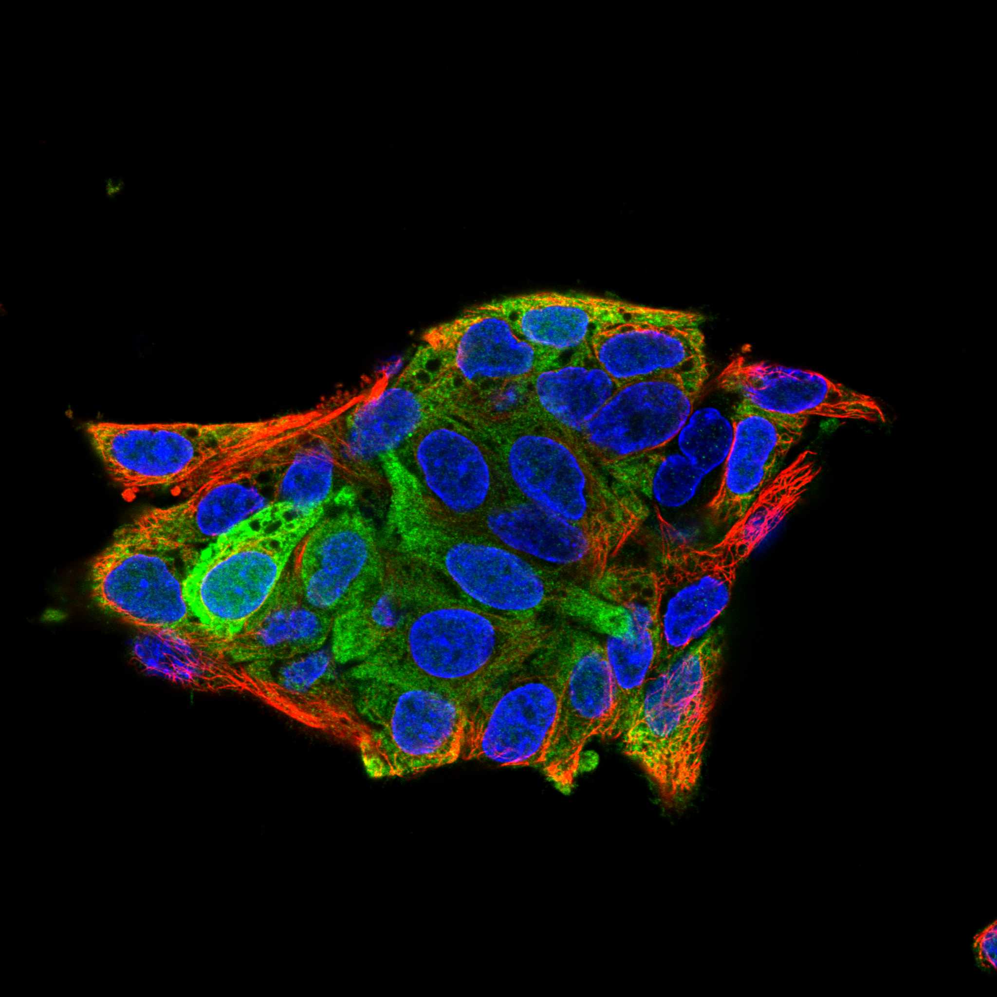
Comprehensive Human Protein Atlas IHC scoring per tissue (reliability: Enhanced — High consistency between antibody staining and RNA expression data.). Rows are taken directly from the HPA tissue chart — click any row's HPA link to view the source.
| Tissue | Cell type | Level | Evidence | Source |
|---|---|---|---|---|
| Placenta | Cytotrophoblasts | High | Protein (IHC) | HPA → |
| Testis | Elongated or late spermatids | High | Protein (IHC) | HPA → |
| Adrenal gland | Glandular cells | Medium | Protein (IHC) | HPA → |
| Skin | Cells in granular layer | Medium | Protein (IHC) | HPA → |
| Tissue | Cell type | Level | Evidence | Source |
|---|---|---|---|---|
| Adipose tissue | Adipocytes | Not detected | Protein (IHC) | HPA → |
| Appendix | Glandular cells | Not detected | Protein (IHC) | HPA → |
| Bone marrow | Hematopoietic cells | Not detected | Protein (IHC) | HPA → |
| Breast | Adipocytes | Not detected | Protein (IHC) | HPA → |
| Bronchus | Respiratory epithelial cells | Not detected | Protein (IHC) | HPA → |
Troubleshoot PEG10 staining by checking retrieval, cell type and subcellular pattern before comparing signal across sections (datasheet A03240-2; HPA tissue IHC; HPA subcellular).
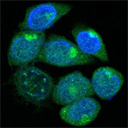
Two anti-PEG10 antibodies have paraffin-section IHC images from human tissue and rat testis (catalog IHC captions); one also has IF data from methanol-fixed HepG2 cells (A03240 IF caption).
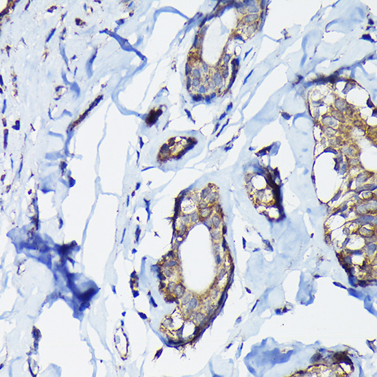
A03240-2 has IHC images of paraffin-embedded rat testis, human breast cancer, and human placenta (A03240-2 IHC captions). A03240 has an IHC image of paraffin-embedded human placenta and an IF image of methanol-fixed HepG2 cells (A03240 IHC and IF captions).
Which to pick: For tissue IHC, choose the rabbit antibody A03240-2 when its rat testis or human tissue examples fit your sample; its captions specify citrate retrieval at pH 6.0 and 1:100 antibody dilution (A03240-2 IHC captions). For IF in cultured cells, choose the mouse monoclonal A03240, which lists IF and shows cytoplasmic staining in methanol-fixed HepG2 cells (A03240 applications and IF caption). For mouse or rat tissue IHC, A03240-2 lists those species, while A03240 lists human only; both IHC captions describe paraffin-embedded sections, but neither reports the tissue fixative (catalog reactivity and IHC captions).