PFKM / ATP-dependent 6-phosphofructokinase, muscle type · IHC design guide

Design Immunohistochemistry for PFKM

Plan chromogenic PFKM staining in paraffin sections using the expected cytoplasmic pattern (HPA tissue IHC). Start with the catalog antibody’s 2–5 μg/ml IHC range (datasheet A00437-2) and interpret tissue comparisons in light of the reported low consistency between staining and RNA data (HPA tissue IHC).

Evidence assembled Oct 2026 · For research use; verify linked source records and product datasheet before use
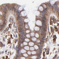
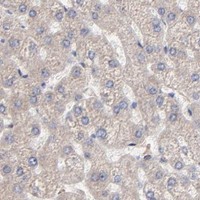
Immunohistochemistry protocol sheet for PFKM (IHC for PFKM): expected localisation General cytoplasmic staining (HPA tissue IHC), antibody A00437-2, validated IHC image, and IHC protocol steps
Printable PFKM IHC protocol sheet — expected localisation General cytoplasmic staining (HPA tissue IHC), antibody A00437-2, controls and protocol steps. Open the full PFKM IHC guide →

PFKM Immunohistochemistry Experimental Design Guide

Expected localisation, validated protocols, controls and antibodies — the at-a-glance facts below, then the full design guide.

Must know before staining
Expected localisation General cytoplasmic staining (HPA tissue IHC)
Staining pattern Cytoplasmic staining in glandular and neuronal cells (HPA tissue IHC)
Antigen retrieval EDTA pH 8.0 HIER, heat-mediated (datasheet A00437-2)
Positive control ⓘ Adrenal gland+4 more · see all
Negative control ⓘ Adipose tissue+1 more · see all
Important caveats
Reasons your staining may differ from the expected pattern.
Fixation Keep fixation consistent across sections (standard IHC practice; not target-specific)
Caveat Antibody staining has low consistency with RNA data (HPA tissue IHC)
Regulation RNA enriched in skeletal muscle and tongue (HPA RNA)
Isoform / epitope 3 isoforms; verify epitope coverage (UniProt)
Section 1

Recommended PFKM IHC & IF Protocols

The catalog antibody’s IHC-P protocol (datasheet A00437-2) is accompanied by published paraffin-section protocols for mouse kidney (PMC12932785) and processed tissues (PMC10795323).

Recommended immunohistochemistry (IHC-P) protocol parameters
SampleParaffin-embedded human endometrioid adenocarcinoma tissue; fixative not specified (datasheet A00437-2)
FixationImage fixative and duration unreported (datasheet A00437-2); verify before use.
Sectioning4–5 µm sections on charged slides (standard)
DeparaffinisationXylene, graded ethanol series to water (standard)
Antigen retrievalHeat retrieval: EDTA pH 8.0 (datasheet A00437-2); 20 min, 95–100 °C (standard)
Peroxidase block3% H2O2, 10 min, room temperature (standard)
Blocking10% goat serum (datasheet A00437-2)
Primary antibodyRabbit anti-PFKM, 2-5 μg/ml (datasheet A00437-2)
Primary incubationOvernight at 4 °C (datasheet A00437-2)
DetectionHRP-conjugated secondary, DAB chromogen (datasheet A00437-2)
CounterstainHematoxylin, blue, dehydrate and mount (standard)
Expected resultPFKM-positive staining in glandular cells of adrenal gland (HPA tissue IHC: High). HPA tissue profile: General cytoplasmic expression. No signal in the no-primary control.
💡Decision noteStart with heat-mediated EDTA pH 8.0 retrieval (datasheet A00437-2); a published protocol also used boiling sodium citrate or EDTA for 20 min (PMC10795323).
Section 2

What Is the Expected PFKM Staining Pattern?

PFKM is a cytoplasmic enzyme with no annotated transmembrane segment (UniProt P08237: location and topology). In paraffin-section IHC, expect cytoplasmic staining in cell populations reported as high by HPA, including glandular cells in colon and appendix, respiratory epithelial cells in bronchus, and neuronal cells in cerebral cortex (HPA: tissue IHC). HPA rates its tissue IHC profile Approved but reports low consistency between antibody staining and RNA expression data (HPA: reliability).

What am I looking at on my slide?
Cytoplasmic chromogen in colon glandular cells, with cellular outlines still readable.This fits a reported high-staining cell population and the expected compartment (HPA: High in colon glandular cells; UniProt P08237: Cytoplasm). Compare cells within the section before treating overall color intensity as a measure of PFKM abundance (standard IHC practice).
Strong nuclear-only or sharply membrane-restricted staining dominates the slide.This does not fit the reported general cytoplasmic IHC profile or the lack of a transmembrane segment (HPA: tissue IHC profile; UniProt P08237: topology). Treat it as a possible staining artefact and check controls before assigning it to PFKM (standard IHC practice).
Adipocytes or esophageal squamous epithelial cells stain strongly while expected positive cells do not.Those cell populations are reported as not detected, so strong selective staining there warrants a specificity check (HPA: adipocytes and esophageal squamous epithelial cells, Not detected). Cross-reactivity is possible; with enzyme-based detection, endogenous activity is another conditional explanation (standard IHC practice).
Color spreads across extracellular spaces and many cell types, obscuring cytoplasmic boundaries.A widespread haze cannot establish the cell-specific cytoplasmic pattern reported for PFKM (HPA: general cytoplasmic expression). Assess the negative control, blocking and wash conditions, and chromogen development before scoring cells (standard IHC practice).
No cellular signal appears in colon glandular cells or cerebral-cortex neuronal cells.Both are reported as high by HPA, so an entirely blank expected-positive region calls for a run-level check (HPA: High in colon glandular cells and cerebral-cortex neuronal cells). Review tissue preservation, retrieval, primary-antibody application and detection controls; a blank slide alone does not identify which step failed (standard IHC practice).
💡Expected PFKM appearanceCall a result consistent with PFKM when clear cytoplasmic staining appears in an HPA high-staining cell population, such as colon glandular cells or cortical neurons; isolated nuclear or membrane-restricted color, or strong staining confined to HPA not-detected cells, is suspect (UniProt P08237: Cytoplasm and topology; HPA: tissue IHC profile and levels).
How each factor affects the staining
Tissue and cell populationHPA reports High staining in several distinct populations, including bone-marrow hematopoietic cells and cerebellar granular-layer cells, but Not detected in adipocytes and esophageal squamous epithelial cells (HPA: tissue IHC). Interpret the named cell population, rather than assigning one expected intensity to every cell in an organ.
Strength of tissue evidenceThe HPA tissue IHC profile is Approved, with low consistency between antibody staining and RNA expression data (HPA: reliability). Use its reported staining levels as interpretation guides, not as proof that every specimen or antibody run must match them exactly.
Isoforms and antibody scopeUniProt lists three PFKM isoforms (UniProt P08237: isoforms 1–3). The supplied record gives no epitope or isoform-reactivity information for the IHC antibody, so staining cannot be assigned to a particular isoform from this evidence.
Tetramer compositionPFKM can form PFK tetramers with subunits whose composition differs by tissue; muscle is described as M4, while liver predominantly contains PFKL L4 (UniProt P08237: subunit). This biology does not by itself predict chromogenic IHC intensity or establish antibody cross-reactivity.
IF/ICC Q&A: Is endoplasmic-reticulum staining expected?HPA reports mainly endoplasmic-reticulum localization in ICC-IF, with additional principal-piece localization, whereas its tissue IHC profile is general cytoplasmic expression (HPA: subcellular ICC-IF; HPA: tissue IHC). Interpret each application against its own reported pattern; the ICC-IF observation does not define an IHC protocol or require an ER-restricted chromogenic result.
Why is my staining missing, weak or wrong?
SituationLikely causeNext action
Expected-positive cells are blank throughout the IHC run.A failed or weak staining run is possible; colon glandular cells and cortical neurons are reported High (HPA: tissue IHC).Check a documented positive section and the run controls, then review retrieval, primary-antibody application and detection in sequence (standard IHC practice). Do not infer PFKM absence from an unvalidated blank run.
Most structures acquire a uniform brown haze.Diffuse color can arise from nonspecific reagent retention or excessive chromogen development (standard IHC practice).Compare the negative control, inspect wash and blocking steps, and assess development time before scoring cytoplasmic cells (standard IHC practice).
Color persists in a section processed without primary antibody.The primary-independent signal may reflect endogenous enzyme activity when enzyme-based chromogenic detection is used (standard IHC practice).Review the detection chemistry and its appropriate endogenous-activity control or blocking step; interpret the tissue section only after control signal is resolved (standard IHC practice).
Strong staining appears mainly in adipocytes or esophageal squamous cells.Both are HPA not-detected populations; the pattern may reflect cross-reactivity or background rather than the reported PFKM profile (HPA: tissue IHC).Compare an expected-positive cell population and negative control in the same run, and confirm cellular localization before assigning the signal to PFKM (standard IHC practice).
A reader scores a nuclear-only or membrane-rim pattern as positive.That distribution conflicts with cytoplasmic localization and the absence of a transmembrane segment (UniProt P08237: location and topology; HPA: tissue IHC profile).Reinspect counterstain and cell boundaries, compare controls, and score only interpretable cellular staining; investigate the discordant pattern separately (standard IHC practice).
An IF image appears ER-enriched while IHC looks broadly cytoplasmic.These are the distinct reported application patterns: ER-mainly in ICC-IF and general cytoplasmic expression in tissue IHC (HPA: subcellular ICC-IF; HPA: tissue IHC).Record the application and compartment with each observation. Judge the paraffin-section IHC result against tissue IHC evidence; consult the separate IF/ICC guide for that application.

Sample controls for PFKM IHC & IF

🧪Run bone marrow first and score staining in hematopoietic cells (HPA: High in bone marrow hematopoietic cells). Run adipose tissue as the negative tissue (HPA: Not detected in adipocytes); on the positive slide, treat unstained non-hematopoietic cells, if present, as tentative internal negative comparators because their PFKM status is not specified by HPA.
Positive control tissue: Adrenal gland (Glandular cells, HPA High)
Negative control tissue: Adipose tissue (HPA Not detected)
ICC-IF cell lines (HPA subcellular resource): HPA ICC-IF images show PFKM in A-431, U-251MG, U2OS, Sperm, with annotated localisation: Endoplasmic reticulum (approved) (HPA subcellular).
Technical controls: Include a no-primary, secondary-only control, a concentration-matched rabbit IgG isotype control, and PFKM knockout tissue or a peptide-block control when a suitable blocking peptide is available (caption: rabbit primary antibody). Quench endogenous peroxidase in bone marrow before HRP/DAB detection because myeloid cells can produce background signal (HPA: bone marrow hematopoietic cells; caption: HRP/DAB detection).
⚠️Feasibility: A target-specific fixation window or fixation effect is unreported, and the selected A00437-2 tissue-IHC caption does not state a fixative (selected-SKU caption: fixative not stated). The caption uses heat retrieval in EDTA at pH 8.0, but it does not establish that PFKM staining depends on that retrieval condition; neither frozen sections nor IF are shown to be easier by the supplied evidence (selected-SKU caption: EDTA retrieval). In bone marrow, endogenous myeloid peroxidase can complicate chromogenic scoring, so interpret staining alongside the peroxidase-quenched controls (HPA: bone marrow hematopoietic cells; caption: HRP/DAB detection).

HPA tissue IHC evidence for PFKM

Comprehensive Human Protein Atlas IHC scoring per tissue (reliability: Approved — Low consistency between antibody staining and RNA expression data.). Rows are taken directly from the HPA tissue chart — click any row's HPA link to view the source.

Positive expression · recommended positive controls

TissueCell typeLevelEvidenceSource
Adrenal gland Glandular cells High Protein (IHC) HPA →
Appendix Glandular cells High Protein (IHC) HPA →
Bone marrow Hematopoietic cells High Protein (IHC) HPA →
Bronchus Respiratory epithelial cells High Protein (IHC) HPA →
Caudate Neuronal cells High Protein (IHC) HPA →

Undetected expression · recommended negative controls

TissueCell typeLevelEvidenceSource
Adipose tissue Adipocytes Not detected Protein (IHC) HPA →
Esophagus Squamous epithelial cells Not detected Protein (IHC) HPA →
Section 3

Advanced PFKM IHC Tips

Troubleshoot PFKM chromogenic IHC in paraffin sections using the catalog antibody’s documented conditions and cell-specific staining controls (caption A00437-2; HPA tissue IHC).

How should I adjust retrieval when PFKM staining is weak?
Start with heat-mediated antigen retrieval in EDTA at pH 8.0 for paraffin sections (datasheet A00437-2). The documented section then received 2 µg/mL primary antibody overnight at 4°C, providing a useful starting condition for comparison (caption A00437-2). Keep section preparation, heating, and cooling consistent across test and control slides so retrieval changes can be assessed fairly (standard IHC practice). If staining remains weak, adjust heating duration on matched sections; consider another buffer only as a fallback after checking tissue preservation and background (standard IHC practice). Judge improvement by the expected cellular distribution, not intensity alone (HPA tissue IHC; standard IHC practice).
Could fixation explain uneven PFKM staining in paraffin sections?
The documented paraffin-section image does not state its fixative, so PFKM-specific fixation sensitivity is unknown (caption A00437-2). Review each specimen’s recorded fixative, fixation interval, and processing history before attributing patchiness to the antibody (standard IHC practice). Compare sections processed together, using the documented EDTA retrieval at pH 8.0 and 2 µg/mL primary antibody as starting conditions (datasheet A00437-2; caption A00437-2). Include an appropriate positive control and assess whether uneven staining follows section folds, damaged tissue, or processing boundaries (standard IHC practice). Neither tissue staining patterns nor PFKM topology establish a target-specific fixation effect (HPA tissue IHC; UniProt P08237 topology).
Should PFKM appear diffusely cytoplasmic or concentrated near the endoplasmic reticulum?
Evaluate chromogenic staining primarily in the cytoplasm, consistent with PFKM’s annotated location and the tissue IHC profile (UniProt P08237 subcellular location; HPA tissue IHC). Cell imaging additionally places PFKM mainly at the endoplasmic reticulum, so a cytoplasmic pattern with regional concentration can warrant closer examination (HPA subcellular). Compare that pattern across intact cells and nearby controls before interpreting a sharply nuclear or extracellular deposit as PFKM (standard IHC practice; UniProt P08237 subcellular location). Tissue IHC and cell imaging sample different preparations, so their apparent patterns need not match pixel for pixel (HPA tissue IHC; HPA subcellular). Record compartment and cell type separately when reviewing slides (standard IHC practice).
Can this stain distinguish PFKM isoforms or other phosphofructokinase subunits?
Do not score an IHC-positive cell as expressing a particular PFKM isoform without epitope-specific evidence: 3 isoforms are listed, but the supplied antibody information does not map its epitope (UniProt P08237 isoforms; caption A00437-2). PFKM can form tetramers with PFKL or PFKP subunits, so enzyme-complex composition cannot be inferred from a PFKM stain alone (UniProt P08237 subunit). PFKM has no annotated transmembrane segment, and its listed modifications include phosphorylation at residues 133, 377, and 667 (UniProt P08237 topology; modified residues). Their effect on antibody binding is unknown without epitope mapping (UniProt P08237 modified residues; caption A00437-2). Use independently validated specificity controls if an isoform-level conclusion matters (standard IHC practice).
How should I investigate PFKM localisation by multiplex immunofluorescence?
Treat IF as a separate assay from this paraffin-section chromogenic IHC workflow; the supplied antibody image documents IHC conditions only (caption A00437-2). Pair PFKM with a validated marker of the expected cell type, such as a neuronal marker when examining cerebral cortex, where neuronal staining is reported high (HPA tissue IHC; standard IF practice). Select spectrally separated fluorophores and consider a far-red channel when tissue autofluorescence obscures shorter-wavelength signal (standard IF practice). For an intracellular epitope, optimize mild permeabilisation because PFKM is cytoplasmic and lacks a transmembrane segment; the epitope’s exact accessibility is unreported (UniProt P08237 subcellular location; topology; caption A00437-2). Include single-label and secondary-only controls when judging colocalisation (standard IF practice).
What causes diffuse brown background in PFKM chromogenic IHC?
First inspect a no-primary control for secondary-antibody staining and endogenous peroxidase activity before calling diffuse brown signal PFKM (standard IHC practice). The documented section used 10% goat-serum block, a peroxidase-conjugated secondary for 30 minutes at 37°C, and DAB development (caption A00437-2). Peroxidase blocking and controlled DAB development are general chromogenic IHC steps, not evidence that PFKM itself creates background (standard IHC practice). Compare primary-antibody concentration, washing, and chromogen development on matched sections when troubleshooting nonspecific staining (standard IHC practice). Assess cellular staining separately from pigment, tissue edges, folds, and damaged areas (standard IHC practice).
How should I quantify PFKM staining across heterogeneous tissue sections? ⚠ ANSWER MARKED FOR VERIFICATION
Score cytoplasmic signal within a prespecified cell population, since tissue IHC describes general cytoplasmic expression and different cell types show different staining levels (HPA tissue IHC). An H-score combines the percentages of cells at intensities 0–3 into a 0–300 scale; record percent positive cells alongside it (standard IHC scoring practice). Normalize cell-based scores to evaluable, viable cells of that population, or report positive-cell density per mm² of defined viable tissue (standard IHC quantification practice). Use the same retrieval, imaging, and scoring thresholds across samples (standard IHC practice). Keep glandular, neuronal, and other cell compartments separate rather than averaging unlike populations (HPA tissue IHC; standard IHC practice).
How can I distinguish convincing PFKM staining from artefact?
A convincing result shows reproducible intracellular staining in intact cells, with a mainly cytoplasmic pattern compatible with the tissue IHC profile (HPA tissue IHC; standard IHC practice). Check whether signal appears in expected populations, such as cerebral-cortex neurons or colon glandular cells, while interpreting adipocytes and esophageal squamous cells cautiously because they were reported as undetected (HPA tissue IHC). Nuclear-only deposits, edge staining, necrotic regions, or signal persisting without primary antibody warrant investigation as artefact (UniProt P08237 subcellular location; standard IHC practice). Exclude endogenous peroxidase contribution with appropriate controls before attributing DAB signal to PFKM (standard IHC practice). Tissue staining has low consistency with RNA expression, so avoid treating one positive field as conclusive validation (HPA tissue IHC).
Boster reagents

Best PFKM / ATP-dependent 6-phosphofructokinase, muscle type IHC Antibodies

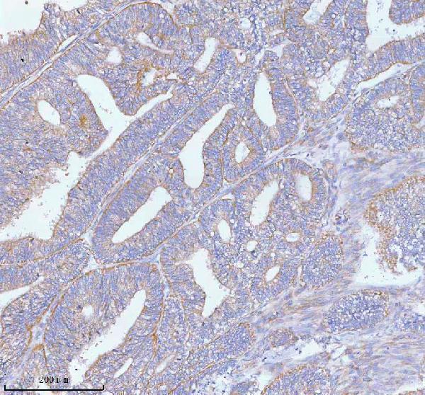
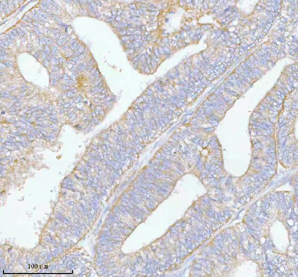
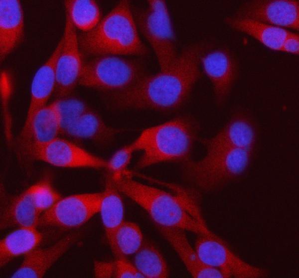
The IHC-validated antibody A00437-2 has paraffin-section images from human endometrioid and rectum adenocarcinoma and an IF image from HELA cells (A00437-2 image captions). Its listed reactivity includes human, mouse and rat (catalog).

Real IHC data IHC analysis of PFKM using anti-PFKM antibody (A00437-2). PFKM was detected in a paraffin-embedded section of human endometrioid adenocarcinoma tissue. Heat mediated antigen retrieval was performed in EDTA buffer (pH 8.0, epitope retrieval solution). The tissue section was blocked with 10% goat serum. The tissue section was then incubated with 2 μg/ml rabbit anti-PFKM Antibody (A00437-2) overnight at 4°C. Peroxidase Conjugated Goat Anti-rabbit IgG was used as secondary antibody and incubated for 30 minutes at 37°C. The tissue section was developed using HRP Conjugated Rabbit IgG Super Vision Assay Kit (Catalog # SV0002) with DAB as the chromogen.
Anti-PFKM Antibody ®
Cat # A00437-2

A00437-2 is the SKU with a rendered card; its IHC figure shows a paraffin section of human endometrioid adenocarcinoma (A00437-2 IHC image caption). Its IF caption shows HELA cells, and the catalog lists IHC and IF applications with human, mouse and rat reactivity (A00437-2 IF image caption; catalog).

Which to pick: Choose A00437-2 for tissue IHC: its own caption documents heat retrieval in EDTA at pH 8.0, primary antibody at 2 μg/ml, and chromogenic detection in a human paraffin section; the fixative is unreported (A00437-2 IHC image caption). Choose A00437-2 for IF/ICC when an image from the intended application matters; M04370-1 is a rabbit monoclonal with IF/ICC listed, but no IF image is supplied for it (A00437-2 IF image caption; M04370-1 catalog). For cross-species work, both SKUs list human, mouse and rat reactivity, while the supplied IHC and IF images for A00437-2 show human samples only (catalog; A00437-2 image captions).

Each figure is that product's own IHC / IF validation image from its datasheet.

References

  1. UniProt Consortium. UniProt entry P08237 (PFKAM_HUMAN, ATP-dependent 6-phosphofructokinase, muscle type).
  2. Human Protein Atlas. PFKM tissue IHC expression (reliability: Approved).
  3. Human Protein Atlas. PFKM subcellular location (ICC-IF): Mainly localized to the endoplasmic reticulum. In addition localized to the principal piece..
  4. Human Protein Atlas. PFKM antibody validation summary (1 antibodies).
  5. The glycolytic enzyme PFKM promotes renal fibrosis by activating the NF-κB pathway via lactate-mediated H3K18 lactylation. Cellular and molecular life sciences : CMLS 2026 — PMC12932785.
  6. USP7 facilitates brain tumor survival upon glucose deprivation by regulating phosphofructokinase muscle-type nuclear translocation in mice. PLoS biology 2026 — PMC12981444.
  7. NAT10 mediated ac4C acetylation driven m(6)A modification via involvement of YTHDC1-LDHA/PFKM regulates glycolysis and promotes osteosarcoma. Cell communication and signaling : CCS 2024 — PMC10795323.
  8. PFKM gene defect and glycogen storage disease GSDVII with misleading enzyme histochemistry. Neurology. Genetics 2015 — PMC4821086.
  9. PubMed PMID:1833270 — UniProt-cited evidence.
  10. PubMed PMID:2526045 — UniProt-cited evidence.
  11. PubMed PMID:2822475 — UniProt-cited evidence.