This website uses cookies to ensure you get the best experience on our website.


- Table of Contents
Plan PFKP chromogenic IHC on paraffin sections using the catalog antibody’s documented conditions. Expect cytoplasmic staining (HPA tissue IHC), and interpret intensity cautiously because tissue staining has low concordance with RNA data (HPA tissue IHC).

Expected localisation, validated protocols, controls and antibodies — the at-a-glance facts below, then the full design guide.
| Expected localisation | General cytoplasmic tissue staining (HPA tissue IHC) | |
| Staining pattern | Cytoplasmic staining in glandular and endothelial cells (HPA tissue IHC) | |
| Antigen retrieval | EDTA pH 8.0 HIER, heat-mediated (datasheet A07337-3) | |
| Positive control | Adrenal gland+4 more · see all | |
| Negative control | Adipose tissue |
| Fixation | Keep fixation consistent across sections (standard IHC practice; not target-specific) | |
| Caveat | Tissue staining has low concordance with RNA data (HPA tissue IHC) | |
| Regulation | Staining regulation is not established (UniProt) | |
| Isoform / epitope | 2 isoforms; check antibody epitope coverage (UniProt) |
The catalog antibody uses EDTA pH 8.0 retrieval (datasheet A07337-3). Three published PFKP IHC protocols provide comparisons (PMC13502873; PMC7259711; PMC7210009).
| Sample | Paraffin-embedded human colon adenocarcinoma tissue; fixative not specified (datasheet A07337-3) |
| Fixation | Image fixative and duration unreported (datasheet A07337-3); verify before use. |
| Sectioning | 4–5 µm sections on charged slides (standard) |
| Deparaffinisation | Xylene, graded ethanol series to water (standard) |
| Antigen retrieval | Heat retrieval: EDTA pH 8.0 (datasheet A07337-3); 20 min, 95–100 °C (standard) |
| Peroxidase block | 3% H2O2, 10 min, room temperature (standard) |
| Blocking | 10% goat serum (datasheet A07337-3) |
| Primary antibody | Rabbit anti-PFKP, 2-5 μg/ml (datasheet A07337-3) |
| Primary incubation | Overnight at 4 °C (datasheet A07337-3) |
| Detection | HRP-conjugated secondary, DAB chromogen (datasheet A07337-3) |
| Counterstain | Hematoxylin, blue, dehydrate and mount (standard) |
| Expected result | PFKP-positive staining in glandular cells of adrenal gland (HPA tissue IHC: High). HPA tissue profile: General cytoplasmic expression. No signal in the no-primary control. |
PFKP is a cytoplasmic enzyme without a transmembrane segment, so interpretable staining should favor cell cytoplasm (UniProt Q01813: subcellular location and topology). HPA reports high IHC staining in several glandular, respiratory epithelial, glial, and endothelial cell populations, with a general cytoplasmic profile (HPA: tissue IHC). Treat these as provisional expectations: HPA rates the tissue staining Uncertain because antibody staining and RNA expression show low consistency (HPA: tissue IHC reliability).
| Cytoplasmic staining in adrenal or breast glandular cells, or bronchial respiratory epithelium (HPA: High in each population). | This fits the reported tissue pattern and PFKP location (HPA: tissue IHC; UniProt Q01813: Cytoplasm). Judge the relevant cells against nearby tissue and controls; intensity alone cannot establish specificity because HPA rates its tissue IHC evidence Uncertain (HPA: reliability). |
| A dominant nuclear, membrane-rim, or extracellular deposit replaces the cytoplasmic pattern. | That distribution conflicts with the reported cytoplasmic location and absence of a transmembrane segment (UniProt Q01813: location and topology). Check morphology and controls before calling it PFKP; misplaced chromogen can reflect nonspecific staining or a detection artifact (general IHC practice). |
| Strong staining appears chiefly in an unexpected cell population, while the reported population is faint. | Compare cell identity and distribution with the HPA tissue entry before scoring (HPA: tissue IHC). Cross-reactivity or endogenous detection activity is possible, but HPA's Uncertain rating means an unfamiliar pattern cannot be dismissed solely because it differs from its reference image (HPA: reliability; general IHC practice). |
| Diffuse color obscures tissue boundaries or coats cells with no clear cytoplasmic contrast. | This is difficult to assign to PFKP's reported cytoplasmic compartment (UniProt Q01813: Cytoplasm). Review no-primary controls, detection chemistry, and wash and blocking conditions; widespread deposition can arise from nonspecific reagent binding or endogenous enzyme activity (general IHC practice). |
| No visible staining occurs in a section containing a reported high-staining population. | Adrenal glandular cells and bronchial respiratory epithelial cells are reported High, but HPA rates the underlying tissue pattern Uncertain (HPA: tissue IHC). First establish that the run and tissue are interpretable; a blank slide alone cannot distinguish low assay sensitivity from a reference-pattern discrepancy (general IHC practice; HPA: reliability). |
| Cell and tissue context | HPA reports High staining in multiple populations, including glandular, respiratory epithelial, glial, and endothelial cells; hepatocytes are Low and adipocytes Not detected (HPA: tissue IHC). Score the named cells within each tissue rather than treating a whole section as uniformly positive or negative (general IHC practice). |
| Evidence strength and antibody choice | The tissue IHC profile is Uncertain, and HPA018257 has an Uncertain IHC status (HPA: reliability; HPA: antibodies). A matching distribution is supportive, not definitive; compare controls and independent evidence when specificity matters (general IHC practice). |
| Protein compartment and processing | UniProt places PFKP in cytoplasm, lists no transmembrane segment or signal peptide, and identifies a chain spanning residues 1–784 (UniProt Q01813: location, topology, processing). These annotations support a cytoplasmic reading; they do not identify an antibody epitope or predict retrieval performance. |
| Isoforms and epitope limits | UniProt lists two PFKP isoforms, but the supplied record does not map the IHC antibody epitope (UniProt Q01813: isoforms; HPA: antibodies). Therefore, a difference between specimens cannot be assigned to isoform recognition from these sources alone. |
| IF/ICC Q&A: where should signal appear? | Expect cytosolic signal: HPA reports a supported cytosol location in ICC-IF and marks HPA056484 ICC Approved (HPA: subcellular; HPA: antibodies). That ICC assessment does not raise the separate tissue IHC profile above Uncertain (HPA: tissue IHC reliability). |
| Situation | Likely cause | Next action |
|---|---|---|
| A reported High cell population shows no chromogenic signal. | The run may lack usable sensitivity, or the chosen specimen may not reproduce HPA's Uncertain tissue pattern (general IHC practice; HPA: reliability). | Check section integrity and the run's controls, then review retrieval and detection settings as general IHC variables; no PFKP-specific retrieval condition is supplied (general IHC practice). |
| Color is concentrated in nuclei or along cell borders. | The distribution disagrees with PFKP's cytoplasmic location and lack of a transmembrane segment (UniProt Q01813: location and topology). | Inspect the corresponding tissue morphology and no-primary control; adjust detection or blocking only if controls point to an assay artifact (general IHC practice). |
| Diffuse background hides the expected cytoplasm. | Nonspecific reagent binding, residual detection activity, or excessive chromogen development can reduce contrast (general IHC practice). | Compare a no-primary control, confirm the detection system's blocking steps, and optimize washes and development time (general IHC practice). |
| Unexpected cells stain more strongly than the reported cell population. | Cross-reactivity or endogenous detection activity may contribute, but HPA's tissue pattern is Uncertain (general IHC practice; HPA: reliability). | Identify the stained cells on the section, compare the relevant HPA tissue entry, and use appropriate negative controls before assigning PFKP positivity (HPA: tissue IHC; general IHC practice). |
| Hepatocytes stain weakly or adipocytes show no signal. | Those findings match the supplied Low and Not detected observations, respectively (HPA: tissue IHC). | Do not use either population alone to declare assay failure; assess a reported High population in a suitable control section while retaining HPA's Uncertain reliability caveat (HPA: tissue IHC; general IHC practice). |
| An IF/ICC image and a paraffin IHC section appear different. | HPA supports cytosolic ICC-IF localization but rates tissue IHC staining Uncertain; the antibody assessments also differ by application (HPA: subcellular; HPA: tissue IHC; HPA: antibodies). | Interpret each image with its own application controls and cell context; use the shared cytoplasmic location as the compartment check (UniProt Q01813: Cytoplasm; general IHC/IF practice). |
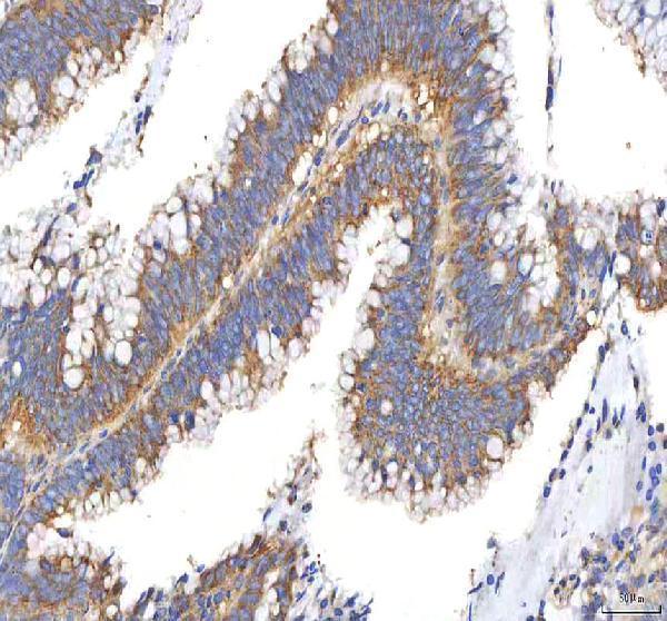
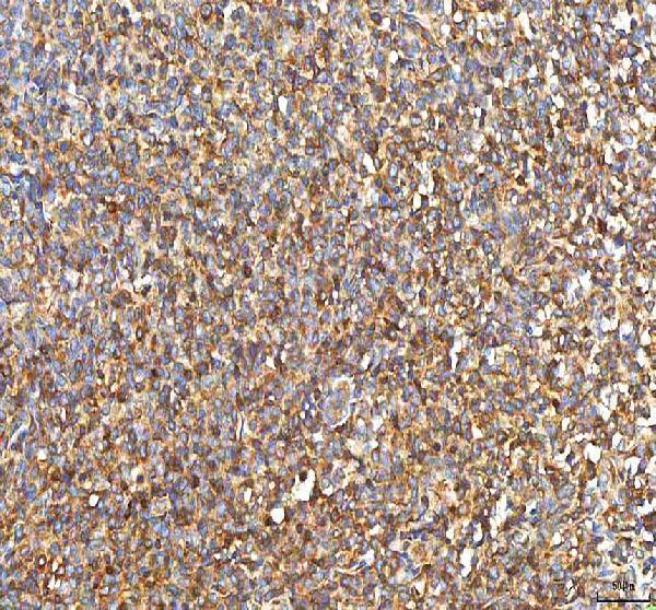
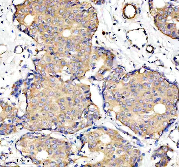
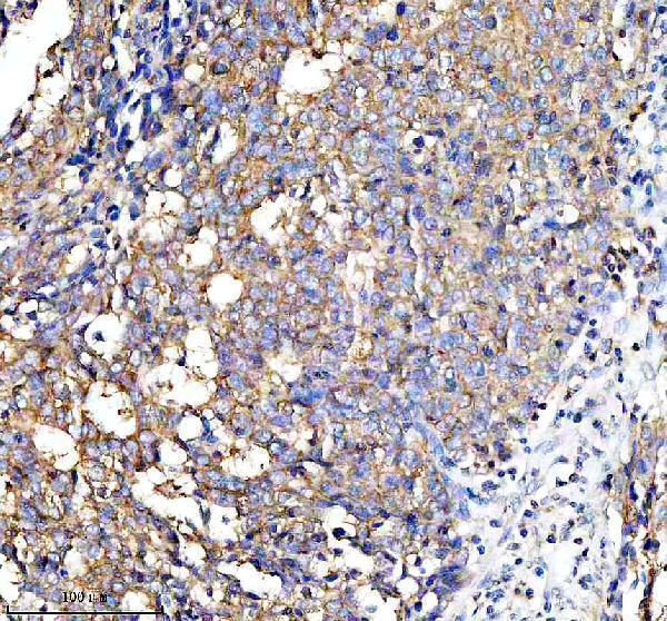


Comprehensive Human Protein Atlas IHC scoring per tissue (reliability: Uncertain — Low consistency between antibody staining and RNA expression data. Pending external verification.). Rows are taken directly from the HPA tissue chart — click any row's HPA link to view the source.
| Tissue | Cell type | Level | Evidence | Source |
|---|---|---|---|---|
| Adrenal gland | Glandular cells | High | Protein (IHC) | HPA → |
| Appendix | Glandular cells | High | Protein (IHC) | HPA → |
| Breast | Glandular cells | High | Protein (IHC) | HPA → |
| Bronchus | Respiratory epithelial cells | High | Protein (IHC) | HPA → |
| Caudate | Glial cells | High | Protein (IHC) | HPA → |
| Tissue | Cell type | Level | Evidence | Source |
|---|---|---|---|---|
| Adipose tissue | Adipocytes | Not detected | Protein (IHC) | HPA → |
PFKP is a cytoplasmic glycolytic enzyme; interpret paraffin-section staining with attention to retrieval, cell identity and assay controls (UniProt Q01813; HPA tissue IHC).
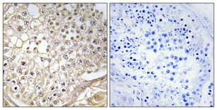
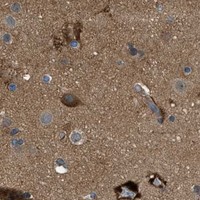
Three PFKP antibodies have human paraffin-section IHC images (catalog image captions). A07337-2 also has mouse and rat IHC images; A07337-2 and A30471 list IF/ICC applications (catalog image captions; catalog applications).
A07337-3 shows IHC in human colon adenocarcinoma; A07337-2 shows IHC in human breast cancer and mouse and rat brain (respective image captions). A30471 shows IHC in human testis and lists IF/ICC among its applications (A30471 image caption; catalog applications).
Which to pick: For human paraffin-section chromogenic IHC, choose A07337-3: its image caption documents EDTA pH 8 retrieval, 2 μg/ml primary antibody and DAB detection (A07337-3 image caption). For IF/ICC or human, mouse and rat IHC, choose polyclonal A07337-2, which lists IF/ICC and has IHC images for all three species (A07337-2 dilution_raw; catalog applications; image captions). A30471 is the polyclonal option that lists monkey reactivity, though its IHC image documents only human paraffin-section testis; fixation is unreported in all three SKUs’ image captions (A30471 dilution_raw; catalog reactivity; image captions).