PGK1 / Phosphoglycerate kinase 1 · IHC design guide

Design Immunohistochemistry for PGK1

Plan chromogenic PGK1 IHC in paraffin sections with the IHC-validated antibody at 1:25 (datasheet M01449-1 IHC-P). Score cytoplasmic and nuclear staining by cell type, using kidney glomeruli as a positive reference (HPA tissue IHC).

Evidence assembled Oct 2026 · For research use; verify linked source records and product datasheet before use
Immunohistochemistry protocol sheet for PGK1 (IHC for PGK1): expected localisation Cytoplasmic and nuclear tissue staining (HPA tissue IHC), antibody M01449-1, validated IHC image, and IHC protocol steps
Printable PGK1 IHC protocol sheet — expected localisation Cytoplasmic and nuclear tissue staining (HPA tissue IHC), antibody M01449-1, controls and protocol steps. Open the full PGK1 IHC guide →

PGK1 Immunohistochemistry Experimental Design Guide

Expected localisation, validated protocols, controls and antibodies — the at-a-glance facts below, then the full design guide.

Must know before staining
Expected localisation Cytoplasmic and nuclear tissue staining (HPA tissue IHC)
Staining pattern Widespread cytoplasmic and nuclear staining (HPA tissue IHC)
Antigen retrieval Citrate pH 6 HIER, heat-mediated (datasheet M01449-1)
Positive control ⓘ Breast+4 more · see all
Negative control ⓘ Smooth muscle
Important caveats
Reasons your staining may differ from the expected pattern.
Fixation Use formaldehyde-fixed paraffin sections (datasheet M01449-1 IHC-P). (selected-SKU IHC image M01449-1)
Caveat Staining may include protein from another gene (HPA tissue IHC)
Regulation Staining intensity varies by cell type (HPA tissue IHC)
Isoform / epitope 2 isoforms; epitope differences unspecified (UniProt)
Section 1

Recommended PGK1 IHC & IF Protocols

Compare the catalog antibody’s IHC-P protocol (datasheet: M01449-1) with two published PGK1 staining protocols (PMC3869792; PMC5787500).

Recommended immunohistochemistry (IHC-P) protocol parameters
SampleFormaldehyde-fixed, paraffin-embedded human kidney tissue (datasheet M01449-1)
FixationImage formalin-fixed; duration unreported (datasheet M01449-1); verify before use.
Sectioning4–5 µm sections on charged slides (standard)
DeparaffinisationXylene, graded ethanol series to water (standard)
Antigen retrievalHeat retrieval: Citrate pH 6 (datasheet M01449-1); 20 min, 95–100 °C (standard)
Peroxidase block3% H2O2, 10 min, room temperature (standard)
Blocking10% normal serum of the secondary host, 30 min, room temperature (standard)
Primary antibodyMouse monoclonal (clone 1086CT10.2.1) anti-PGK1, 1:25 (datasheet M01449-1)
Primary incubation1 hours at 37°C (datasheet M01449-1)
DetectionHRP-polymer secondary, DAB chromogen 5–10 min (standard)
CounterstainHematoxylin, blue, dehydrate and mount (standard)
Expected resultPGK1-positive staining in glandular cells of breast (HPA tissue IHC: High). HPA tissue profile: Ubiquitous cytoplasmic and nuclear expression. No signal in the no-primary control.
💡Decision noteStart with citrate pH 6 heat retrieval (datasheet: M01449-1); the endometrial study also used citrate heat retrieval but did not report its pH (PMC5787500).
Section 2

What Is the Expected PGK1 Staining Pattern?

PGK1 is annotated in the cytosol and, under hypoxic conditions, the mitochondrial matrix; it has no transmembrane segment (UniProt P00558). For tissue IHC, expect widespread cytoplasmic and nuclear staining (HPA: tissue IHC profile), including strong staining in several glandular cell populations and kidney glomerular cells (HPA: High). Interpret specificity cautiously: HPA rates the tissue profile Approved with medium agreement between staining and RNA data and warns that the antibodies may target proteins from multiple genes (HPA: tissue IHC reliability).

What am I looking at on my slide?
Cytoplasmic staining, sometimes with nuclear staining, is strongest in breast or cervix glandular cells and kidney glomerular cells.This fits the reported tissue pattern (HPA: ubiquitous cytoplasmic and nuclear expression; High in these cells). Assess the named cell population, rather than treating every cell in a positive tissue as equally stained (HPA: cell-level IHC ratings). The pattern is supportive, though the cross-gene warning limits attribution of every stained structure to PGK1 (HPA: tissue IHC caution).
Staining outlines cell membranes or extracellular structures while the expected intracellular staining is absent.A dominant surface or extracellular pattern does not fit the reported cytoplasmic and nuclear tissue profile (HPA: tissue IHC profile) or the absence of a transmembrane segment (UniProt P00558 topology). Treat it as suspect and compare it with control sections and the IHC-validated antibody's documented pattern (general IHC practice).
Smooth muscle cells stain strongly, especially if neighboring known-positive cells show little signal.HPA reports PGK1 as not detected in smooth muscle cells (HPA: Smooth muscle). Check cell identification and investigate nonspecific binding or endogenous detection activity with appropriate controls (general IHC practice). This reference pattern does not make every individual smooth muscle cell an absolute negative control, given the reported cross-gene caution (HPA: tissue IHC caution).
Brown signal spreads across tissue, empty spaces, or the whole section without clear cell boundaries.Such diffuse signal is difficult to assign to the reported intracellular pattern (HPA: tissue IHC profile). Inspect a no-primary control, washing, blocking, and the detection chemistry before scoring cells (general IHC practice). A stained background alone cannot establish PGK1 localization, particularly when the tissue assessment already carries a cross-gene warning (HPA: tissue IHC caution).
A known-positive population, such as breast glandular cells, has no visible stain.That result conflicts with the reported high staining in that population (HPA: Breast glandular cells, High). First verify that the expected cells are present and that the chromogenic run worked, using a matched positive control (general IHC practice). Review the catalog antibody's IHC-P instructions before interpreting a blank slide as absent PGK1 (general IHC practice).
💡Expected PGK1 appearanceCall a section pattern-consistent when identified positive cells show clear intracellular cytoplasmic staining, with possible nuclear staining, and high signal in the cited cell populations (HPA: tissue IHC profile; High ratings); dominant membrane or extracellular staining, or signal confined to blank areas, is suspect (UniProt P00558 topology; general IHC practice).
How each factor affects the staining
Antibody validation and attributionFour listed antibodies have Approved IHC status (HPA: HPA045385, HPA073644, HPA073656, CAB010065). The tissue profile has medium agreement with RNA data and a warning about targeting proteins from multiple genes (HPA: tissue IHC reliability). An Approved pattern therefore warrants cell-level interpretation and appropriate controls; it is not proof that each positive cell contains only PGK1-derived signal.
Compartment and hypoxic targetingUniProt places PGK1 in cytosol and mitochondrial matrix and describes hypoxia-associated mitochondrial targeting (UniProt P00558). HPA tissue IHC reports cytoplasmic and nuclear staining (HPA: tissue IHC profile). Do not infer a mitochondrial shift from an ordinary chromogenic section alone; assessing organelle colocalization requires a suitable localization assay (general IHC practice).
Choice of comparison cellsHPA rates glandular cells in breast, cervix, fallopian tube, gallbladder, and parathyroid gland as High, as well as kidney glomerular cells and lymph-node non-germinal-center cells (HPA: tissue IHC ratings). Smooth muscle cells are Not detected (HPA: Smooth muscle). Compare the specified cells within a section; a tissue name alone does not identify which cells should be scored.
Testis interpretationHPA rates elongated or late spermatids High (HPA: Testis), while UniProt describes expression mainly in spermatogonia and localization to the sperm principal piece (UniProt P00558). These records address different observations and should be kept distinct. Identify the germ-cell stage before comparing testis staining with either description.
IF/ICC question: What pattern is reported?HPA approves localization to the sperm mid piece, principal piece, and end piece in its ICC-IF record (HPA: subcellular summary). That record also warns that the supporting antibodies target proteins from multiple genes (HPA: subcellular caution). Use this as localization context, not as a paraffin-section staining rule.
Why is my staining missing, weak or wrong?
SituationLikely causeNext action
No stain in a section expected to contain high-staining cells.The expected cell population may be absent from the field, or the staining run may have failed (HPA: cell-level High ratings; general IHC practice).Locate the named cells on a counterstained section and run a matched positive control; review the catalog antibody's IHC-P instructions and the detection steps (general IHC practice).
All cells look uniformly positive, obscuring differences between populations.Excess background, strong detection, or insufficient discrimination during scoring can obscure cell-specific staining (general IHC practice).Compare a no-primary control, inspect blank areas, and review primary-antibody and detection conditions using the catalog IHC-P guidance (general IHC practice). Reassess named populations against HPA's cell-level ratings (HPA: tissue IHC ratings).
Smooth muscle appears strongly positive.The cell identity may be mistaken, or nonspecific binding or endogenous detection activity may contribute (HPA: Smooth muscle, Not detected; general IHC practice).Confirm the cells morphologically and compare control sections. If using enzyme-based chromogenic detection, check the relevant endogenous-activity control before assigning the signal to PGK1 (general IHC practice).
Signal is predominantly membranous or extracellular.This distribution conflicts with the reported intracellular profile and lack of a transmembrane segment (HPA: tissue IHC profile; UniProt P00558 topology).Check the no-primary control and tissue morphology, then compare with intracellular staining in a documented positive population (general IHC practice; HPA: High-rated populations).
Nuclear staining appears alongside cytoplasmic staining.Nuclear staining is included in HPA's tissue IHC profile, despite UniProt's cytosol and mitochondrial-matrix localization annotations (HPA: tissue IHC profile; UniProt P00558).Record nuclear and cytoplasmic staining separately. Do not reject an otherwise matching section solely for nuclear signal; retain the cross-gene attribution caveat (HPA: tissue IHC profile and caution).
Testis staining seems inconsistent with a chosen reference.The references emphasize different stages: late spermatids in tissue IHC and mainly spermatogonia in UniProt's tissue description (HPA: Testis, High; UniProt P00558).Identify the stained germ-cell stage and report the observation against each source separately. Avoid treating either statement as a stage-independent positive-control rule (HPA: Testis; UniProt P00558).

Sample controls for PGK1 IHC & IF

🧪Run kidney first and expect staining in glomerular cells (HPA: High in kidney cells in glomeruli); use smooth muscle as the negative tissue (HPA: Not detected in smooth muscle cells). On the kidney slide, treat cells without visible signal as internal background references, but do not designate a kidney cell type as PGK1-negative from these rows (HPA: kidney row identifies glomerular cells only).
Positive control tissue: Breast (Glandular cells, HPA High)
Negative control tissue: Smooth muscle (HPA Not detected)
ICC-IF cell lines (HPA subcellular resource): HPA ICC-IF images show PGK1 in A-431, U2OS, Sperm, with annotated localisation: Mid piece (approved), Principal piece (approved), End piece (approved) (HPA subcellular).
Technical controls: Include no-primary (secondary-only) and concentration-matched isotype controls matched to the primary antibody’s host species and clonality, plus a PGK1 knockout sample or validated peptide block as a biological specificity control (standard IHC practice). For kidney chromogenic staining, check endogenous peroxidase and endogenous biotin background because the selected caption uses a biotinylated secondary antibody (selected-SKU tissue-IHC caption: biotinylated secondary; standard IHC practice).
⚠️Feasibility: Paraffin-section IHC is supported by the selected kidney example, which uses heat-mediated citrate retrieval at pH 6; whether PGK1 staining depends on that retrieval condition is unreported (selected-SKU tissue-IHC caption). A target-specific fixation window or fixation effect is unreported, and the fixative is unreported in the selected-SKU fixative field (selected-SKU tissue-IHC caption: fixative field). The supplied evidence does not establish that frozen sections or IF are easier; kidney endogenous biotin may complicate interpretation with the caption’s biotinylated secondary (selected-SKU tissue-IHC caption; standard IHC practice).

HPA tissue IHC evidence for PGK1

Comprehensive Human Protein Atlas IHC scoring per tissue (reliability: Approved — Medium consistency between antibody staining and RNA expression data. Caution, targets protein from more than one gene.). Rows are taken directly from the HPA tissue chart — click any row's HPA link to view the source.

Positive expression · recommended positive controls

TissueCell typeLevelEvidenceSource
Breast Glandular cells High Protein (IHC) HPA →
Cervix Glandular cells High Protein (IHC) HPA →
Fallopian tube Glandular cells High Protein (IHC) HPA →
Gallbladder Glandular cells High Protein (IHC) HPA →
Kidney Cells in glomeruli High Protein (IHC) HPA →

Undetected expression · recommended negative controls

TissueCell typeLevelEvidenceSource
Smooth muscle Smooth muscle cells Not detected Protein (IHC) HPA →
Section 3

Advanced PGK1 IHC Tips

Use matched controls and compartment-aware scoring to troubleshoot PGK1 staining in paraffin sections; interpret IF findings separately.

Which retrieval conditions should I try first for weak PGK1 staining?
Start with heat-mediated citrate retrieval at pH 6 for paraffin PGK1 IHC (datasheet M01449-1). Deparaffinise sections fully, then keep heating and cooling conditions identical across the test and control slides (standard IHC practice). The selected kidney example used citrate retrieval, followed by 3% BSA blocking for 30 minutes and primary antibody at 1:25 for 1 hour at 37°C (selected IHC caption M01449-1). If staining remains weak, compare a longer retrieval time on serial sections while checking morphology; only then consider another buffer as a documented fallback experiment (standard IHC practice).
Could fixation be masking PGK1 in my paraffin sections?
PGK1-specific sensitivity to fixation is unknown because no controlled fixation comparison is supplied (supplied evidence). Use promptly fixed, consistently processed tissue and record fixative, tissue thickness and fixation duration for each specimen (standard IHC practice). If archived sections vary, compare similarly processed sections side by side with the same citrate pH 6 retrieval and antibody conditions (datasheet M01449-1; standard IHC practice). A change in staining across those sections alone cannot establish a PGK1-specific fixation effect; assess morphology and a matched positive control before changing fixation or retrieval conditions (standard IHC practice).
How should I evaluate cytoplasmic, nuclear and punctate PGK1 staining?
Expect substantial cytosolic staining, while mitochondrial matrix localisation can occur under hypoxic conditions (UniProt P00558 subcellular). Score cytoplasmic and nuclear signals separately: HPA describes both in tissue, but cautions that its antibodies can detect proteins from more than one gene (HPA tissue IHC). In kidney, examine glomerular cells as an internal positive reference, since their staining is reported as high; smooth muscle cells provide a contrasting site reported as not detected (HPA tissue IHC). Treat isolated nuclear or punctate signal as provisional until its distribution repeats across sections and an independent antibody or control supports it (standard IHC practice; HPA tissue IHC caution).
Could epitope choice explain inconsistent PGK1 staining between antibodies?
PGK1 has 2 annotated isoforms, no annotated domains and no transmembrane segment; the supplied record does not locate the catalog antibody’s epitope (UniProt P00558 record). It also lists 26 modified residues, so an epitope near a modification is a hypothesis to test, not an established cause of staining differences (UniProt P00558 record). Compare antibodies against known, distinct epitopes on adjacent sections using matched retrieval and detection conditions when epitope information is available (standard IHC practice). Do not assign an isoform or modification from chromogenic intensity alone; seek epitope documentation and an independent specificity control (standard IHC practice).
How can IF help assess a puzzling PGK1 IHC pattern?
Use IF as a separate localisation check, multiplexing PGK1 with a marker that identifies the expected cell population, such as glomerular cells in kidney or late spermatids in testis (HPA tissue IHC; standard IF practice). Choose a far-red fluorophore where tissue autofluorescence is lower, and inspect unstained and secondary-only controls in every channel (standard IF practice). Permeabilise to access cytosolic PGK1; assessing the mitochondrial matrix may require stronger membrane permeabilisation, which should be titrated against morphology (UniProt P00558 subcellular; standard IF practice). Compare compartment patterns with IHC, while accounting for HPA’s warning that its localisation data involve antibodies targeting multiple genes (HPA subcellular).
What controls distinguish PGK1 staining from chromogenic background?
Run a no-primary control and inspect background in the same compartments used for PGK1 scoring (standard IHC practice). The selected kidney example used an undiluted biotinylated secondary antibody, 3% BSA blocking for 30 minutes and primary antibody at 1:25 (selected IHC caption M01449-1). With biotin-based detection, check for endogenous biotin; with peroxidase and DAB detection, include a peroxidase block and assess pigment or precipitate separately (standard IHC practice). If diffuse colour persists, titrate the primary antibody, strengthen washing and compare serial sections before attributing the signal to widely expressed PGK1 (standard IHC practice; HPA tissue IHC).
How should I score PGK1 staining across tissue samples? ⚠ ANSWER MARKED FOR VERIFICATION
Define the cell population and compartment before scoring, because HPA reports widespread cytoplasmic and nuclear staining and low tissue specificity (HPA tissue IHC). For chromogenic sections, record the percentage of positive cells and staining intensity, or calculate an H-score from 0–300 using intensity categories 0–3 (standard IHC practice). Normalise positive counts to evaluable nucleated cells in the selected compartment, or report positive-cell density per mm² of viable tissue when cell counts are impractical (standard IHC practice). Apply one threshold across batches, exclude necrosis and edge artefacts, and compare matched controls processed with the same retrieval and detection conditions (standard IHC practice).
When is a PGK1-positive pattern biologically credible?
A credible pattern includes cytosolic staining and, in appropriate conditions, possible mitochondrial localisation (UniProt P00558 subcellular). Kidney glomerular cells and testis late spermatids are reported as highly stained, whereas smooth muscle cells are reported as not detected in HPA tissue IHC (HPA tissue IHC). Interpret nuclear staining cautiously: HPA reports it but warns that its antibodies can target proteins from more than one gene (HPA tissue IHC). Reject staining concentrated at section edges, necrotic regions or sites of endogenous peroxidase activity unless it persists with morphology intact and suitable no-primary, detection and independent-antibody controls (standard IHC practice).
Boster reagents

Best PGK1 / Phosphoglycerate kinase 1 IHC Antibodies

Anti-PGK1 antibodies have IHC images from human paraffin sections and IF/ICC images from HepG2 and U87 cells (catalog image captions); the listed reagents include human, mouse, and rat reactivity (catalog applications and reactivity).

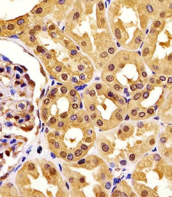
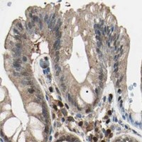
Real IHC data M01449-1 staining PGK1 in human kidney tissue sections by Immunohistochemistry (IHC-P -paraformaldehyde-fixed, paraffin-embedded sections). Tissue was fixed with formaldehyde and blocked with 3% BSA for 0. 5 hour at room temperature; antigen retrieval was by heat mediation with a citrate buffer (pH6). Samples were incubated with primary antibody (1/25) for 1 hours at 37°C. A undiluted biotinylated goat polyvalent antibody was used as the secondary antibody.
Anti-PGK1 Antibody
Cat # M01449-1
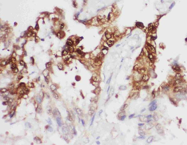
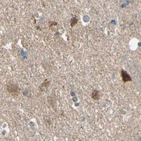
Real IHC data IHC analysis of PGK1 using anti-PGK1 antibody (PA2045). PGK1 was detected in a paraffin-embedded section of Human Lung Cancer tissue. Heat mediated antigen retrieval was performed in EDTA buffer (pH 8.0, epitope retrieval solution). The tissue section was blocked with 10% goat serum. The tissue section was then incubated with 1 μg/ml rabbit anti-PGK1 Antibody (PA2045) overnight at 4°C. Peroxidase Conjugated Goat Anti-rabbit IgG was used as secondary antibody and incubated for 30 minutes at 37°C. The tissue section was developed using HRP Conjugated Rabbit IgG Super Vision Assay Kit (Catalog # SV0002) with DAB as the chromogen.
Anti-Phosphoglycerate kinase 1 PGK1 Antibody ®
Cat # PA2045
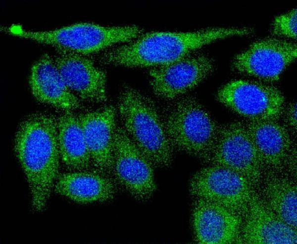
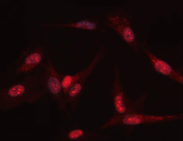
Real IF data ICC staining PGK1 in HepG2 cells (green). The nuclear counter stain is DAPI (blue). Cells were fixed in paraformaldehyde, permeabilised with 0.25% Triton X100/PBS.
Anti-Phosphoglycerate kinase 1 PGK1 Antibody
Cat # A01449
Real IF data IF analysis of PGK1 using anti-PGK1 antibody (PB9774). PGK1 was detected in an immunocytochemical section of U87 cells. Enzyme antigen retrieval was performed using IHC enzyme antigen retrieval reagent (AR0022) for 15 mins. The cells were blocked with 10% goat serum. And then incubated with 5 μg/mL rabbit anti-PGK1 Antibody (PB9774) overnight at 4°C. Cy3 Conjugated Goat Anti-Rabbit IgG (BA1032) was used as secondary antibody at 1:500 dilution and incubated for 30 minutes at 37°C. Visualize using a fluorescence microscope and filter sets appropriate for the label used.
Anti-PGK1 Antibody ®
Cat # PB9774

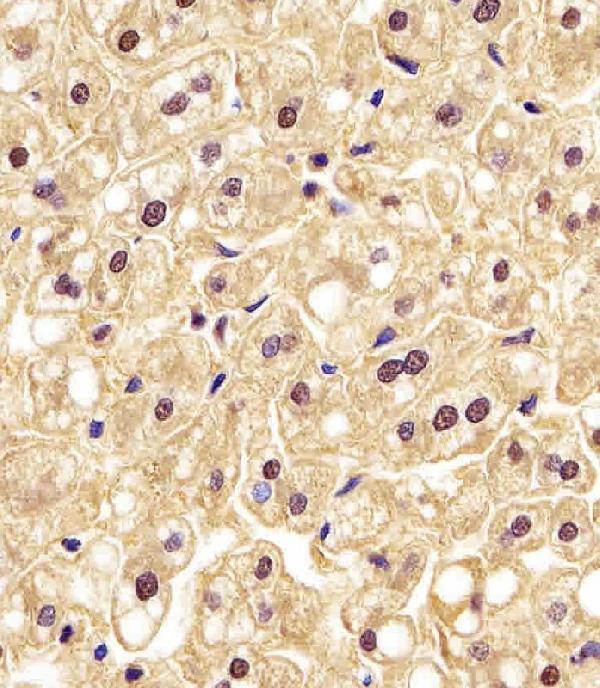
M01449-1 has IHC images from human kidney and liver paraffin sections, while PA2045 has an IHC image from a human lung cancer paraffin section (catalog image captions). A01449 has an ICC image from HepG2 cells, and PB9774 has an IF image from U87 cells (catalog image captions).

Which to pick: For tissue IHC, choose M01449-1 for formaldehyde-fixed paraffin sections with citrate retrieval (M01449-1 image caption), or PA2045 for paraffin sections with EDTA retrieval; PA2045’s fixative is unreported (PA2045 image caption). For IF/ICC, choose A01449 for the HepG2 example or PB9774 for the U87 example; both list IF and ICC applications (catalog applications and image captions). For cross-species tissue IHC, PA2045 lists human, mouse, and rat reactivity, while the monoclonal M01449-1 lists human and mouse reactivity (catalog reactivity and clone fields).

Each figure is that product's own IHC / IF validation image from its datasheet.

References

  1. UniProt Consortium. UniProt entry P00558 (PGK1_HUMAN, Phosphoglycerate kinase 1).
  2. Human Protein Atlas. PGK1 tissue IHC expression (reliability: Approved).
  3. Human Protein Atlas. PGK1 subcellular location (ICC-IF): Localized to the mid piece, principal piece and end piece. Caution: Based on antibodies targeting proteins from multiple genes..
  4. Human Protein Atlas. PGK1 antibody validation summary (4 antibodies).
  5. Hypoxia-induced PGK1 expression promotes esophageal squamous cell carcinoma progression via stimulating MYH9-mediated GSK3β/β-catenin signalling. Clinical and translational medicine 2025 — PMC12177104.
  6. PGK1 as predictor of CXCR4 expression, bone marrow metastases and survival in neuroblastoma. PloS one 2013 — PMC3869792.
  7. Phosphoglycerate kinase 1 a promoting enzyme for peritoneal dissemination in gastric cancer. International journal of cancer 2010 — PMC2811232.
  8. PGK1 and GRP78 overexpression correlates with clinical significance and poor prognosis in Chinese endometrial cancer patients. Oncotarget 2018 — PMC5787500.
  9. PubMed PMID:6188151 — UniProt-cited evidence.
  10. PubMed PMID:2995995 — UniProt-cited evidence.
  11. PubMed PMID:14702039 — UniProt-cited evidence.