This website uses cookies to ensure you get the best experience on our website.


- Table of Contents
Plan PGM3 chromogenic IHC using the observed cytoplasmic and nuclear tissue pattern (HPA tissue IHC). Compare strongly stained prostate glandular cells with an appropriate negative control, and interpret lung macrophage staining in light of the differing tissue reports (HPA tissue IHC; UniProt).

Expected localisation, validated protocols, controls and antibodies — the at-a-glance facts below, then the full design guide.
| Expected localisation | Cytoplasmic and nuclear staining (HPA tissue IHC) | |
| Staining pattern | High in prostate glandular cells; cytoplasmic and nuclear overall (HPA tissue IHC) | |
| Antigen retrieval | EDTA pH 8.0 HIER, heat-mediated (datasheet A02052-1) | |
| Positive control | Cerebellum+4 more · see all | |
| Negative control | Adipose tissue+4 more · see all |
| Fixation | Keep fixation conditions consistent across sections. (standard IHC practice; not target-specific) | |
| Caveat | Lung macrophages stain despite the reported absence in lung (HPA tissue IHC; UniProt) | |
| Regulation | Specific expression regulation unreported (UniProt) | |
| Isoform / epitope | Three isoforms; epitope differences unreported (UniProt) |
The catalog antibody protocol uses EDTA pH 8.0 heat retrieval (datasheet A02052-1). One published brain tumor IHC protocol provides a citrate retrieval comparison (PMC12007565).
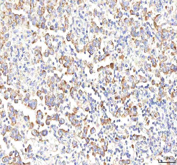
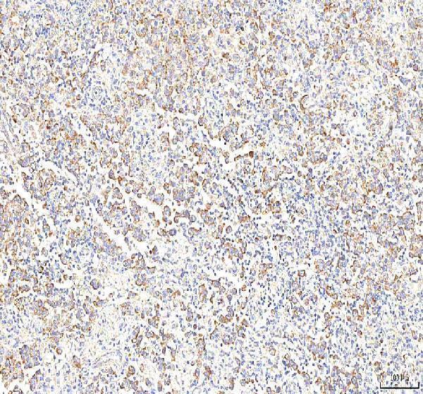
| Sample | Paraffin-embedded human testicular seminoma tissue; fixative not specified (datasheet A02052-1) |
| Fixation | Image fixative and duration unreported (datasheet A02052-1); verify before use. |
| Sectioning | 4–5 µm sections on charged slides (standard) |
| Deparaffinisation | Xylene, graded ethanol series to water (standard) |
| Antigen retrieval | Heat retrieval: EDTA pH 8.0 (datasheet A02052-1); 20 min, 95–100 °C (standard) |
| Peroxidase block | 3% H2O2, 10 min, room temperature (standard) |
| Blocking | 10% goat serum (datasheet A02052-1) |
| Primary antibody | Rabbit anti-PGM3, 2-5 μg/ml (datasheet A02052-1) |
| Primary incubation | Overnight at 4 °C (datasheet A02052-1) |
| Detection | HRP-conjugated secondary, DAB chromogen (datasheet A02052-1) |
| Counterstain | Hematoxylin, blue, dehydrate and mount (standard) |
| Expected result | PGM3-positive staining in processes in granular layer of cerebellum (HPA tissue IHC: High). HPA tissue profile: Cytoplasmic and nuclear expression in several tissues, most abundant in prostate and placenta. No signal in the no-primary control. |
PGM3 staining should be assessed in both cytoplasm and nucleus, with the clearest tissue examples in prostate glandular cells, duodenal Brunner glands, small intestinal Paneth cells and cerebellar granular-layer processes (HPA tissue IHC). PGM3 has no transmembrane segment (UniProt O95394 topology). HPA rates its tissue IHC profile Enhanced, while reporting medium agreement with RNA data and pending external verification (HPA tissue IHC).
| Cytoplasmic and nuclear staining appears in prostate glandular cells, with a tissue pattern that follows the gland boundaries (HPA tissue IHC). | This fits HPA's reported cytoplasmic and nuclear expression and high staining in prostate glandular cells (HPA tissue IHC). Judge the relevant cells and compartments together; staining intensity alone does not establish specificity (general IHC practice). |
| A slide shows only sharp membrane outlines, without convincing cytoplasmic or nuclear staining. | Membrane-only staining does not match the reported tissue pattern or the approved nucleoplasm and cytosol locations (HPA tissue IHC; HPA subcellular). Treat it as a possible artefact and review controls before calling the tissue positive (general IHC practice). |
| A supposedly negative cell population stains strongly, while the expected positive cells do not. | For example, adipocytes in adipose tissue are reported as not detected (HPA tissue IHC). Staining there may reflect cross-reactivity or endogenous detection activity; compare a matched control and the expected cell population (general IHC practice). |
| Brown reaction product spreads evenly across cells, stroma and blank areas of the section. | This diffuse background cannot be assigned to the cell-specific PGM3 pattern reported by HPA (HPA tissue IHC). Review blocking, detection reagent exposure and wash stringency, and check a control lacking primary antibody (general IHC practice). |
| A prostate section shows no signal in glandular cells, even though the tissue is intact. | Prostate glandular cells are a reported high staining population (HPA tissue IHC). First check section quality, antibody and detection controls, and the documented retrieval conditions; absence of staining alone does not prove that PGM3 is absent (general IHC practice). |
| Cell population and tissue | Expression is cell dependent: Paneth cells and Brunner glands stain high, whereas liver hepatocytes stain low and adipose adipocytes are not detected (HPA tissue IHC). Score the named cells, rather than assigning one intensity to an entire organ (general IHC practice). |
| Lung interpretation | UniProt describes PGM3 as found in many tissues except lung (UniProt O95394 tissue specificity), while HPA reports medium staining in lung macrophages (HPA tissue IHC). Record the stained cell type and source; the two statements should not be treated as identical measurements. |
| Antibody evidence | Two listed antibodies have Enhanced IHC validation and one has Supported IHC validation (HPA antibodies). HPA's overall tissue profile still has medium agreement with RNA data and awaits external verification (HPA tissue IHC); interpret unexpected staining with appropriate controls (general IHC practice). |
| Isoforms and epitope coverage | UniProt lists 3 PGM3 isoforms (UniProt O95394 isoforms). The supplied records do not identify the IHC antibody epitope or establish which isoforms it detects; do not infer isoform-specific staining from a positive section. |
| Fixation sensitivity | Target-specific effects of fixation or antigen retrieval on PGM3 staining are unreported in the supplied UniProt and HPA records. Document the conditions used when comparing sections (general IHC practice). |
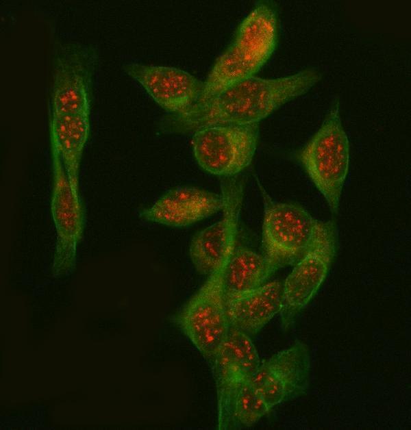
| IF/ICC Q&A: where should signal appear? | In IF/ICC, look for nucleoplasm and cytosol signal; both locations are approved, with images listed for A-431, U-251MG and U2OS (HPA subcellular). This localization answer does not establish an IF/ICC protocol or transfer IHC antibody validation to IF/ICC (HPA antibodies). |
| Situation | Likely cause | Next action |
|---|---|---|
| The expected prostate glandular-cell signal is absent (HPA tissue IHC). | An assay or section problem is possible; HPA's high prostate observation does not identify a particular technical failure (HPA tissue IHC). | Check tissue integrity and positive and detection controls; review the antibody conditions and retrieval record before interpreting the negative result (general IHC practice). |
| Only nuclei or only cytoplasm stain in an otherwise positive tissue. | The result incompletely matches the reported cytoplasmic and nuclear tissue pattern (HPA tissue IHC); relative signal can vary by cell and assay (general IHC practice). | Compare the named positive cells with a matched control, then review counterstain and chromogen development before calling the compartment restriction biological (general IHC practice). |
| Membrane rims dominate the staining. | A membrane-only pattern conflicts with approved nucleoplasm and cytosol localization and with the absence of a transmembrane segment (HPA subcellular; UniProt O95394 topology). | Inspect background and detection controls and reassess localization in the expected cell population before scoring the membrane signal as PGM3 (general IHC practice). |
| Brown signal appears broadly in tissue and in the no-primary control. | Signal in a control lacking primary antibody suggests endogenous detection activity or nonspecific detection reagent staining (general IHC practice). | Review the detection system's endogenous activity block, blocking steps and washes; compare the corrected control with the cell-specific HPA pattern (general IHC practice; HPA tissue IHC). |
| Adipocytes stain strongly, but nearby cells provide no convincing expected pattern. | Adipose adipocytes are reported as not detected; strong staining there raises concern for nonspecific signal (HPA tissue IHC). | Compare a matched no-primary control and a known-positive tissue, then reassess antibody conditions before accepting the adipocyte signal (general IHC practice; HPA tissue IHC). |
| A lung section gives an apparently conflicting result. | UniProt's tissue statement excludes lung, whereas HPA reports medium lung macrophage staining (UniProt O95394 tissue specificity; HPA tissue IHC). | Record whether macrophages specifically stain and report which source supports the interpretation; use matched controls to investigate unexpected staining (HPA tissue IHC; general IHC practice). |


Comprehensive Human Protein Atlas IHC scoring per tissue (reliability: Enhanced — Medium consistency between antibody staining and RNA expression data. Pending external verification.). Rows are taken directly from the HPA tissue chart — click any row's HPA link to view the source.
| Tissue | Cell type | Level | Evidence | Source |
|---|---|---|---|---|
| Cerebellum | Processes in granular layer | High | Protein (IHC) | HPA → |
| Duodenum | Glands of Brunner | High | Protein (IHC) | HPA → |
| Prostate | Glandular cells | High | Protein (IHC) | HPA → |
| Small intestine | Paneth cells | High | Protein (IHC) | HPA → |
| Lung | Macrophages | Medium | Protein (IHC) | HPA → |
| Tissue | Cell type | Level | Evidence | Source |
|---|---|---|---|---|
| Adipose tissue | Adipocytes | Not detected | Protein (IHC) | HPA → |
| Adrenal gland | Glandular cells | Not detected | Protein (IHC) | HPA → |
| Breast | Adipocytes | Not detected | Protein (IHC) | HPA → |
| Bronchus | Respiratory epithelial cells | Not detected | Protein (IHC) | HPA → |
| Caudate | Glial cells | Not detected | Protein (IHC) | HPA → |
Troubleshoot PGM3 staining by checking retrieval, cell type, and subcellular pattern before interpreting chromogenic signal.
A02052-1 has documented IHC on human paraffin sections and IF on HeLa cells and rat paraffin sections (catalog image captions). Human, mouse, and rat reactivity is listed (catalog: A02052-1).
A02052-1 will render with IHC data from a human testicular seminoma paraffin section (IHC image caption: A02052-1). Its IF images show HeLa cells and a rat brain paraffin section; the catalog lists ICC and IF applications and human, mouse, and rat reactivity (IF image captions and catalog: A02052-1).
Which to pick: For tissue IHC, choose A02052-1: its caption documents EDTA retrieval at pH 8.0 and 2 μg/ml antibody on a human paraffin section; the fixative is unreported (IHC image caption: A02052-1). For IF/ICC, the same SKU has HeLa cell and rat brain section images at 5 μg/ml (IF image captions: A02052-1). For cross-species work, A02052-1 lists human, mouse, and rat reactivity, although the supplied IHC image documents human tissue only (catalog and IHC image caption: A02052-1).