This website uses cookies to ensure you get the best experience on our website.


- Table of Contents
Plan PGRMC1 staining in paraffin sections using the observed cytoplasmic tissue pattern (HPA tissue IHC). Compare cells within each tissue and account for the reported secreted variant when interpreting signal (HPA tissue IHC).

Expected localisation, validated protocols, controls and antibodies — the at-a-glance facts below, then the full design guide.
| Expected localisation | Cytoplasmic tissue staining (HPA tissue IHC) | |
| Staining pattern | Cytoplasmic staining across diverse tissue cell types (HPA tissue IHC) | |
| Antigen retrieval | EDTA pH 8.0 HIER, heat-mediated (datasheet PB9775) | |
| Positive control | Cerebellum+4 more · see all | |
| Negative control | Heart muscle+1 more · see all |
| Fixation | Keep fixation consistent across paraffin sections. (standard IHC practice; not target-specific) | |
| Caveat | A secreted variant may complicate tissue staining (HPA tissue IHC) | |
| Regulation | Liver-enhanced RNA; staining may vary (HPA tissue RNA + IHC) | |
| Isoform / epitope | 2 isoforms; epitope map unspecified (UniProt) |
The catalog antibody’s IHC-P protocol is paired with published staining details for tissue sections and canine tissue sections (PMC3203473; PMC4908248).
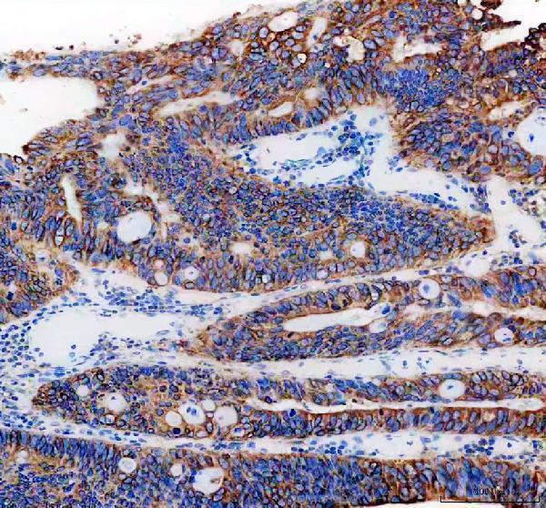
| Sample | Paraffin-embedded human colon cancer tissue; fixative not specified (datasheet PB9775) |
| Fixation | Image fixative and duration unreported (datasheet PB9775); verify before use. |
| Sectioning | 4–5 µm sections on charged slides (standard) |
| Deparaffinisation | Xylene, graded ethanol series to water (standard) |
| Antigen retrieval | Heat retrieval: EDTA pH 8.0 (datasheet PB9775); 20 min, 95–100 °C (standard) |
| Peroxidase block | 3% H2O2, 10 min, room temperature (standard) |
| Blocking | 10% goat serum (datasheet PB9775) |
| Primary antibody | Rabbit anti-PGRMC1, 2-5μg/ml (datasheet PB9775) |
| Primary incubation | Overnight at 4 °C (datasheet PB9775) |
| Detection | HRP-conjugated secondary, DAB chromogen (datasheet PB9775) |
| Counterstain | Hematoxylin, blue, dehydrate and mount (standard) |
| Expected result | PGRMC1-positive staining in gLUC cells - cytoplasm/membrane of cerebellum (HPA tissue IHC: High). HPA tissue profile: Ubiquitous cytoplasmic expression. No signal in the no-primary control. |
In paraffin sections, expect predominantly cytoplasmic PGRMC1 staining in endometrial stromal cells, liver cholangiocytes, kidney Bowman’s capsule cells and testicular Leydig cells (HPA: High in each). A membrane-associated pattern is biologically plausible because PGRMC1 has one transmembrane segment and a cytoplasmic region (UniProt O00264 topology). HPA calls the tissue profile ubiquitous cytoplasmic expression, with Enhanced reliability but only medium agreement between staining and RNA data (HPA: tissue IHC).
| Distinct cytoplasmic staining in endometrial stromal cells or cholangiocytes, with tissue structure still clear. | This fits high staining reported in those cell populations (HPA: High in endometrial stroma; High in cholangiocytes). Compare cells within the section: HPA reports a broadly cytoplasmic profile, so a positive call should rest on recognizable cellular staining rather than color anywhere in the tissue (HPA: ubiquitous cytoplasmic expression; general IHC interpretation). |
| Signal appears mainly nuclear, without convincing cytoplasmic staining, in an otherwise interpretable section. | Treat nuclear-only chromogenic staining as discordant with the expected tissue pattern and investigate assay background before scoring it as PGRMC1 (HPA: ubiquitous cytoplasmic expression). HPA reports nucleoli as an additional location in ICC-IF, but that finding alone does not establish nuclear-only staining as the expected paraffin IHC result (HPA: subcellular ICC-IF). |
| Strong stain dominates cardiomyocytes or vaginal squamous epithelium while expected positive cells are faint. | These populations are reported as not detected, so this distribution raises concern for cross-reactivity or endogenous detection activity (HPA: Not detected in cardiomyocytes and vaginal squamous epithelial cells; general IHC practice). Recheck the same run against a known-positive tissue; HPA staining levels describe observed patterns, not an absolute exclusion of expression in every specimen (HPA: tissue IHC). |
| Uniform color covers stroma, lumens, edges or many cell types and obscures cellular boundaries. | This is difficult to score as a cytoplasmic PGRMC1 pattern (HPA: ubiquitous cytoplasmic expression; general IHC interpretation). Excess detection background, incomplete blocking or retained reagent can produce nonspecific color in chromogenic IHC (general IHC practice). Assess reagent controls and tissue morphology before interpreting weak cell-associated signal (general IHC practice). |
| No staining appears in endometrial stromal cells, cholangiocytes or Leydig cells. | An absent signal in a suitable positive-control section conflicts with HPA’s high staining in those cells (HPA: High in endometrial stroma, cholangiocytes and Leydig cells). First evaluate section integrity, reagent performance and detection controls (general IHC practice). A failed positive control makes a negative result in the test specimen inconclusive (general IHC interpretation). |
| Which cells provide useful pattern anchors? | HPA reports high staining in endometrial stroma, cholangiocytes, Bowman’s capsule, Leydig cells, seminal-vesicle glandular cells and skin fibrohistiocytic cells; adipocytes are medium, while cardiomyocytes and vaginal squamous epithelial cells are not detected (HPA: tissue IHC). Choose a control with identifiable positive cells and assess those cells specifically (general IHC practice). |
| How should membrane or unusual compartment staining be weighed? | PGRMC1 has a transmembrane segment at residues 25–43 and a cytoplasmic region at 44–195 (UniProt O00264 topology). UniProt also places it at microsome, smooth ER and mitochondrial outer membranes, and notes cell-membrane localization under serum-starved conditions (UniProt O00264 subcellular location). These locations do not make nuclear-only paraffin staining an expected result (HPA: tissue IHC; general IHC interpretation). |
| Do variant or secreted-protein annotations affect scoring? | UniProt lists two isoforms and a secreted location (UniProt O00264 isoforms and subcellular location). HPA notes that at least one protein variant is secreted and that protein and RNA locations may differ (HPA: Enhanced reliability description). Interpret extracellular staining cautiously and do not require RNA and chromogenic staining to match cell for cell (HPA: tissue IHC). |
| What does antibody validation establish? | Two listed rabbit polyclonal antibodies, HPA002877 and HPA064724, have Enhanced IHC validation (HPA: antibody validation). Enhanced means the pattern is supported by independent antibodies or orthogonal data (HPA: IHC validation summary). It supports the reported pattern; it does not provide a dilution, retrieval condition or guarantee specificity in every specimen (HPA: supplied antibody data; general IHC interpretation). |
| IF/ICC Q&A: what localization can be used for a cross-check? | HPA reports endoplasmic reticulum as the main approved ICC-IF location, with nucleoli, sperm mid piece and principal piece as additional approved locations (HPA: subcellular ICC-IF). Use those observations to interpret an IF/ICC image; they do not specify a paraffin IHC retrieval or detection protocol (HPA: subcellular ICC-IF; general IHC practice). |
| Situation | Likely cause | Next action |
|---|---|---|
| Known-positive cells are unstained. | The positive control has failed, leaving a test-tissue negative call unresolved (HPA: High in endometrial stroma and cholangiocytes; general IHC interpretation). | Check section integrity, antibody application, detection reagents and chromogen with the run controls; then repeat the affected run (general IHC practice). Do not assign a PGRMC1-negative result until the expected positive cells stain (HPA: tissue IHC; general IHC interpretation). |
| Staining is weak in a reported high population. | Assay performance or specimen condition may limit visible signal; HPA’s high category does not prescribe intensity for every section (HPA: tissue IHC; general IHC interpretation). | Compare a concurrently processed positive control and tissue morphology, then review the catalog antibody’s IHC-P instructions for dilution and retrieval if available (general IHC practice). No PGRMC1-specific fixation sensitivity is established by these sources (HPA: supplied tissue IHC; UniProt O00264). |
| Brown color appears across lumens or blank tissue spaces. | Acellular color is inconsistent with HPA’s cytoplasmic cellular profile and may reflect nonspecific background (HPA: ubiquitous cytoplasmic expression; general IHC practice). | Inspect reagent and negative controls; review blocking, washes and detection steps before lowering the threshold for a positive call (general IHC practice). Score recognizable cells rather than deposited color alone (general IHC interpretation). |
| Cardiomyocytes or vaginal squamous cells stain strongly. | This conflicts with HPA’s not-detected observations in those cell types; cross-reactivity or endogenous detection activity is possible (HPA: heart muscle and vagina IHC; general IHC practice). | Check a reagent-control section and the expected positive-cell pattern in the same run; investigate endogenous activity appropriate to the detection chemistry (general IHC practice). Treat the unexpected stain as unresolved until those checks pass (general IHC interpretation). |
| Only nuclei stain in paraffin sections. | Nuclear-only staining conflicts with the reported ubiquitous cytoplasmic tissue pattern, although HPA separately reports nucleoli in ICC-IF (HPA: tissue IHC; HPA: subcellular ICC-IF). | Review morphology and reagent controls, and compare with cytoplasmic staining in a known-positive population before assigning specificity (general IHC practice; HPA: High in endometrial stroma). |
| Protein staining and RNA abundance appear to disagree. | HPA reports medium consistency between antibody staining and RNA data and notes a secreted variant; protein and RNA locations can differ (HPA: Enhanced reliability description). | Base the IHC call on cell-resolved staining, controls and the reported protein pattern; describe any RNA discrepancy without treating it alone as assay failure (HPA: tissue IHC; general IHC interpretation). |
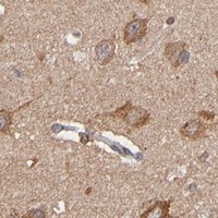


Comprehensive Human Protein Atlas IHC scoring per tissue (reliability: Enhanced — Medium consistency between antibody staining and RNA expression data. At least one protein variant secreted, tissue location of RNA and protein might differ and correlation is complex.). Rows are taken directly from the HPA tissue chart — click any row's HPA link to view the source.
| Tissue | Cell type | Level | Evidence | Source |
|---|---|---|---|---|
| Cerebellum | GLUC cells - cytoplasm/membrane | High | Protein (IHC) | HPA → |
| Endometrium | Cells in endometrial stroma | High | Protein (IHC) | HPA → |
| Kidney | Bowman's capsule | High | Protein (IHC) | HPA → |
| Liver | Cholangiocytes | High | Protein (IHC) | HPA → |
| Seminal vesicle | Glandular cells | High | Protein (IHC) | HPA → |
Troubleshoot PGRMC1 staining in paraffin sections by checking retrieval, controls, cellular localisation and scoring before interpreting chromogenic signal.
PB9775 has real IHC images from human paraffin sections and is listed for human IF (PB9775 IHC captions; catalog applications). Mouse and rat reactivity is listed (catalog reactivity).
PB9775 is listed for IHC and IF, with human, mouse and rat reactivity (catalog applications/reactivity). Its IHC images show human colon and renal cancer paraffin sections; no IF image is supplied (PB9775 IHC/IF image captions).
Which to pick: For tissue IHC, choose PB9775: its own captions document human paraffin sections, while the fixative is unreported (PB9775 IHC captions). For human IF, PB9775 is listed at 5 μg/ml; ICC validation is unreported (catalog dilution/applications). For mouse or rat work, PB9775 has listed reactivity, but the supplied IHC images and application-specific dilutions concern human samples (catalog reactivity/dilution; PB9775 IHC captions).