PHF6 / PHD finger protein 6 · IHC design guide

Design Immunohistochemistry for PHF6

Plan PHF6 chromogenic IHC in paraffin sections around its widespread nuclear tissue pattern (HPA tissue IHC). Start with the IHC-validated antibody at 2–5 μg/ml (datasheet A03065-1), and compare strongly staining cells with populations where staining was undetected (HPA tissue IHC).

Evidence assembled Oct 2026 · For research use; verify linked source records and product datasheet before use
Immunohistochemistry protocol sheet for PHF6 (IHC for PHF6): expected localisation Nuclear staining in tissue sections (HPA tissue IHC), antibody A03065-1, validated IHC image, and IHC protocol steps
Printable PHF6 IHC protocol sheet — expected localisation Nuclear staining in tissue sections (HPA tissue IHC), antibody A03065-1, controls and protocol steps. Open the full PHF6 IHC guide →

PHF6 Immunohistochemistry Experimental Design Guide

Expected localisation, validated protocols, controls and antibodies — the at-a-glance facts below, then the full design guide.

Must know before staining
Expected localisation Nuclear staining in tissue sections (HPA tissue IHC)
Staining pattern Widespread nuclear staining across many cell types (HPA tissue IHC)
Antigen retrieval EDTA pH 8.0 HIER, heat-mediated (datasheet A03065-1)
Positive control ⓘ Appendix+4 more · see all
Negative control ⓘ Liver+2 more · see all
Important caveats
Reasons your staining may differ from the expected pattern.
Fixation Keep fixation consistent across paraffin sections (standard IHC practice; not target-specific)
Caveat Some glandular cell populations have no detectable staining (HPA tissue IHC)
Regulation Low tissue specificity (HPA tissue RNA)
Isoform / epitope 5 isoforms; antibody epitope coverage is unknown (UniProt)
Section 1

Recommended PHF6 IHC & IF Protocols

The catalog antibody’s IHC-P protocol uses EDTA pH 8.0 retrieval (datasheet A03065-1). One published PHF6 protocol provides an alternative for paraffin sections (PMC10901781).

Recommended immunohistochemistry (IHC-P) protocol parameters
SampleParaffin-embedded human pancreas ductal adenocarcinoma tissue; fixative not specified (datasheet A03065-1)
FixationImage fixative and duration unreported (datasheet A03065-1); verify before use.
Sectioning4–5 µm sections on charged slides (standard)
DeparaffinisationXylene, graded ethanol series to water (standard)
Antigen retrievalHeat retrieval: EDTA pH 8.0 (datasheet A03065-1); 20 min, 95–100 °C (standard)
Peroxidase block3% H2O2, 10 min, room temperature (standard)
Blocking10% goat serum (datasheet A03065-1)
Primary antibodyRabbit anti-PHF6, 2-5 μg/ml (datasheet A03065-1)
Primary incubationOvernight at 4 °C (datasheet A03065-1)
DetectionHRP-conjugated secondary, DAB chromogen (datasheet A03065-1)
CounterstainHematoxylin, blue, dehydrate and mount (standard)
Expected resultPHF6-positive staining in germinal center cells of appendix (HPA tissue IHC: High). HPA tissue profile: Ubiquitous nuclear expression. No signal in the no-primary control.
💡Decision noteStart with heat-mediated EDTA pH 8.0 retrieval (datasheet A03065-1); consider the published pH 9.0 retrieval if optimizing staining (PMC10901781).
Section 2

What Is the Expected PHF6 Staining Pattern?

PHF6 is principally nuclear, particularly nucleolar, with reported centromere and kinetochore localization (UniProt Q8IWS0). In paraffin-section IHC, expect nuclear staining across many cell types (HPA tissue IHC: ubiquitous nuclear expression). Germinal center cells in appendix and Purkinje cells in cerebellum are examples with High staining (HPA tissue IHC). HPA rates the tissue profile Enhanced, with medium consistency between antibody staining and RNA expression (HPA tissue IHC).

What am I looking at on my slide?
Nuclear staining, potentially accentuated in nucleoli, in appendix germinal center cells or cerebellar Purkinje cells.This fits PHF6 localization and two reported High cell populations (UniProt Q8IWS0; HPA tissue IHC). Score the stained cells and compartment together: intensity alone cannot establish that a chromogenic signal is PHF6. A distinct nucleolar component is plausible, but its visibility may vary across sections (UniProt Q8IWS0).
Predominantly cytoplasmic or membrane-associated chromogen, with little nuclear signal.Treat this as a discordant pattern requiring a control check: PHF6 is nuclear and has no annotated transmembrane segment (UniProt Q8IWS0). Check whether pigment, precipitate, or nonspecific antibody binding better explains the apparent location (general IHC practice). An isolated extranuclear signal should not be scored as the expected pattern.
Strong nuclear staining in cholangiocytes, or in parathyroid or prostate glandular cells.These specific populations were reported as Not detected in the HPA tissue survey (HPA tissue IHC). Review morphology and assay controls for cross-reactivity or endogenous detection activity (general IHC practice). The HPA result describes observations under its assay conditions; it is not proof that every cell in those tissues lacks PHF6.
Diffuse staining over nuclei and surrounding tissue, without a clear cell-by-cell pattern.The expected HPA pattern is nuclear, so broadly distributed color is difficult to interpret as PHF6 (HPA tissue IHC). Check background in a no-primary control, then review blocking, washing, chromogen development, and section quality (general IHC practice). Avoid assigning cell-specific intensity scores until the background is resolved.
No convincing nuclear signal in a reported High population.A blank appendix germinal center or cerebellar Purkinje population conflicts with the reported examples (HPA tissue IHC). First confirm that the relevant cells are present and identifiable; then check the IHC staining run and controls (general IHC practice). A negative result in one section alone cannot establish biological absence.
💡Expected PHF6 appearanceA positive result is cell-associated nuclear staining, potentially nucleolar, in a reported High population such as appendix germinal center cells; diffuse extranuclear color or strong staining in HPA Not detected cells is a false-positive candidate to investigate (UniProt Q8IWS0; HPA tissue IHC).
How each factor affects the staining
Tissue and cell selectionPHF6 has low tissue specificity by RNA and a ubiquitous nuclear IHC profile, yet staining levels differ by cell population (HPA tissue IHC). Use the reported cell types when interpreting a section: appendix germinal center cells are High, while endometrial glandular cells are Low (HPA tissue IHC).
Strength of tissue evidenceThe tissue profile is rated Enhanced, but antibody staining and RNA show only medium consistency (HPA tissue IHC). The listed antibody HPA001023 also has Enhanced IHC validation (HPA antibodies). These ratings support the broad pattern; they do not validate every individual stained cell or settle an unexpected result in another preparation.
Protein forms and epitope uncertaintyUniProt lists 5 PHF6 isoforms, a chain spanning residues 2–365, and modified residues including phosphoserines (UniProt Q8IWS0). The payload gives no antibody epitope or isoform-specific IHC result. Do not infer which form produces a particular stain or predict an effect of those modifications on antibody binding.
IF/ICC Q&AQ: What localization should an IF/ICC image show? A: Nucleoplasm and nucleoli are the supported main locations (HPA subcellular ICC-IF). This observation can guide compartment interpretation; it does not specify an IF protocol or override the tissue-specific IHC observations (HPA subcellular ICC-IF; HPA tissue IHC).
Why is my staining missing, weak or wrong?
SituationLikely causeNext action
Known-positive cells are blank while the counterstain and tissue morphology are intact.The assay may have failed to produce detectable nuclear signal; the selected population may also be absent from the section (general IHC practice).Verify cell identity against an HPA High example, then review the antibody's IHC-P instructions, antigen retrieval, detection reagents, and a same-run positive control (HPA tissue IHC; general IHC practice). No PHF6-specific retrieval condition is supplied.
Only cytoplasm or cell borders stain.That compartment conflicts with PHF6's nuclear localization and lack of a transmembrane segment (UniProt Q8IWS0). Pigment or nonspecific deposition may mimic cell-associated signal (general IHC practice).Inspect the deposit at higher magnification and compare a no-primary control; reassess blocking, washing, and chromogen development before interpreting the stain (general IHC practice).
A nominally negative cell population stains strongly.The signal conflicts with an HPA Not detected call for that specific population, though that call is assay-dependent (HPA tissue IHC).Confirm the cell type on the section and compare positive and no-primary controls. If the signal persists, treat it as unresolved rather than automatically reclassifying the population (general IHC practice).
The entire section has diffuse color or nuclear detail is obscured.Widespread background can arise from nonspecific binding, endogenous detection activity, or excess chromogen development (general IHC practice).Use an appropriate no-primary control and review blocking, washing, and detection conditions; score PHF6 only after nuclei can be distinguished from background (general IHC practice; HPA tissue IHC: nuclear profile).
Nuclear signal is present, but no separate nucleolar focus is visible.HPA ICC-IF supports both nucleoplasm and nucleoli, while UniProt describes particularly nucleolar localization; neither source requires every paraffin-section nucleus to display a distinct focus (HPA subcellular ICC-IF; UniProt Q8IWS0).Record the observed nuclear pattern and cell type, then compare with a known-positive section in the same run; avoid calling the stain negative solely because nucleoli are indistinct (general IHC practice).
Staining varies between reported positive cell types.The HPA survey includes High, Low, and Not detected cell populations despite its ubiquitous nuclear summary (HPA tissue IHC).Score each cell population separately and compare like with like across sections. Use HPA cell-specific observations as context, while accounting for the profile's medium antibody–RNA consistency (HPA tissue IHC; general IHC practice).

Sample controls for PHF6 IHC & IF

🧪Run appendix first and expect PHF6 staining in germinal center cells (HPA: Appendix germinal center cells High); use liver cholangiocytes as the negative comparator (HPA: Liver cholangiocytes Not detected). No appendix cell type is established as PHF6-negative in the supplied evidence, so use cells outside germinal centers to assess background without assigning them a negative result (HPA: Appendix germinal center cells High; UniProt: ubiquitously expressed).
Positive control tissue: Appendix (Germinal center cells, HPA High)
Negative control tissue: Liver (HPA Not detected)
ICC-IF cell lines (HPA subcellular resource): HPA ICC-IF images show PHF6 in A-431, U-251MG, U2OS, KOLF2.1J, with annotated localisation: Nucleoplasm (supported), Nucleoli (supported) (HPA subcellular).
Technical controls: Include a no-primary, secondary-only control, a matched rabbit IgG isotype control, and PHF6-knockout tissue or cells as a biological negative (caption: rabbit anti-PHF6; standard IHC practice). Quench endogenous peroxidase and check for background in the appendix section before interpreting DAB staining (standard chromogenic IHC practice).
⚠️Feasibility: No target-specific fixation window or fixation effect is reported, and the selected IHC caption leaves the fixative unreported (selected-SKU tissue-IHC caption: fixative not stated). Heat retrieval in EDTA at pH 8.0 was used for the paraffin-section example, but its necessity has not been established (selected-SKU tissue-IHC caption: heat-mediated EDTA retrieval). The supplied evidence does not establish whether frozen sections or IF/ICC are easier; in appendix, distinguish true cellular staining from endogenous peroxidase background (HPA: Appendix germinal center cells High; standard chromogenic IHC practice).

HPA tissue IHC evidence for PHF6

Comprehensive Human Protein Atlas IHC scoring per tissue (reliability: Enhanced — Medium consistency between antibody staining and RNA expression data.). Rows are taken directly from the HPA tissue chart — click any row's HPA link to view the source.

Positive expression · recommended positive controls

TissueCell typeLevelEvidenceSource
Appendix Germinal center cells High Protein (IHC) HPA →
Cerebellum Purkinje cells High Protein (IHC) HPA →
Cervix Squamous epithelial cells High Protein (IHC) HPA →
Colon Endocrine cells High Protein (IHC) HPA →
Duodenum Enterocytes High Protein (IHC) HPA →

Undetected expression · recommended negative controls

TissueCell typeLevelEvidenceSource
Liver Cholangiocytes Not detected Protein (IHC) HPA →
Parathyroid gland Glandular cells Not detected Protein (IHC) HPA →
Prostate Glandular cells Not detected Protein (IHC) HPA →
Section 3

Advanced PHF6 IHC Tips

Troubleshoot PHF6 staining by checking retrieval, nuclear localisation, controls and scoring against the documented IHC result and expression profile.

Which retrieval conditions should I use when PHF6 nuclear staining is weak?
Start with heat-mediated retrieval in EDTA, pH 8.0 (datasheet A03065-1). The documented paraffin-section result used this retrieval before 2 μg/ml primary antibody overnight at 4°C (datasheet A03065-1). If nuclear staining remains weak, check heating consistency and section integrity before changing retrieval conditions (standard IHC practice). Compare any retrieval adjustment on adjacent sections with the same antibody concentration and detection steps, because changes in several steps obscure the cause of improvement (standard IHC practice). Score nucleoplasmic and nucleolar signal separately when assessing recovery (HPA subcellular; standard IHC practice).
Could fixation explain weak or uneven PHF6 staining?
The selected tissue caption identifies a paraffin-embedded section but does not state its fixative, so PHF6-specific fixation sensitivity is unknown (datasheet A03065-1). Record the fixative, fixation duration and tissue thickness for each specimen, then compare sections processed under matched conditions (standard IHC practice). Assess whether weak nuclear staining tracks with poorly preserved morphology or varies between the surface and interior of a specimen (standard IHC practice). Keep retrieval at EDTA, pH 8.0 and the primary at 2 μg/ml while investigating fixation, so the comparison uses documented staining conditions (datasheet A03065-1; standard IHC practice).
What staining pattern is consistent with PHF6, and how should cytoplasmic signal be assessed?
Expect a predominantly nuclear pattern, with nucleoplasmic and nucleolar staining supported by subcellular imaging (UniProt Q8IWS0 localisation; HPA subcellular). PHF6 is also annotated at the centromere and kinetochore, but diffuse cytoplasmic DAB alone does not match the principal reported location (UniProt Q8IWS0 localisation; standard IHC interpretation). Review nuclear boundaries with the counterstain and compare adjacent cells before calling a faint punctum nucleolar (standard IHC practice). A high-signal reference can include cerebellar Purkinje cells or appendix germinal center cells, while liver cholangiocytes are reported as undetected (HPA tissue IHC). Investigate persistent cytoplasmic staining with detection controls (standard IHC practice).
Can isoforms or epitope accessibility account for discrepant PHF6 staining?
PHF6 has 5 annotated isoforms, so an antibody result cannot establish which isoform is present unless its epitope coverage is known (UniProt Q8IWS0 isoforms; standard IHC interpretation). Check the catalog antibody’s immunogen or mapped epitope against each isoform before attributing patchy staining to alternative splicing (UniProt Q8IWS0 isoforms; standard IHC practice). PHF6 also has annotated phosphorylation sites at residues 138, 145, 155 and 183, but their effect on this antibody’s tissue staining is unreported (UniProt Q8IWS0 modified residues; datasheet A03065-1). Compare matched sections under the documented retrieval and primary conditions before assigning an epitope-based explanation (datasheet A03065-1; standard IHC practice).
How can IF help resolve an uncertain PHF6 IHC pattern?
Use IF as a separate localisation check: supported PHF6 locations are the nucleoplasm and nucleoli (HPA subcellular). Multiplex PHF6 with a marker for the expected cell population, selecting the marker from the tissue being examined, and inspect both channels at single-cell resolution (standard IF practice). Choose a fluorophore channel with low tissue autofluorescence and include an unstained section to measure background in that channel (standard IF practice). Because PHF6 is nuclear and has no transmembrane segment, use permeabilisation compatible with access to intracellular epitopes, then verify that nuclear morphology remains intact (UniProt Q8IWS0 topology and localisation; standard IF practice).
How do I reduce diffuse DAB signal without losing PHF6 nuclei?
The documented tissue stain used 10% goat serum, 2 μg/ml rabbit primary overnight at 4°C, and a peroxidase-linked secondary for 30 minutes at 37°C (datasheet A03065-1). If background is high, compare a no-primary section and inspect whether staining follows tissue edges, folds or damaged areas (standard IHC practice). Block endogenous peroxidase for the chromogenic workflow and confirm that DAB development is stopped consistently across sections (standard IHC practice). Adjust blocking, washes or primary concentration one variable at a time, while checking that expected nuclear staining remains detectable (UniProt Q8IWS0 localisation; standard IHC practice).
What is a defensible way to quantify PHF6 in paraffin sections? ⚠ ANSWER MARKED FOR VERIFICATION
Define the tissue compartment and cell population before scoring, because HPA reports differing PHF6 levels among cell types (HPA tissue IHC; standard IHC practice). Record the percentage of positive nuclei and nuclear intensity, then calculate an H-score if intensity categories are reproducible (standard IHC practice). For spatial comparisons, count PHF6-positive nuclei per mm² of viable tissue and report the total nuclei or viable area used as the denominator (standard IHC practice). Keep retrieval at EDTA, pH 8.0 and hold imaging and threshold settings constant within a comparison; exclude folds and necrosis by a predefined rule (datasheet A03065-1; standard IHC practice).
How can I distinguish genuine PHF6 signal from staining artefacts?
Prioritise staining that follows nuclei and, where resolved, nucleoli, because these are supported PHF6 locations (UniProt Q8IWS0 localisation; HPA subcellular). Check cell identity: HPA reports high staining in duodenal enterocytes and ovarian follicle cells, but no detected staining in liver cholangiocytes (HPA tissue IHC). Signal restricted to section edges, folds or necrotic areas warrants review as an artefact, especially when nearby intact nuclei lack the same pattern (standard IHC practice). Use a no-primary control to assess detection background and an endogenous-peroxidase control when unexplained DAB persists (standard IHC practice). Treat HPA’s reported medium staining–RNA consistency as a limit on expression-based conclusions (HPA tissue IHC).
Boster reagents

Best PHF6 / PHD finger protein 6 IHC Antibodies

Anti-PHF6 antibodies have pictured IHC data from human paraffin tissue and IF data from human cells (catalog image captions); A03065-1 also lists mouse and rat IHC reactivity (catalog applications).

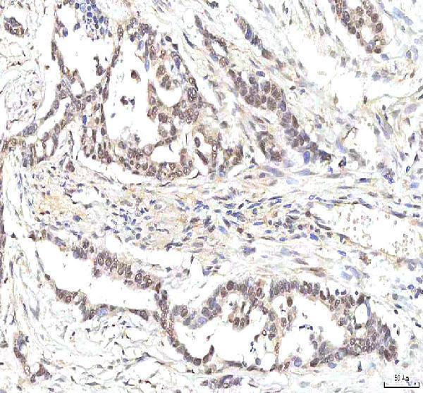
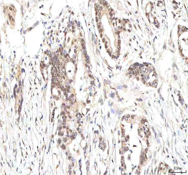
Real IHC data IHC analysis of PHF6 using anti-PHF6 antibody (A03065-1). PHF6 was detected in a paraffin-embedded section of human pancreas ductal adenocarcinoma tissue. Heat mediated antigen retrieval was performed in EDTA buffer (pH 8.0, epitope retrieval solution). The tissue section was blocked with 10% goat serum. The tissue section was then incubated with 2 μg/ml rabbit anti-PHF6 Antibody (A03065-1) overnight at 4°C. Peroxidase Conjugated Goat Anti-rabbit IgG was used as secondary antibody and incubated for 30 minutes at 37°C. The tissue section was developed using HRP Conjugated Rabbit IgG Super Vision Assay Kit (Catalog # SV0002) with DAB as the chromogen.
Anti-PHF6 Antibody ®
Cat # A03065-1
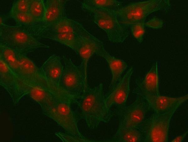
Real IF data Immunofluorescent analysis of 4% paraformaldehyde-fixed, 0.1% Triton X-100 permeabilized HeLa (human cervical epithelial adenocarcinoma cell line) cells labeling PHF6 with M03065 at 1/25 dilution, followed by Dylight® 488-conjugated goat anti-rabbit IgG secondary antibody at 1/200 dilution (green). Immunofluorescence image showing nucleus staining on HeLa cell line. Cytoplasmic actin is detected with Dylight® 554 Phalloidin at 1/100 dilution (red).
Anti-PHF6 Antibody (C-Term)
Cat # M03065

A03065-1 has pictured IHC results from human pancreas ductal adenocarcinoma, colon adenocarcinoma and glioblastoma paraffin sections, plus IF results from A549 cells (A03065-1 image captions). M03065 has pictured nuclear IF staining in HeLa cells and lists IF, but not IHC, among its applications (M03065 image caption; catalog applications).

Which to pick: Choose A03065-1 for paraffin-section IHC: its pictured sections used EDTA retrieval at pH 8.0 and 2 μg/ml primary antibody (A03065-1 IHC captions); the tissue fixative is unreported (A03065-1 IHC captions). For cell IF, both SKUs have pictured results, while A03065-1 also lists ICC as an application; M03065 is listed as polyclonal and has pictured nuclear staining in HeLa cells (catalog applications; M03065 dilution record; image captions). For mouse or rat IHC, choose A03065-1 based on its listed IHC reactivity; its pictured IHC results are from human tissue (A03065-1 dilution record; IHC captions).

Each figure is that product's own IHC / IF validation image from its datasheet.

References

  1. UniProt Consortium. UniProt entry Q8IWS0 (PHF6_HUMAN, PHD finger protein 6).
  2. Human Protein Atlas. PHF6 tissue IHC expression (reliability: Enhanced).
  3. Human Protein Atlas. PHF6 subcellular location (ICC-IF): Localized to the nucleoplasm and nucleoli..
  4. Human Protein Atlas. PHF6 antibody validation summary (1 antibodies).
  5. Systematic Pan-Cancer Analysis Identifies PHF6 as an Immunological and Prognostic Biomarker. Diagnostics (Basel, Switzerland) 2025 — PMC12785632.
  6. Characterization of a Mouse Model of Börjeson-Forssman-Lehmann Syndrome. Cell reports 2018 — PMC6261530.
  7. Molecular and clinical analyses of PHF6 mutant myeloid neoplasia provide their pathogenesis and therapeutic targeting. Nature communications 2024 — PMC10901781.
  8. PHF6 recruits BPTF to promote HIF-dependent pathway and progression in YAP-high breast cancer. Journal of translational medicine 2023 — PMC10040131.
  9. PubMed PMID:12415272 — UniProt-cited evidence.
  10. PubMed PMID:11347906 — UniProt-cited evidence.
  11. PubMed PMID:14702039 — UniProt-cited evidence.