This website uses cookies to ensure you get the best experience on our website.


- Table of Contents
Plan PHGDH staining in paraffin sections using its reported cytoplasmic and occasional nuclear tissue pattern (HPA tissue IHC). The guide covers fixation, antigen retrieval, antibody incubation, chromogenic detection and interpretation of the tissue evidence.

Expected localisation, validated protocols, controls and antibodies — the at-a-glance facts below, then the full design guide.
| Expected localisation | General cytoplasmic staining; nuclear in several tissues (HPA tissue IHC) | |
| Staining pattern | Glandular and other cells: cytoplasmic, sometimes nuclear (HPA tissue IHC) | |
| Antigen retrieval | EDTA pH 8.0 HIER, heat-mediated (datasheet A03355-1) | |
| Positive control | Appendix+4 more · see all | |
| Negative control | None in HPA (detected in all 45 tissues); use no-primary + isotype controls |
| Fixation | Matched tissue-IHC evidence does not establish the fixation claim. Validate the specimen-specific method before use. (selected-SKU IHC image A03355-1) | |
| Caveat | Antibody staining and RNA show only medium consistency (HPA tissue IHC) | |
| Regulation | Tissue-level regulation is not annotated (UniProt) | |
| Isoform / epitope | No isoforms annotated; the mature chain spans residues 2–533 (UniProt) |
The catalog antibody protocol uses EDTA pH 8.0 retrieval (datasheet: A03355-1). Published PHGDH IHC protocols below provide additional sample preparation, detection, and scoring context (PMC8871196; PMC4907894; PMC3107855).
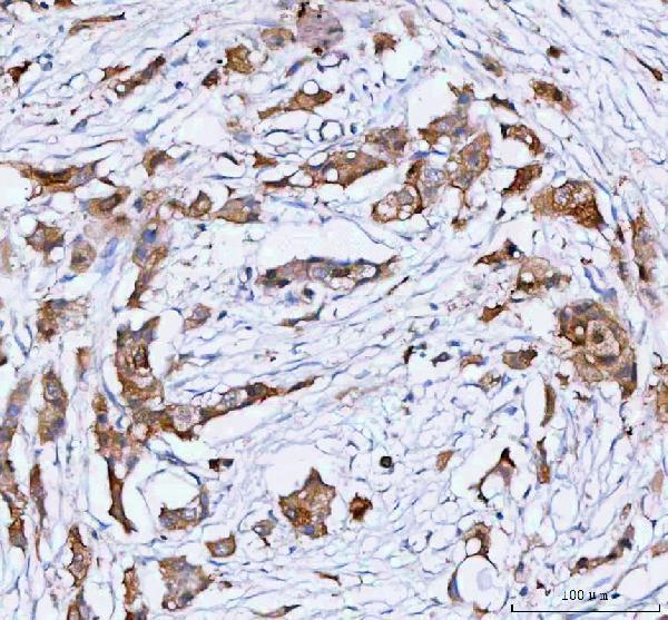
| Sample | Paraffin-embedded human breast cancer tissue; fixative not specified (datasheet A03355-1) |
| Fixation | Image fixative and duration unreported (datasheet A03355-1); verify before use. |
| Sectioning | 4–5 µm sections on charged slides (standard) |
| Deparaffinisation | Xylene, graded ethanol series to water (standard) |
| Antigen retrieval | Heat retrieval: EDTA pH 8.0 (datasheet A03355-1); 20 min, 95–100 °C (standard) |
| Peroxidase block | 3% H2O2, 10 min, room temperature (standard) |
| Blocking | 10% goat serum (datasheet A03355-1) |
| Primary antibody | Rabbit anti-PHGDH, 2-5 μg/ml (datasheet A03355-1) |
| Primary incubation | Overnight at 4 °C (datasheet A03355-1) |
| Detection | HRP-conjugated secondary, DAB chromogen (datasheet A03355-1) |
| Counterstain | Hematoxylin, blue, dehydrate and mount (standard) |
| Expected result | PHGDH-positive staining in glandular cells of appendix (HPA tissue IHC: High). HPA tissue profile: General cytoplasmic expression with nuclear expression in several tissues. No signal in the no-primary control. |
PHGDH staining in tissue is generally cytoplasmic, with nuclear staining in several tissues (HPA tissue IHC). Expect strong staining in selected glandular, basal, glial and endothelial cells, depending on the tissue (HPA tissue IHC). HPA rates the tissue profile Enhanced, while noting medium agreement with RNA data and pending external verification (HPA tissue IHC). PHGDH has no annotated transmembrane segment (UniProt O43175 topology).
| Clear cytoplasmic staining in appendix or breast glandular cells, with possible nuclear staining (HPA tissue IHC). | This matches PHGDH’s reported tissue pattern; both cell identity and compartment matter when judging a positive slide (HPA tissue IHC). Compare intensity with a low-staining cell population on the same run where available, while allowing for tissue-to-tissue variation (HPA tissue IHC; standard IHC practice). |
| Signal appears mainly in extracellular material, section edges or debris, without the expected cellular pattern. | This does not match the reported general cytoplasmic and occasional nuclear tissue pattern (HPA tissue IHC). Check morphology and controls before scoring it as PHGDH; edge and debris staining can reflect staining artefact (standard IHC practice). |
| Strong staining appears in an unexpected cell population while the expected cells stain weakly. | Question specificity when staining conflicts with the cell-level reference pattern (HPA tissue IHC). Primary-antibody cross-reactivity is possible; chromogen in a no-primary control instead points toward endogenous detection activity or another detection-stage background source (standard IHC practice). |
| A uniform haze covers cells and surrounding tissue, obscuring cellular boundaries. | Diffuse background prevents a reliable compartment or cell-type call (standard IHC practice). Inspect a no-primary control and review blocking, washes and detection conditions before comparing the slide with HPA’s cellular pattern (HPA tissue IHC; standard IHC practice). |
| No cellular signal appears in a section containing appendix or breast glandular cells. | Those cells are reported as High in HPA tissue IHC, so an entirely blank positive control calls the run into question (HPA tissue IHC). Check tissue integrity, antigen retrieval, antibody use and detection controls before treating the result as biological absence (standard IHC practice). |
| Cell-level tissue reference | HPA reports High staining in appendix and breast glandular cells, bronchus basal cells, caudate glial cells and cerebral-cortex endothelial cells (HPA tissue IHC). It reports Low staining in lung alveolar cells and heart cardiomyocytes, among others (HPA tissue IHC). Choose comparisons by cell type, since a whole-section average can hide the relevant population (standard IHC practice). |
| Compartment and topology | Tissue IHC reports general cytoplasmic staining and nuclear staining in several tissues (HPA tissue IHC). PHGDH has no annotated transmembrane segment (UniProt O43175 topology). A membrane-like outline alone therefore needs cellular and control-based review; it should not override the tissue IHC reference (HPA tissue IHC; standard IHC practice). |
| Strength of the reference evidence | The tissue profile is rated Enhanced but has medium consistency with RNA expression and awaits external verification (HPA tissue IHC). Among listed antibodies, CAB003681 has Enhanced IHC validation; HPA021241, HPA024031 and CAB068216 are Approved for IHC (HPA antibodies). Interpret disagreement cautiously and record which antibody produced the slide (standard IHC practice). |
| Fixation and retrieval evidence | Target-specific fixation sensitivity and an optimal retrieval condition are unreported in the supplied UniProt and HPA records. For an IHC-P run, follow the IHC-validated antibody’s documented starting conditions when available, then assess the positive tissue and controls together (standard IHC practice). |
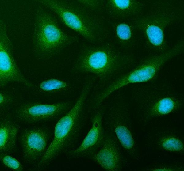
| IF/ICC Q: Where might PHGDH fluorescence appear? | A: HPA ICC-IF reports approved nucleoplasm, plasma-membrane and cytosol localization, plus mitotic-spindle and primary-cilium localization (HPA subcellular ICC-IF). Those cell-image observations add context; they do not establish that every compartment should be visible in a paraffin tissue IHC section (HPA tissue IHC; HPA subcellular ICC-IF). |
| Situation | Likely cause | Next action |
|---|---|---|
| Expected High cells are blank, and the run’s positive control is also blank. | A shared processing or detection failure is possible; appendix or breast glandular cells are reported High (HPA tissue IHC; standard IHC practice). | Verify the antibody, retrieval, detection reagents and counterstained tissue integrity against the documented IHC-P workflow; rerun a suitable positive control before interpreting absence (standard IHC practice). |
| Expected cells are blank, but the run’s positive control stains appropriately. | The sampled cells may differ from the reference population, or the section may lack interpretable target cells (HPA tissue IHC; standard IHC practice). | Recheck cell identity and morphology; compare an HPA High population in a matched run before assigning a negative score (HPA tissue IHC; standard IHC practice). |
| Chromogen persists in the no-primary control. | Endogenous detection activity or detection-stage background can produce signal without primary antibody (standard IHC practice). | Review the control’s distribution and apply the blocking or detection controls appropriate to the chosen chromogen system; score PHGDH only after background is controlled (standard IHC practice). |
| Unexpected cells stain strongly, while the no-primary control is clean. | Primary-antibody cross-reactivity is possible, although PHGDH intensity varies by cell type and tissue (HPA tissue IHC; standard IHC practice). | Compare the cell identity and compartment with HPA tissue IHC; where feasible, check the pattern with a separately validated PHGDH antibody (HPA antibodies; standard IHC practice). |
| A diffuse haze obscures the expected cellular pattern. | Background from the staining workflow can mask interpretable cytoplasmic staining (HPA tissue IHC; standard IHC practice). | Inspect the no-primary control, then review blocking, antibody dilution, washing and chromogen development using the documented workflow (standard IHC practice). |
| Staining is confined to section edges, folds or damaged areas. | Local section artefact can mimic a positive signal without reproducing the reported cell-level pattern (HPA tissue IHC; standard IHC practice). | Evaluate intact tissue away from the affected area, compare the control section and repeat staining on a usable section if the relevant cells cannot be scored (standard IHC practice). |


Comprehensive Human Protein Atlas IHC scoring per tissue (reliability: Enhanced — Medium consistency between antibody staining and RNA expression data. Pending external verification.). Rows are taken directly from the HPA tissue chart — click any row's HPA link to view the source.
| Tissue | Cell type | Level | Evidence | Source |
|---|---|---|---|---|
| Appendix | Glandular cells | High | Protein (IHC) | HPA → |
| Breast | Glandular cells | High | Protein (IHC) | HPA → |
| Bronchus | Basal cells | High | Protein (IHC) | HPA → |
| Caudate | Glial cells | High | Protein (IHC) | HPA → |
| Cerebral cortex | Endothelial cells | High | Protein (IHC) | HPA → |
| Tissue | Cell type | Level | Evidence | Source |
|---|---|---|---|---|
| None in HPA: PHGDH is detected in all 45 scored tissues. Use a no-primary (secondary-only) and an isotype control instead. | ||||
Troubleshoot PHGDH staining in paraffin sections by checking retrieval, controls, cellular localization and scoring before comparing results across specimens.
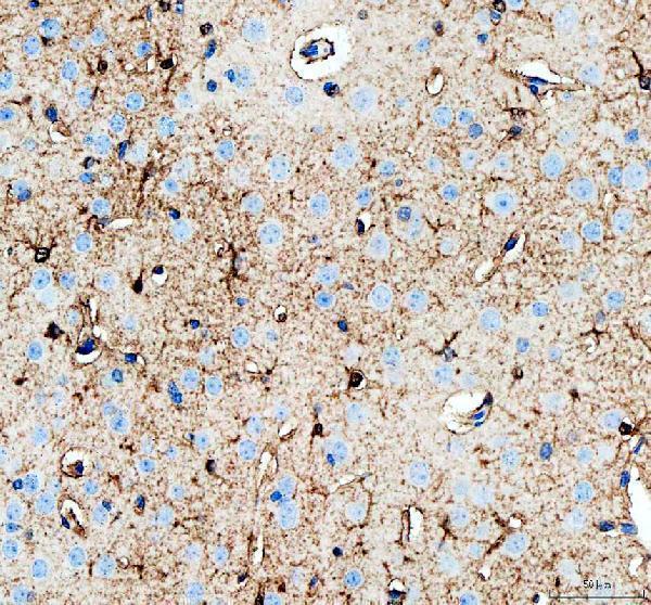
The catalog antibody has IHC images from paraffin sections of human breast cancer, mouse brain and rat brain (catalog IHC captions), plus IF data from U2OS cells (catalog IF caption).
A03355-1 has IHC images from paraffin sections of human breast cancer, mouse brain and rat brain (catalog IHC captions). A03355-1 is listed for IF/ICC, with an IF image from U2OS cells (catalog applications; catalog IF caption).
Which to pick: Choose A03355-1 for paraffin-section IHC: its captions document EDTA retrieval at pH 8.0 and 2 μg/ml primary antibody in human breast cancer, mouse brain and rat brain sections (A03355-1 IHC captions); the fixative is unreported (A03355-1 IHC captions). The same SKU is listed for IF/ICC and has a U2OS IF image using 5 μg/ml primary antibody (catalog applications; A03355-1 IF caption). For work across human, mouse and rat, A03355-1 lists reactivity with all three species; its clonality is unreported (catalog reactivity; catalog clone field).