This website uses cookies to ensure you get the best experience on our website.


- Table of Contents
Plan paraffin section staining for PICALM using the IHC-validated antibody and its documented dilution of 0.5–1 μg/mL (datasheet A02053-1). Compare cytoplasmic and membranous staining with tissue controls informed by the Human Protein Atlas profile (HPA tissue IHC).

Expected localisation, validated protocols, controls and antibodies — the at-a-glance facts below, then the full design guide.
| Expected localisation | Cytoplasmic and membranous tissue staining (HPA tissue IHC) | |
| Staining pattern | Widespread cytoplasmic/membranous; highest in GI, kidney, placenta (HPA tissue IHC) | |
| Antigen retrieval | EDTA pH 8.0 HIER, heat-mediated (datasheet A02053-1) | |
| Positive control | Adrenal gland+4 more · see all | |
| Negative control | Skeletal muscle+1 more · see all |
| Fixation | Keep fixation consistent across sections. (standard IHC practice; not target-specific) | |
| Caveat | Staining and RNA show medium concordance (HPA tissue IHC) | |
| Regulation | Broad expression across examined tissues (UniProt) | |
| Isoform / epitope | 5 isoforms; verify antibody epitope coverage (UniProt) |
Compare the catalog antibody’s IHC-P protocol (datasheet: A02053-1) with four published PICALM IHC methods (PMC4148152; PMC3949918; PMC9063212; PMC4482781).
| Sample | Paraffin-embedded human Ovarian cancer tissue; fixative not specified (datasheet A02053-1) |
| Fixation | Image fixative and duration unreported (datasheet A02053-1); verify before use. |
| Sectioning | 4–5 µm sections on charged slides (standard) |
| Deparaffinisation | Xylene, graded ethanol series to water (standard) |
| Antigen retrieval | Heat retrieval: EDTA pH 8.0 (datasheet A02053-1); 20 min, 95–100 °C (standard) |
| Peroxidase block | 3% H2O2, 10 min, room temperature (standard) |
| Blocking | 10% goat serum (datasheet A02053-1) |
| Primary antibody | Rabbit anti-PICALM, 0.5-1μg/ml (datasheet A02053-1) |
| Primary incubation | Overnight at 4 °C (datasheet A02053-1) |
| Detection | Streptavidin-biotin complex (SABC), DAB chromogen (datasheet A02053-1) |
| Counterstain | Hematoxylin, blue, dehydrate and mount (standard) |
| Expected result | PICALM-positive staining in glandular cells of adrenal gland (HPA tissue IHC: High). HPA tissue profile: Ubiquitous cytoplasmic and membranous expression, most abundant in gastrointestinal tract, kidney and placenta. No signal in the no-primary control. |
PICALM should appear mainly in cytoplasmic and membranous compartments, consistent with its coated-pit and vesicle localisation and lack of a transmembrane segment (UniProt Q13492). Expect broad tissue staining, with prominent gastrointestinal, kidney and placental expression (HPA: tissue IHC). Interpret intensity by cell type: several glandular and endocrine populations stain strongly, while muscle cells may be unstained (HPA: tissue IHC). HPA rates the tissue pattern Enhanced, with medium consistency between staining and RNA data (HPA: tissue IHC).
| Cytoplasmic and membranous signal in glandular or endocrine cells, with nearby tissue structures distinguishable. | This fits the reported broad pattern; strong glandular staining occurs in adrenal gland, breast, endometrium, fallopian tube and gallbladder, while endocrine cells stain strongly in appendix, colon and duodenum (HPA: tissue IHC). Judge the relevant cell population rather than treating every cell in a section as an equal positive control. |
| Predominantly nuclear staining, or a uniform outline of every cell with little cytoplasmic detail. | A nuclear-only or uniformly outlined pattern warrants an artefact check against the expected cytoplasmic and membranous tissue pattern (HPA: tissue IHC). Nuclear localisation is possible in some cells through interaction with PIMREG, so isolated nuclear signal is not sufficient by itself to establish nonspecific staining (UniProt Q13492). |
| Strong chromogen in skeletal myocytes or smooth muscle cells, especially when expected positive cells are weak. | These cell types are reported as not detected in the sampled HPA tissues (HPA: tissue IHC). Check cell identification and compare a no-primary control: unexpected colour may reflect cross-reactivity or, with enzyme-based detection, endogenous activity (standard IHC practice). A single discrepant field cannot establish either cause. |
| Diffuse colour across cells and extracellular areas, obscuring compartment boundaries. | That distribution does not resolve the reported cytoplasmic and membranous pattern (HPA: tissue IHC). Review the no-primary control, wash and blocking steps, and chromogen development (standard IHC practice). Interpret apparent low-level positivity only after background is sufficiently controlled to identify individual cells. |
| No detectable signal in an HPA-reported high-staining glandular or endocrine population. | First verify that the expected cells are present in the section; high staining refers to specified cell types, not every structure in the named organ (HPA: tissue IHC). If those cells are present, review the IHC-P detection workflow and antibody use before calling the sample biologically negative (standard IHC practice). |
| Tissue and cell selection | PICALM is expressed across examined tissues (UniProt Q13492), but HPA tissue IHC varies by cell population: high staining is documented in selected glandular and endocrine cells, whereas skeletal and smooth muscle cells are reported as not detected (HPA: tissue IHC). Choose controls by the named cell population and assess that population on the slide. |
| Compartment and topology | PICALM is associated with the cell membrane, coated pits, Golgi area and clathrin-coated vesicles, and has no transmembrane segment (UniProt Q13492). Its membrane-associated signal need not resemble a continuous surface rim; read punctate or regional cytoplasmic staining alongside the tissue pattern (UniProt Q13492; HPA: tissue IHC). |
| Interpretation across antibodies | HPA lists both HPA019053 and HPA019061 as IHC Enhanced (HPA: antibody validation). The tissue profile is also rated Enhanced, but its staining and RNA data have medium consistency (HPA: tissue IHC). Treat a matching pattern as supporting evidence, while checking unexpected cell types or compartments against controls. |
| Isoforms and epitope coverage | UniProt records five PICALM isoforms and an ENTH domain at residues 14–145 (UniProt Q13492). The supplied evidence does not map the catalog antibody epitope or establish isoform-specific IHC staining. Do not attribute a tissue difference to an isoform, phosphorylation site or epitope without separate validation. |
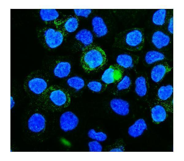
| IF/ICC Q&A: What should fluorescence show? | HPA reports vesicles as the enhanced main ICC-IF location, with images from A-431, U-251MG, U2OS and NIH 3T3 (HPA: subcellular). Expect a vesicular signal when assessing IF/ICC, while interpreting that application on its own guide page; the tissue IHC pattern describes paraffin-section chromogenic staining (HPA: tissue IHC). |
| Situation | Likely cause | Next action |
|---|---|---|
| A proposed positive control is faint or negative. | The sampled field may lack the specific high-staining cell population, or a low-staining population may have been selected (HPA: tissue IHC). | Confirm cell identity and use a section containing an HPA-listed high-staining glandular or endocrine population; then check the IHC-P staining and detection steps (HPA: tissue IHC; standard IHC practice). |
| Myocytes appear strongly positive. | HPA reports skeletal myocytes and smooth muscle cells as not detected; unexpected colour could arise from nonspecific antibody binding or endogenous enzyme activity in chromogenic detection (HPA: tissue IHC; standard IHC practice). | Check the no-primary control and cell identity. If an enzyme-based system is used, review its endogenous-activity control before interpreting the myocyte signal (standard IHC practice). |
| Colour is widespread and cell boundaries cannot be resolved. | Diffuse background can obscure the expected cytoplasmic and membranous distribution (HPA: tissue IHC; standard IHC practice). | Compare with a no-primary control and review blocking, washing and chromogen development; score only fields where individual cells and compartments remain distinguishable (standard IHC practice). |
| The signal appears exclusively nuclear. | Nuclear localisation can occur in some cells, but the reported tissue IHC profile is predominantly cytoplasmic and membranous (UniProt Q13492; HPA: tissue IHC). | Recheck the same slide for the expected cytoplasmic pattern, compare controls and avoid calling nuclear-only staining a confirmed PICALM pattern without further validation (standard IHC practice). |
| A strong signal forms a continuous edge around nearly every cell. | PICALM associates with coated pits and vesicles and lacks a transmembrane segment; an uninterrupted rim alone does not establish the reported compartment pattern (UniProt Q13492; HPA: tissue IHC). | Inspect higher magnification for cytoplasmic or vesicular detail, compare the no-primary control and judge staining within the relevant cell types (HPA: tissue IHC; standard IHC practice). |
| A positive tissue remains unstained after cell identity is confirmed. | The evidence supplied does not establish a PICALM-specific fixation or retrieval failure; absent staining can reflect a problem elsewhere in the IHC-P workflow (standard IHC practice). | Check that the antibody and detection reagents were applied correctly, that the control run developed, and that retrieval followed the catalog antibody's IHC-P instructions (standard IHC practice). |
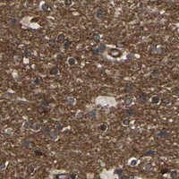


Comprehensive Human Protein Atlas IHC scoring per tissue (reliability: Enhanced — Medium consistency between antibody staining and RNA expression data.). Rows are taken directly from the HPA tissue chart — click any row's HPA link to view the source.
| Tissue | Cell type | Level | Evidence | Source |
|---|---|---|---|---|
| Adrenal gland | Glandular cells | High | Protein (IHC) | HPA → |
| Appendix | Endocrine cells | High | Protein (IHC) | HPA → |
| Breast | Glandular cells | High | Protein (IHC) | HPA → |
| Colon | Endocrine cells | High | Protein (IHC) | HPA → |
| Duodenum | Endocrine cells | High | Protein (IHC) | HPA → |
Use the documented paraffin-section workflow as a starting point, then assess staining by compartment, cell type and matched controls (datasheet A02053-1; HPA tissue IHC).
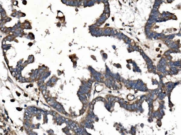
A02053-1 has IHC data from a human ovarian cancer paraffin section and IF data from A431 cells; its listed reactivity includes human, mouse and rat (catalog image captions; catalog reactivity).
A02053-1 will render with IHC from a human ovarian cancer paraffin section and IF from A431 cells (A02053-1 image captions). M02053 will render as a human-reactive IHC and IF/ICC antibody, with no image-based sample validation in the payload (M02053 applications, reactivity and image captions).
Which to pick: For tissue IHC, choose A02053-1 when a demonstrated paraffin-section workflow matters: its IHC caption reports EDTA pH 8 retrieval and 1 μg/ml primary antibody, but does not report the fixative (A02053-1 IHC caption). For IF/ICC, A02053-1 has an A431-cell IF image; M02053 is a monoclonal alternative listed for human IF/ICC and IHC, without a sample image in this payload (A02053-1 IF caption; M02053 catalog applications, clone and image captions). For mouse or rat samples, A02053-1 is the listed cross-species choice, although its supplied IHC and IF images show human samples (A02053-1 catalog reactivity and image captions).