This website uses cookies to ensure you get the best experience on our website.


- Table of Contents
Plan paraffin-section IHC for PIP5K1A using its cytoplasmic tissue staining profile (HPA tissue IHC). This guide pairs tissue controls with the catalog antibody’s validated staining conditions (datasheet A07416-3).

Expected localisation, validated protocols, controls and antibodies — the at-a-glance facts below, then the full design guide.
| Expected localisation | Cytoplasmic staining in most tissues (HPA tissue IHC) | |
| Staining pattern | Cytoplasmic staining across most tissues (HPA tissue IHC) | |
| Antigen retrieval | EDTA pH 8.0 HIER, heat-mediated (datasheet A07416-3) | |
| Positive control | Appendix+4 more · see all | |
| Negative control | Adipose tissue+4 more · see all |
| Fixation | Keep fixation consistent across sections. (standard IHC practice; not target-specific) | |
| Caveat | Heart expression contrasts with absent cardiomyocyte staining (UniProt; HPA tissue IHC) | |
| Regulation | Calcium-linked membrane recruitment (UniProt) | |
| Isoform / epitope | 4 isoforms; check epitope coverage (UniProt) |
The catalog antibody’s IHC-P protocol is paired with one published PIP5K1A tissue microarray protocol (PMC13193874).
| Sample | Paraffin-embedded mouse brain tissue; fixative not specified (datasheet A07416-3) |
| Fixation | Image fixative and duration unreported (datasheet A07416-3); verify before use. |
| Sectioning | 4–5 µm sections on charged slides (standard) |
| Deparaffinisation | Xylene, graded ethanol series to water (standard) |
| Antigen retrieval | Heat retrieval: EDTA pH 8.0 (datasheet A07416-3); 20 min, 95–100 °C (standard) |
| Peroxidase block | 3% H2O2, 10 min, room temperature (standard) |
| Blocking | 10% goat serum (datasheet A07416-3) |
| Primary antibody | Rabbit anti-PIP5K1A, 2-5 μg/ml (datasheet A07416-3) |
| Primary incubation | Overnight at 4 °C (datasheet A07416-3) |
| Detection | HRP-conjugated secondary, DAB chromogen (datasheet A07416-3) |
| Counterstain | Hematoxylin, blue, dehydrate and mount (standard) |
| Expected result | PIP5K1A-positive staining in lymphoid tissue of appendix (HPA tissue IHC: High). HPA tissue profile: Cytoplasmic expression in most tissues. No signal in the no-primary control. |
PIP5K1A should show mainly cytoplasmic staining in tissue IHC, with membrane-associated signal possible in suitable cells (HPA tissue IHC; UniProt Q99755 localization). Strong examples include germinal center cells in lymph node and tonsil, lymphoid tissue in appendix, and cells in testicular seminiferous ducts (HPA: High). The protein has no transmembrane segment (UniProt Q99755 topology). HPA rates the tissue staining Approved, with medium consistency against RNA expression (HPA tissue IHC).
| Clear cytoplasmic signal in germinal center cells, appendix lymphoid tissue, or cells in seminiferous ducts; nearby structures remain distinguishable. | This matches the strongest supplied tissue observations (HPA: High in these cell populations). Compare staining within each tissue compartment; HPA's Approved rating and medium RNA consistency support a plausible pattern but do not make every positive cell specific (HPA tissue IHC). |
| A membrane rim accompanies cytoplasmic staining, or a limited nuclear component appears in otherwise plausible cells. | Membrane and nuclear localization are biologically compatible with PIP5K1A (UniProt Q99755 localization). HPA ICC-IF supports plasma membrane and cytosol, with additional nucleoplasm (HPA subcellular). Tissue IHC is summarized as cytoplasmic, so use the tissue pattern and controls before interpreting a minor second compartment (HPA tissue IHC). |
| Staining is confined to a compartment unrelated to the supplied localizations, with no credible cytoplasmic or membrane-associated signal. | Treat an isolated, incompatible pattern as a possible artefact and review controls and tissue morphology (UniProt Q99755 localization; standard IHC practice). Nuclear signal alone needs care: nuclear localization is reported, including speckles, although HPA's tissue IHC profile is cytoplasmic (UniProt Q99755 localization; HPA tissue IHC). |
| An unexpected cell population stains strongly while the expected positive population is weak or absent. | Consider cross-reactivity or, for enzyme-based detection, endogenous activity (standard IHC practice). Cell identity matters: HPA reports lung macrophages as Low and heart cardiomyocytes as Not detected, despite broader tissue-level expression statements in UniProt (HPA tissue IHC; UniProt Q99755 tissue specificity). |
| Color spreads across cells, stroma, or section edges without a discernible intracellular pattern, or a known-positive cell population has no signal. | Diffuse staining limits localization and may reflect detection background; absent staining requires a check of the antibody and IHC workflow (standard IHC practice). Use an HPA High population as a reference, then assess morphology and controls before calling the specimen negative (HPA tissue IHC; standard IHC practice). |
| Tissue and cell selection (HPA tissue IHC) | HPA reports High staining in lymph node and tonsil germinal center cells, appendix lymphoid tissue, and cells in testicular seminiferous ducts. It reports Not detected in adipocytes and heart cardiomyocytes, among other listed populations. Choose comparisons by cell type within the section (HPA tissue IHC). |
| Strength of tissue evidence (HPA tissue IHC) | The tissue profile is Approved, with medium consistency between antibody staining and RNA expression; HPA lists low RNA tissue specificity. These ratings support pattern review but leave individual unusual staining results to be checked against controls and morphology (HPA tissue IHC; standard IHC practice). |
| Location and topology (UniProt Q99755; HPA subcellular) | PIP5K1A is reported at the cell membrane, in cytoplasm, and in the nucleus, including speckles; it has no transmembrane segment (UniProt Q99755 localization and topology). HPA ICC-IF supports plasma membrane and cytosol, with additional nucleoplasm; its tissue IHC summary emphasizes cytoplasm (HPA subcellular; HPA tissue IHC). |
| Protein forms and antibody scope (UniProt Q99755) | UniProt lists four isoforms, a single chain spanning residues 1–562, no signal peptide or propeptide, and no listed glycosylation sites (UniProt Q99755). The supplied record does not locate this antibody's epitope, so these features cannot establish which isoforms it recognizes or predict retrieval behavior. |
| IF/ICC question: should its image match paraffin IHC? (HPA subcellular) | IF/ICC may resolve plasma membrane, cytosol, and additional nucleoplasm in the imaged cell lines A-431, U-251MG, and U2OS (HPA subcellular). Paraffin tissue IHC has a cytoplasmic summary and cell-specific staining levels; compare each application with its own evidence (HPA tissue IHC; HPA subcellular). |
| Situation | Likely cause | Next action |
|---|---|---|
| No signal in an HPA High reference population. | The staining run may have failed, or the chosen section may lack an intact, identifiable reference population (HPA tissue IHC; standard IHC practice). | Confirm the expected cells on the counterstained section and review the catalog antibody's IHC-P conditions, retrieval, dilution, and detection controls before interpreting other tissue as negative (standard IHC practice). |
| A test population is negative, but the HPA High reference stains as expected. | The test population may genuinely show little or no detectable staining; HPA lists several cell populations as Not detected or Low (HPA tissue IHC). | Score the named cell type rather than the organ as a whole, preserve the successful reference result, and report the observed localization and intensity (HPA tissue IHC; standard IHC practice). |
| Only nuclei stain strongly across many unrelated cell types. | Nuclear PIP5K1A is possible, but widespread nuclear-only tissue staining differs from HPA's cytoplasmic IHC summary (UniProt Q99755 localization; HPA tissue IHC). | Check whether expected cytoplasmic staining appears in an HPA High reference and compare negative controls before assigning the nuclear signal to PIP5K1A (HPA tissue IHC; standard IHC practice). |
| An HPA Not detected cell population shows prominent signal. | The observation conflicts with that cell-specific HPA entry; cross-reactivity or endogenous detection activity is possible (HPA tissue IHC; standard IHC practice). | Verify the cell identity and morphology, then examine a negative reagent control; if enzyme-based detection is used, check its endogenous-activity control (standard IHC practice). |
| Background obscures cell borders and subcellular localization. | Non-specific binding or detection background can make staining uninterpretable (standard IHC practice). | Review blocking, washing, antibody dilution, and detection controls under the chosen IHC workflow; reassess only when intracellular staining can be separated from background (standard IHC practice). |
| Heart tissue appears positive overall, but cardiomyocytes do not stain. | UniProt reports high expression in heart, while HPA specifically scores cardiomyocytes as Not detected by tissue IHC; the statements measure different scopes (UniProt Q99755 tissue specificity; HPA tissue IHC). | Identify which heart cell population carries the signal and report that observation without assigning it to cardiomyocytes or overriding the cell-specific HPA result (HPA tissue IHC; standard IHC practice). |
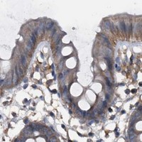
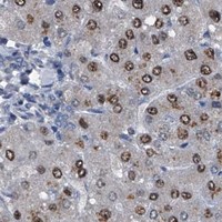
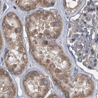


Comprehensive Human Protein Atlas IHC scoring per tissue (reliability: Approved — Medium consistency between antibody staining and RNA expression data.). Rows are taken directly from the HPA tissue chart — click any row's HPA link to view the source.
| Tissue | Cell type | Level | Evidence | Source |
|---|---|---|---|---|
| Appendix | Lymphoid tissue | High | Protein (IHC) | HPA → |
| Lymph node | Germinal center cells | High | Protein (IHC) | HPA → |
| Testis | Cells in seminiferous ducts | High | Protein (IHC) | HPA → |
| Tonsil | Germinal center cells | High | Protein (IHC) | HPA → |
| Adrenal gland | Glandular cells | Medium | Protein (IHC) | HPA → |
| Tissue | Cell type | Level | Evidence | Source |
|---|---|---|---|---|
| Adipose tissue | Adipocytes | Not detected | Protein (IHC) | HPA → |
| Duodenum | Glandular cells | Not detected | Protein (IHC) | HPA → |
| Endometrium | Cells in endometrial stroma | Not detected | Protein (IHC) | HPA → |
| Esophagus | Squamous epithelial cells | Not detected | Protein (IHC) | HPA → |
| Fallopian tube | Glandular cells | Not detected | Protein (IHC) | HPA → |
Use the catalog antibody’s paraffin-section result as the starting point, then assess staining by compartment and cell type (datasheet A07416-3; UniProt Q99755; HPA tissue IHC).
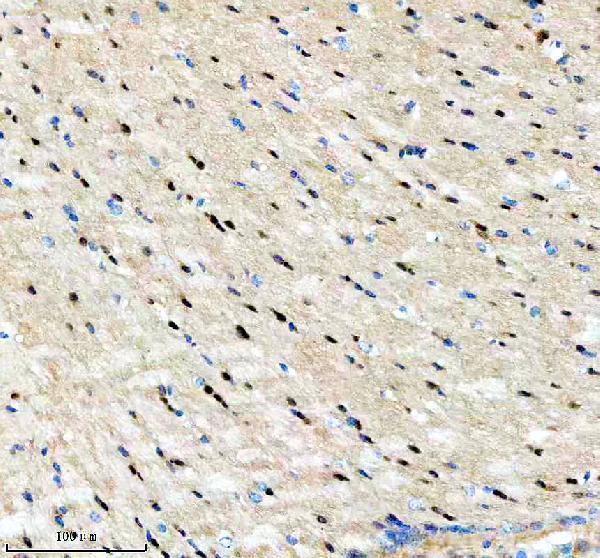
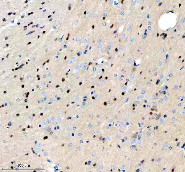
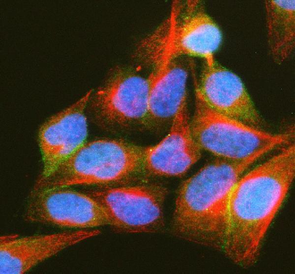
The IHC-validated antibody has paraffin-section images from human liver and liver cancer, mouse brain, and rat brain, plus an IF image from HeLa cells (catalog image captions).
A07416-3 has IHC images from paraffin sections of mouse and rat brain and human liver and liver cancer (A07416-3 IHC captions). A07416-3 also has an IF image from HeLa cells and lists IHC, ICC, and IF among its applications (A07416-3 IF caption; catalog applications).
Which to pick: Choose A07416-3 for tissue IHC on paraffin sections; its captions document EDTA retrieval at pH 8.0 and primary antibody at 2 μg/ml (A07416-3 IHC captions). Choose the same SKU for IF/ICC based on its HeLa cell IF image and listed ICC/IF applications (A07416-3 IF caption; catalog applications). For cross-species work, it lists human, mouse, and rat reactivity and has IHC images from all three species (A07416-3 catalog reactivity; IHC captions); its clone and the tissue fixative are unreported (A07416-3 catalog record; IHC captions).