PITX1 / Pituitary homeobox 1 · IHC design guide

Design Immunohistochemistry for PITX1

Plan PITX1 chromogenic IHC in paraffin sections around variable nuclear staining in subsets of cells (HPA tissue IHC). Use esophageal squamous epithelium, where staining is high, as a positive reference and adipocytes, where staining was not detected, as a comparison (HPA tissue IHC).

Evidence assembled Oct 2026 · For research use; verify linked source records and product datasheet before use
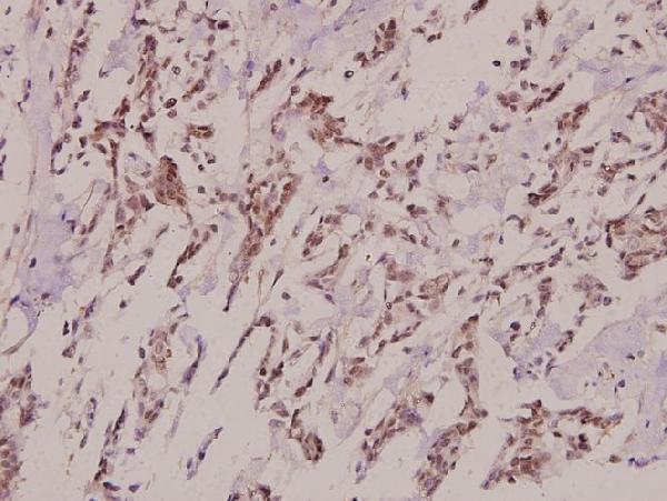
Immunohistochemistry protocol sheet for PITX1 (IHC for PITX1): expected localisation Nuclei in subsets of tissue cells (HPA tissue IHC), antibody A02993-1, validated IHC image, and IHC protocol steps
Printable PITX1 IHC protocol sheet — expected localisation Nuclei in subsets of tissue cells (HPA tissue IHC), antibody A02993-1, controls and protocol steps. Open the full PITX1 IHC guide →

PITX1 Immunohistochemistry Experimental Design Guide

Expected localisation, validated protocols, controls and antibodies — the at-a-glance facts below, then the full design guide.

Must know before staining
Expected localisation Nuclei in subsets of tissue cells (HPA tissue IHC)
Staining pattern Variable nuclear staining in subsets across most tissues (HPA tissue IHC)
Antigen retrieval Tris-EDTA pH 9.0 HIER, 95–98 °C, 20 min (rule: nuclear antigen)
Positive control ⓘ Esophagus+4 more · see all
Negative control ⓘ Adipose tissue+4 more · see all
Important caveats
Reasons your staining may differ from the expected pattern.
Fixation Keep fixation conditions consistent between samples. (standard IHC practice; not target-specific)
Caveat Presumed off-target staining may complicate scoring (HPA tissue IHC)
Regulation No expression regulator annotated (UniProt)
Isoform / epitope 0 isoforms; one 1–314 chain (UniProt)
Section 1

Recommended PITX1 IHC & IF Protocols

Compare the catalog antibody’s IHC-P protocol with published PITX1 staining protocols for paraffin-embedded specimens (PMC3919858) and skin tissue (PMC6424612).

Recommended immunohistochemistry (IHC-P) protocol parameters
SampleParaffin-embedded human breast carcinoma tissue; fixative not specified (datasheet A02993-1)
FixationImage fixative and duration unreported (datasheet A02993-1); verify before use.
Sectioning4–5 µm sections on charged slides (standard)
DeparaffinisationXylene, graded ethanol series to water (standard)
Antigen retrievalHeat-induced epitope retrieval in Tris-EDTA buffer, pH 9.0, 20 min at 95–98 °C (standard rule: nuclear antigen)
Peroxidase block3% H2O2, 10 min, room temperature (standard)
Blocking10% normal serum of the secondary host, 30 min, room temperature (standard)
Primary antibodyRabbit anti-PITX1, 1:50-1:200 (datasheet A02993-1)
Primary incubationOvernight at 4 °C (standard)
DetectionHRP-polymer secondary, DAB chromogen 5–10 min (standard)
CounterstainHematoxylin, blue, dehydrate and mount (standard)
Expected resultPITX1-positive staining in squamous epithelial cells of esophagus (HPA tissue IHC: High). HPA tissue profile: Nuclear expression in subsets of cells in most tissues at variable levels. No signal in the no-primary control.
💡Decision noteStart with Tris-EDTA pH 9.0 HIER at 95–98 °C for 20 min (page antigen-retrieval rule); the published protocols used pH 6 conditions (PMC3919858; PMC6424612).
Section 2

What Is the Expected PITX1 Staining Pattern?

PITX1 is a nuclear transcription factor with no transmembrane segment (UniProt P78337: function, location, topology). In paraffin tissue sections, expect staining in subsets of cells, including strong staining of esophageal squamous epithelium (HPA tissue IHC: High). HPA rates tissue staining Approved but reports low consistency with RNA expression and presumed off-target binding; interpret an unexpected positive cautiously (HPA tissue IHC: reliability).

What am I looking at on my slide?
Distinct nuclear stain in some esophageal squamous epithelial cells.This matches a strong positive tissue and the expected compartment (HPA tissue IHC: High in esophageal squamous epithelial cells; UniProt P78337: nucleus). Score the fraction of stained cells as well as intensity: HPA describes expression in subsets, so uniform staining is not required (HPA tissue IHC: profile).
Nuclear staining in breast glandular cells or skin keratinocytes is weaker than in esophagus.A difference in intensity can fit the reference pattern: HPA calls these cell populations Medium and esophageal squamous epithelial cells High (HPA tissue IHC: breast, skin, esophagus). Compare the relevant cells within each section; a tissue-wide average can hide a positive subset (HPA tissue IHC: profile).
Predominantly cytoplasmic or membrane-like stain, with little nuclear signal.Treat this as a localization mismatch: PITX1 is nuclear and has no transmembrane segment (UniProt P78337: location, topology). Check morphology and the matched negative control before assigning it to PITX1; diffuse chromogen or nonspecific antibody signal can mimic cellular staining (general IHC practice).
Strong staining of adipocytes or heart cardiomyocytes.These cell types are listed as Not detected, so investigate cross-reactivity or endogenous detection activity (HPA tissue IHC: adipose tissue, heart muscle; general IHC practice). A discordant result deserves particular scrutiny because HPA reports presumed off-target binding and low staining–RNA consistency (HPA tissue IHC: reliability).
A broad, low-level haze covers cells and surrounding tissue.Background that does not resolve to nuclei cannot establish PITX1 positivity (UniProt P78337: nucleus; general IHC practice). Compare with a no-primary control and inspect whether the haze follows tissue edges, damaged areas, or every compartment (general IHC practice).
💡Expected PITX1 appearanceCall a section positive when a subset of the relevant cells shows distinct nuclear staining, especially strong esophageal squamous epithelial staining; widespread cytoplasmic, membrane-like, or cell-type-discordant stain is suspect (UniProt P78337: location, topology; HPA tissue IHC: profile, esophagus, reliability).
How each factor affects the staining
Compartment and cell selectionPITX1 is a nuclear, DNA-binding transcription factor (UniProt P78337: function, location). HPA tissue IHC reports variable nuclear staining in cell subsets, so identify the cell population before scoring intensity (HPA tissue IHC: profile).
Tissue reference strengthEsophageal squamous epithelial cells are High; breast glandular cells, skin keratinocytes, and cervical squamous epithelial cells are Medium (HPA tissue IHC: positive tissues). Adipocytes and cardiomyocytes are Not detected (HPA tissue IHC: negative tissues). These observations guide comparison, not a universal positivity threshold (HPA tissue IHC: profile).
Evidence and antibody confidenceThe listed antibody, HPA008743, is Approved for IHC, while the tissue profile is also Approved with a low-consistency and presumed off-target caveat (HPA antibodies: HPA008743; HPA tissue IHC: reliability). Resolve surprising staining with controls and cell-level interpretation (general IHC practice).
IF/ICC localization?On its separate IF/ICC guide page, account for HPA's supported nucleolar localization in A-431 and U2OS images (HPA subcellular ICC-IF: main location, image cell lines). The paraffin IHC tissue profile describes nuclear staining in subsets; do not require visibly isolated nucleoli in chromogenic sections (HPA tissue IHC: profile; HPA subcellular ICC-IF: main location).
Fixation sensitivityTarget-specific effects of fixation or retrieval on PITX1 staining are unreported in the supplied UniProt and HPA records (UniProt P78337; HPA tissue IHC). Record the section and retrieval conditions when comparing runs (general IHC practice).
Why is my staining missing, weak or wrong?
SituationLikely causeNext action
No nuclear signal in esophageal squamous epithelium.The section may have a detection or staining-run failure; esophageal squamous epithelial cells are a High reference population (HPA tissue IHC: esophagus; general IHC practice).Check tissue preservation and the run's positive control, then review primary-antibody and detection steps before interpreting a negative result (general IHC practice). Do not infer PITX1-specific retrieval or fixation sensitivity from this outcome.
Signal appears only in cytoplasm or along cell borders.That distribution conflicts with nuclear PITX1 and its lack of a transmembrane segment (UniProt P78337: location, topology).Compare the same cells with the no-primary control, examine nuclear counterstain alignment, and reassess whether the signal is background or cross-reactivity (general IHC practice).
Adipocytes or cardiomyocytes stain strongly.HPA lists both as Not detected and flags presumed off-target binding in its tissue assessment (HPA tissue IHC: adipose tissue, heart muscle, reliability).Inspect matched controls for endogenous detection activity, confirm the cell identity, and require corroboration before scoring these cells PITX1-positive (general IHC practice).
Most nuclei stain uniformly across a section.This exceeds HPA's description of nuclear expression in subsets at variable levels; it may reflect excess background or off-target staining (HPA tissue IHC: profile, reliability; general IHC practice).Compare a reference positive tissue and a no-primary control from the same run; score cell populations separately and review staining conditions if controls show haze (general IHC practice).
Diffuse brown haze makes nuclei hard to score.Nonspecific binding or endogenous detection activity can obscure chromogenic IHC localization (general IHC practice).Review blocking, washing, detection controls, and counterstain balance; call positivity only where chromogen is clearly localized to nuclei (general IHC practice; UniProt P78337: nucleus).
IF/ICC shows bright nucleoli while IHC looks broadly nuclear.The two HPA records describe different views: supported nucleolar ICC-IF localization and nuclear staining in tissue subsets (HPA subcellular ICC-IF: main location; HPA tissue IHC: profile).Interpret each result against its own assay and cell context; use the separate IF/ICC guide for that assay and score paraffin IHC by identifiable nuclear staining in the expected cells (HPA subcellular ICC-IF; HPA tissue IHC: profile).

Sample controls for PITX1 IHC & IF

🧪Run esophagus first: squamous epithelial cells should stain (HPA: High in esophageal squamous epithelial cells). Use adipose tissue as the negative tissue (HPA: Not detected in adipocytes); within the esophagus section, nuclei without specific staining provide internal negative comparators (standard IHC practice).
Positive control tissue: Esophagus (Squamous epithelial cells, HPA High)
Negative control tissue: Adipose tissue (HPA Not detected)
ICC-IF cell lines (HPA subcellular resource): HPA ICC-IF images show PITX1 in A-431, U2OS, with annotated localisation: Nucleoli (supported) (HPA subcellular).
Technical controls: Include a no-primary, secondary-only control; a nonimmune IgG control matched to the primary antibody’s host species and polyclonal or monoclonal format; and, if available, PITX1 knockout tissue or a validated peptide block (standard IHC practice). For chromogenic detection, block endogenous peroxidase and, if using avidin–biotin detection, endogenous biotin (standard IHC practice).
⚠️Feasibility: A target-specific fixation window and antigen-retrieval dependence are unreported in the supplied evidence; the selected A02993-1 paraffin-section breast carcinoma caption also leaves the fixative unreported (selected-SKU tissue-IHC caption). Frozen-section suitability is unreported, while ICC-IF images exist for A-431 and U2OS with supported nucleolar localization; this does not establish that IF is easier than paraffin IHC (HPA: subcellular). No esophagus-specific artefact is reported in the supplied evidence.

HPA tissue IHC evidence for PITX1

Comprehensive Human Protein Atlas IHC scoring per tissue (reliability: Approved — Low consistency between antibody staining and RNA expression data. Presumed off target binding observed and disregarded.). Rows are taken directly from the HPA tissue chart — click any row's HPA link to view the source.

Positive expression · recommended positive controls

TissueCell typeLevelEvidenceSource
Esophagus Squamous epithelial cells High Protein (IHC) HPA →
Bone marrow Hematopoietic cells Medium Protein (IHC) HPA →
Breast Glandular cells Medium Protein (IHC) HPA →
Cervix Squamous epithelial cells Medium Protein (IHC) HPA →
Lymph node Germinal center cells Medium Protein (IHC) HPA →

Undetected expression · recommended negative controls

TissueCell typeLevelEvidenceSource
Adipose tissue Adipocytes Not detected Protein (IHC) HPA →
Adrenal gland Glandular cells Not detected Protein (IHC) HPA →
Caudate Glial cells Not detected Protein (IHC) HPA →
Cerebellum Cells in granular layer Not detected Protein (IHC) HPA →
Epididymis Glandular cells Not detected Protein (IHC) HPA →
Section 3

Advanced PITX1 IHC Tips

Troubleshoot PITX1 staining in paraffin sections by checking retrieval, nuclear localisation, antibody specificity, and cell type before scoring chromogenic signal.

How should I retrieve PITX1 in paraffin sections when nuclear staining is weak?
Use Tris-EDTA pH 9.0 heat-induced epitope retrieval at 95–98 °C for 20 min before PITX1 chromogenic IHC (page retrieval rule: nuclear antigen). Allow sections to cool in the retrieval solution, then wash consistently before blocking; these are general workflow steps rather than measured PITX1-specific requirements (standard IHC practice). If staining remains weak, compare a modestly longer retrieval with the stated method on adjacent sections while monitoring tissue damage (standard IHC practice). Evaluate the change in nuclei of squamous epithelial cells in esophagus, which are reported as high staining, and retain a negative control to detect retrieval-related background (HPA tissue IHC: esophagus; standard IHC practice).
Could fixation explain weak or uneven PITX1 staining?
The selected antibody caption describes paraffin-embedded human breast carcinoma stained at 1:100, but does not state the fixative, so PITX1-specific fixation sensitivity is unknown (A02993-1 tissue-IHC caption). Record each specimen’s fixative and fixation duration, and compare sections processed alike before attributing a staining difference to PITX1 abundance (standard IHC practice). Check whether weak nuclei track with poorly preserved morphology or uneven processing across the section (standard IHC practice). Use the stated Tris-EDTA pH 9.0 retrieval for a controlled repeat, changing one processing variable at a time (page retrieval rule: nuclear antigen; standard IHC practice). Nuclear localisation and annotated phosphorylation do not establish a fixation effect (UniProt P78337: localisation and modified residues).
Should PITX1 staining appear throughout the nucleus or only in nucleoli?
Prioritise interpretable nuclear chromogen in tissue sections: PITX1 is listed as nuclear, and tissue IHC reports nuclear expression in subsets of cells (UniProt P78337: subcellular location; HPA tissue IHC: profile). Supported nucleolar localisation comes from subcellular IF evidence and does not require every paraffin-section nucleus to show a discrete nucleolus (HPA subcellular: nucleoli; standard IHC practice). Compare staining with a nuclear counterstain and score only nuclei with intact morphology and clear cell boundaries (standard IHC practice). Predominantly diffuse cytoplasmic or extracellular staining warrants review of retrieval, primary-antibody concentration, and detection controls before calling it PITX1 (UniProt P78337: nucleus; standard IHC practice). The protein has no annotated transmembrane segment (UniProt P78337: topology).
How do I assess whether an apparent PITX1 epitope problem is real?
UniProt lists 0 annotated PITX1 isoforms and a single 1–314 chain; the supplied evidence does not identify this antibody’s epitope (UniProt P78337: isoforms and processing; A02993-1 tissue-IHC caption). Thus, an unexpected tissue pattern alone cannot establish isoform-selective staining or epitope masking (standard IHC interpretation). PITX1 has annotated phosphorylation at serines 46 and 48 and N-terminal acetylation, but their effects on this antibody are unreported (UniProt P78337: modified residues). Compare adjacent sections under the stated retrieval conditions and include primary-omission and independently validated antibody controls when available (page retrieval rule: nuclear antigen; standard IHC practice). Interpret agreement in the expected nuclei alongside morphology and cell identity (HPA tissue IHC: profile; standard IHC practice).
How can IF help resolve uncertain PITX1 IHC localisation?
Use IF as a complementary localisation check when chromogenic PITX1 staining is ambiguous, while keeping the paraffin IHC result as the result being evaluated (standard IHC/IF practice). Multiplex PITX1 with a squamous epithelial marker in esophageal sections to test whether signal lies in the expected cell population (HPA tissue IHC: high in esophageal squamous epithelial cells; standard IF practice). Choose a far-red fluorophore and inspect an unstained channel when tissue autofluorescence obscures signal (standard IF practice). For cellular IF, permeabilise sufficiently to access the nuclear antigen; PITX1 has no transmembrane segment, and nucleolar localisation is supported in subcellular IF data (UniProt P78337: topology and nucleus; HPA subcellular: nucleoli; standard IF practice).
What should I check when PITX1 chromogen is widespread or granular?
Run a primary-omission control and examine whether chromogen persists in the same structures; persistent signal points to detection-related background rather than antibody-dependent PITX1 staining (standard IHC practice). Include a peroxidase block before an HRP/DAB workflow and check wash and blocking steps if diffuse staining remains (standard IHC practice). Titrate the antibody around the caption’s 1:100 condition on comparable paraffin sections, without treating that image condition as an established optimum for every specimen (A02993-1 tissue-IHC caption; standard IHC practice). PITX1 is nuclear, so dominant extracellular or diffuse cytoplasmic precipitate should trigger scrutiny (UniProt P78337: nucleus; standard IHC practice). HPA reports presumed off-target binding in its tissue assessment, making a plausible cell pattern alone insufficient for specificity (HPA tissue IHC: reliability description).
How should I quantify PITX1-positive cells across tissue regions? ⚠ ANSWER MARKED FOR VERIFICATION
Define the tissue compartment and cell population before counting, because HPA reports variable nuclear expression in subsets of cells (HPA tissue IHC: profile). Score the percentage of evaluable nuclei at each intensity and calculate an H-score as 1 × weak% + 2 × moderate% + 3 × strong%, or report percentage positive with a prespecified threshold (standard IHC scoring practice). Normalise positive counts to the number of intact nuclei in the same region; for spatial comparisons, report positive nuclei per mm² of evaluable tissue (standard IHC quantification practice). Exclude necrosis, folds, and section edges consistently, and keep acquisition and scoring criteria fixed across specimens (standard IHC practice). Report cell type and region with each value so mixed cellular composition does not obscure the result (standard IHC practice).
When is a PITX1-positive nucleus convincing rather than an artefact?
A convincing result has intracellular nuclear staining in morphologically intact cells, with a distribution compatible with the sampled tissue and appropriate controls (UniProt P78337: nucleus; HPA tissue IHC: profile; standard IHC practice). High staining in esophageal squamous epithelial cells offers a reference pattern, whereas adipocytes are reported as not detected in HPA tissue IHC (HPA tissue IHC: esophagus and adipose tissue). Review unexpected cytoplasmic signal, staining confined to cut edges or necrotic areas, and chromogen persisting in a primary-omission control before assigning positivity (standard IHC practice). Endogenous peroxidase can produce misleading DAB signal unless the detection workflow controls it (standard IHC practice). Because HPA notes low staining–RNA consistency and presumed off-target binding, corroborate consequential findings with an independent specificity control (HPA tissue IHC: reliability description; standard IHC practice).
Boster reagents

Best PITX1 / Pituitary homeobox 1 IHC Antibodies

The catalog lists PITX1 antibodies for IHC and IF across Human, Mouse, and Rat reactivity (catalog applications/reactivity); the supplied image documents IHC in paraffin-embedded human breast carcinoma (A02993-1 image caption).

Real IHC data Immunohistochemistry (IHC) analyzes of Pitx1 (E130) pAb in paraffin-embedded human breast carcinoma tissue at 1:100.
Anti-Pitx1 (E130) Antibody
Cat # A02993-1

Only A02993-1 renders a card; it lists IHC and WB applications and Human, Mouse, and Rat reactivity (card payload: applications/reactivity). Its own IHC image shows paraffin-embedded human breast carcinoma at 1:100 (A02993-1 image caption).

Which to pick: For paraffin-section tissue IHC, pick A02993-1: its own image documents human breast carcinoma at 1:100, with no fixative reported (A02993-1 image caption). For IF/ICC, A02993-2 lists both applications and Human/Mouse reactivity; A02993 lists IF and Human/Mouse/Rat reactivity, but neither has a supplied IF image (catalog applications/reactivity/image alts). For cross-species tissue IHC, A02993-1 is polyclonal and lists Human/Mouse/Rat reactivity, while its own image documents human tissue only (A02993-1 dilution_raw/reactivity/image caption).

Each figure is that product's own IHC / IF validation image from its datasheet.

References

  1. UniProt Consortium. UniProt entry P78337 (PITX1_HUMAN, Pituitary homeobox 1).
  2. Human Protein Atlas. PITX1 tissue IHC expression (reliability: Approved).
  3. Human Protein Atlas. PITX1 subcellular location (ICC-IF): Localized to the nucleoli..
  4. Human Protein Atlas. PITX1 antibody validation summary (1 antibodies).
  5. The transcription factor PITX1 cooperates with super-enhancers to regulate the expression of DUSP4 and inhibit pyroptosis in pulmonary artery smooth muscle cells. Respiratory research 2025 — PMC12004679.
  6. miR-19b regulates hTERT mRNA expression through targeting PITX1 mRNA in melanoma cells. Scientific reports 2015 — PMC4314654.
  7. PITX1 is a reliable biomarker for predicting prognosis in patients with oral epithelial dysplasia. Oncology letters 2014 — PMC3919858.
  8. Hypersensitive IFN Responses in Lupus Keratinocytes Reveal Key Mechanistic Determinants in Cutaneous Lupus. Journal of immunology (Baltimore, Md. : 1950) 2019 — PMC6424612.
  9. PubMed PMID:9070926 — UniProt-cited evidence.
  10. PubMed PMID:9337397 — UniProt-cited evidence.
  11. PubMed PMID:14702039 — UniProt-cited evidence.