PIWIL4 / Piwi-like protein 4 · IHC design guide

Design Immunohistochemistry for PIWIL4

Plan PIWIL4 staining in paraffin sections using the catalog antibody’s IHC protocol. Use testis spermatogonia as a strong positive reference and assess nuclear and cytoplasmic staining (HPA tissue IHC).

Evidence assembled Oct 2026 · For research use; verify linked source records and product datasheet before use
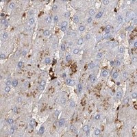
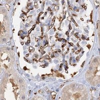
Immunohistochemistry protocol sheet for PIWIL4 (IHC for PIWIL4): expected localisation Nuclear and cytoplasmic staining (HPA tissue IHC), antibody A06797-1, validated IHC image, and IHC protocol steps
Printable PIWIL4 IHC protocol sheet — expected localisation Nuclear and cytoplasmic staining (HPA tissue IHC), antibody A06797-1, controls and protocol steps. Open the full PIWIL4 IHC guide →

PIWIL4 Immunohistochemistry Experimental Design Guide

Expected localisation, validated protocols, controls and antibodies — the at-a-glance facts below, then the full design guide.

Must know before staining
Expected localisation Nuclear and cytoplasmic staining (HPA tissue IHC)
Staining pattern Strong nuclear and cytoplasmic staining in testis spermatogonia (HPA tissue IHC)
Antigen retrieval EDTA pH 8.0 HIER, heat-mediated (datasheet A06797-1)
Positive control ⓘ Testis+4 more · see all
Negative control ⓘ Lymph node
Important caveats
Reasons your staining may differ from the expected pattern.
Fixation Keep fixation consistent across sections (standard IHC practice; not target-specific)
Caveat Staining has medium consistency with RNA expression (HPA tissue IHC)
Regulation Most abundant in testis spermatogonia (HPA tissue IHC)
Isoform / epitope 3 isoforms; epitope coverage is unknown (UniProt)
Section 1

Recommended PIWIL4 IHC & IF Protocols

The catalog antibody’s IHC-P protocol is accompanied by published PIWIL4 chromogenic IHC methods for macaque testis, human breast, and rat embryo and testis sections (PMC13079449; PMC7600338; PMC6557511).

Recommended immunohistochemistry (IHC-P) protocol parameters
SampleParaffin-embedded human placenta tissue; fixative not specified (datasheet A06797-1)
FixationImage fixative and duration unreported (datasheet A06797-1); verify before use.
Sectioning4–5 µm sections on charged slides (standard)
DeparaffinisationXylene, graded ethanol series to water (standard)
Antigen retrievalHeat retrieval: EDTA pH 8.0 (datasheet A06797-1); 20 min, 95–100 °C (standard)
Peroxidase block3% H2O2, 10 min, room temperature (standard)
Blocking10% goat serum (datasheet A06797-1)
Primary antibodyRabbit anti-PIWIL4, 2-5 μg/ml (datasheet A06797-1)
Primary incubationOvernight at 4 °C (datasheet A06797-1)
DetectionStreptavidin-biotin complex (SABC), DAB chromogen (datasheet A06797-1)
CounterstainHematoxylin, blue, dehydrate and mount (standard)
Expected resultPIWIL4-positive staining in spermatogonia cells of testis (HPA tissue IHC: High). HPA tissue profile: Nuclear and cytoplasmic expression in several tissues, most abundant in testis. No signal in the no-primary control.
💡Decision noteStart with EDTA pH 8.0 heat retrieval (datasheet A06797-1). The breast paper lists PIWIL4 at pH 9 but describes citrate pH 6 in its general procedure (PMC7600338).
Section 2

What Is the Expected PIWIL4 Staining Pattern?

PIWIL4 staining in paraffin sections should be nuclear and cytoplasmic, with the strongest reported signal in testicular spermatogonia (HPA: tissue IHC, High in spermatogonia). Expression appears in several tissues; HPA rates the tissue profile Enhanced while describing medium consistency between staining and RNA data (HPA: tissue IHC). PIWIL4 is annotated in the nucleus and cytoplasm and has no transmembrane segment (UniProt: Q7Z3Z4 localization and topology).

What am I looking at on my slide?
Nuclear and cytoplasmic signal is strongest in testicular spermatogonia; other listed cell populations show weaker signal (HPA: tissue IHC).This matches the reported IHC pattern: High in spermatogonia and Medium in, among others, bone marrow hematopoietic cells and adipocytes (HPA: tissue IHC). Judge intensity within the identified cell population, since a whole-section average can obscure a localized positive population (general IHC practice).
Signal is concentrated at cell borders or other compartments outside the nucleus and cytoplasm (UniProt: Q7Z3Z4 localization).A predominantly membranous pattern conflicts with the annotated nuclear and cytoplasmic localization and absence of a transmembrane segment (UniProt: Q7Z3Z4). Check morphology and the negative control before assigning the signal to PIWIL4 (general IHC practice). Nuclear and cytoplasmic proportions can differ across cells; this record gives no required ratio.
Strong signal appears in lymph-node germinal center cells, where PIWIL4 was not detected (HPA: tissue IHC).Treat this as a specificity concern, including possible cross-reactivity or endogenous detection activity, rather than proof of new expression (HPA: germinal center cells, Not detected; general IHC practice). Check the no-primary control and assess which cells carry the signal (general IHC practice).
Chromogenic signal forms a diffuse haze across cells and surrounding tissue (general IHC practice).Diffuse background makes nuclear and cytoplasmic localization difficult to score (general IHC practice). Check a no-primary control, blocking, washes and detection conditions; those checks identify general assay background, not a documented PIWIL4-specific fixation effect (general IHC practice).
Testicular spermatogonia lack detectable signal despite suitable tissue morphology (HPA: High in spermatogonia).A negative result in this reported high-staining population makes the run inconclusive for target detection (HPA: tissue IHC; general IHC practice). Verify the antibody's IHC validation, its prescribed assay conditions and the detection controls before interpreting other negative cells (HPA: antibody validation; general IHC practice).
💡Expected PIWIL4 appearanceCall a positive result when testicular spermatogonia show strong nuclear and cytoplasmic staining (HPA: High in spermatogonia; UniProt: Q7Z3Z4 localization); dominant membrane staining or similarly strong germinal-center staining warrants a false-positive check (UniProt: Q7Z3Z4 topology; HPA: germinal center cells, Not detected).
How each factor affects the staining
Tissue and cell selection (HPA: tissue IHC)Testicular spermatogonia provide the clearest reported high-signal population; several other listed cell populations are Medium, while lymph-node germinal center cells are Not detected (HPA: tissue IHC). Use cell identity when choosing and reading comparison sections (general IHC practice).
Antibody validation (HPA: antibody records)The tissue profile is Enhanced but has medium staining–RNA consistency (HPA: tissue IHC). HPA057508 is Enhanced for IHC; HPA036588 is Supported for IHC; HPA036587 has Supported ICC status without an IHC status in this payload (HPA: antibody records). Match evidence to the assay being interpreted.
Topology and processing (UniProt: Q7Z3Z4)PIWIL4 has no transmembrane segment or annotated signal peptide and is listed as one chain spanning residues 1–852 (UniProt: Q7Z3Z4). These annotations support checking a dominant surface-only pattern; they do not identify an antibody epitope or establish antigen-retrieval requirements.
Isoforms and epitope coverage (UniProt: Q7Z3Z4)Three isoforms are listed (UniProt: Q7Z3Z4). Their presence alone does not show which isoforms an antibody detects. If results differ between antibodies, consult their documented immunogens or epitope information before attributing the difference to isoforms (general IHC practice).
IF/ICC Q: What localization is reported? (HPA: subcellular ICC-IF)A: HPA reports nucleoplasm as Supported and mitochondria as Approved in ICC-IF images from NB4, PC-3 and U2OS (HPA: subcellular ICC-IF). Those observations inform the separate IF/ICC guide; they do not make mitochondrial staining a required pattern in paraffin-section IHC.
Why is my staining missing, weak or wrong?
SituationLikely causeNext action
No staining in testicular spermatogonia (HPA: High in spermatogonia).The expected positive population has failed to appear; the observation alone does not identify whether antibody, detection or tissue handling caused it (HPA: tissue IHC; general IHC practice).Confirm spermatogonia are present, check the antibody's IHC status and follow its documented IHC-P conditions; inspect run controls before scoring other tissues as negative (HPA: antibody records; general IHC practice).
Diffuse signal prevents nuclear and cytoplasmic scoring (UniProt: Q7Z3Z4 localization).General assay background can obscure cellular localization (general IHC practice). This payload provides no PIWIL4-specific fixation-sensitivity finding.Inspect the no-primary control, review blocking and washing, and adjust detection conditions within the validated assay workflow (general IHC practice).
A predominantly membrane-like outline is scored as PIWIL4 (UniProt: Q7Z3Z4 topology).That distribution conflicts with the annotated nuclear and cytoplasmic locations and absence of a transmembrane segment (UniProt: Q7Z3Z4).Recheck cell boundaries and counterstained nuclei, compare the negative control, and verify with an IHC-validated antibody if the pattern persists (general IHC practice; HPA: antibody records).
Lymph-node germinal center cells stain strongly (HPA: Not detected).The discrepancy raises concern for cross-reactivity or endogenous detection activity; staining alone cannot distinguish them (HPA: tissue IHC; general IHC practice).Check a no-primary control and endogenous-activity blocking appropriate to the detection chemistry, then compare the cell pattern with testicular spermatogonia (general IHC practice; HPA: High in spermatogonia).
All cells appear similarly intense across a section (general IHC practice).A uniform result obscures the cell-specific contrast reported by HPA, including High spermatogonia and Medium or undetected comparison populations (HPA: tissue IHC).Review detection strength and background controls, then rescore identifiable cells separately instead of assigning a single intensity to the whole section (general IHC practice).
Antibodies give different IHC patterns (general IHC practice).Their assay evidence differs: HPA057508 is Enhanced for IHC, HPA036588 is Supported, and HPA036587 has no IHC status here (HPA: antibody records). Epitope coverage is unspecified in this payload.Prioritize the antibody with documented IHC validation, check each antibody's own assay documentation, and compare staining in the same identified cell populations (HPA: antibody records; general IHC practice).

Sample controls for PIWIL4 IHC & IF

🧪Run testis first and require staining in spermatogonia (High; HPA: High in spermatogonia cells). Run lymph node as the negative tissue, assessing germinal center cells (HPA: Not detected in germinal center cells); on the testis slide, use cells outside the identified spermatogonia as a background comparison without assuming a particular cell type is PIWIL4-negative (HPA: High in spermatogonia cells).
Positive control tissue: Testis (Spermatogonia cells, HPA High)
Negative control tissue: Lymph node (HPA Not detected)
ICC-IF cell lines (HPA subcellular resource): HPA ICC-IF images show PIWIL4 in NB4, PC-3, U2OS, with annotated localisation: Nucleoplasm (supported), Mitochondria (approved) (HPA subcellular).
Technical controls: Include a no-primary, secondary-only control and a concentration-matched rabbit IgG isotype control (caption: rabbit primary antibody; standard IHC practice); use PIWIL4 knockout material, if available, as a biological specificity control (standard IHC practice). Block endogenous peroxidase and assess endogenous biotin background when using the reported biotin–SABC/DAB detection method (caption: biotinylated secondary, SABC and DAB; standard IHC practice).
⚠️Feasibility: A target-specific fixation window or fixation effect is unreported, and the selected A06797-1 tissue-IHC caption does not state its fixative (caption: fixative not stated). The caption uses heat retrieval in EDTA at pH 8.0, but retrieval dependency is unestablished (caption: EDTA heat retrieval). The supplied evidence does not establish whether frozen sections or IF are easier; for testis IHC, check peroxidase and biotin background under the reported detection method (caption: biotin–SABC/DAB; standard IHC practice).

HPA tissue IHC evidence for PIWIL4

Comprehensive Human Protein Atlas IHC scoring per tissue (reliability: Enhanced — Medium consistency between antibody staining and RNA expression data.). Rows are taken directly from the HPA tissue chart — click any row's HPA link to view the source.

Positive expression · recommended positive controls

TissueCell typeLevelEvidenceSource
Testis Spermatogonia cells High Protein (IHC) HPA →
Adipose tissue Adipocytes Medium Protein (IHC) HPA →
Adrenal gland Glandular cells Medium Protein (IHC) HPA →
Appendix Glandular cells Medium Protein (IHC) HPA →
Bone marrow Hematopoietic cells Medium Protein (IHC) HPA →

Undetected expression · recommended negative controls

TissueCell typeLevelEvidenceSource
Lymph node Germinal center cells Not detected Protein (IHC) HPA →
Section 3

Advanced PIWIL4 IHC Tips

Troubleshoot PIWIL4 staining in paraffin sections by checking retrieval, cell type, compartment and detection controls before interpreting signal intensity.

How should I optimize antigen retrieval when PIWIL4 staining is weak?
Start with heat-mediated antigen retrieval in EDTA buffer at pH 8.0 for A06797-1 paraffin sections (datasheet A06797-1). The documented placenta workflow then uses 10% goat serum and 2 μg/ml primary antibody overnight at 4°C (catalog caption A06797-1). If staining remains weak, compare retrieval times on adjacent sections while holding antibody concentration and DAB development constant; excess heating can damage morphology and raise background (standard IHC practice). Consider another buffer only as a labelled fallback, and check testis spermatogonia against lymph-node germinal-center cells to distinguish retrieval failure from expected staining differences (HPA tissue IHC; standard IHC practice).
Could fixation explain variable PIWIL4 staining between paraffin blocks?
Target-specific PIWIL4 sensitivity to fixation is unknown from the supplied evidence, and the A06797-1 placenta caption does not state its fixative (catalog caption A06797-1). Record fixative, fixation duration, processing history and section age for each block before comparing staining intensities (standard IHC practice). Run blocks together with EDTA retrieval at pH 8.0 and 2 μg/ml primary antibody overnight at 4°C, matching the documented staining conditions (datasheet A06797-1; catalog caption A06797-1). If block age tracks with signal loss, repeat staining on freshly cut adjacent sections and compare morphology and background before attributing the difference to PIWIL4 abundance (standard IHC practice).
Should PIWIL4 staining be nuclear, cytoplasmic or both?
Assess nuclear and cytoplasmic staining separately: both locations are reported for PIWIL4 in tissues and in the protein record (HPA tissue IHC; UniProt Q7Z3Z4). HPA subcellular observations specify supported nucleoplasm and approved mitochondria locations, so a granular cytoplasmic pattern deserves closer inspection rather than automatic rejection (HPA subcellular). PIWIL2 is required for nuclear localisation by similarity, making nuclear intensity potentially context dependent, but this does not establish a diagnostic staining pattern in a given section (UniProt Q7Z3Z4). Compare the same cell type across sections, using nuclear counterstain and matched exposure or DAB development, and report each compartment explicitly (standard IHC practice).
How can isoforms or epitope choice affect a PIWIL4 IHC result?
PIWIL4 has 3 listed isoforms and contains a PAZ domain at residues 272–384 and a Piwi domain at 546–838 (UniProt Q7Z3Z4). The supplied A06797-1 caption gives staining conditions but no immunogen sequence, so its isoform coverage and epitope location cannot be established here (catalog caption A06797-1). When samples disagree, request the antibody epitope information and compare it with the isoforms present in the material before interpreting a negative section (standard IHC practice). Use an independent antibody targeting a documented different region, with appropriate controls, to investigate discordant nuclear or cytoplasmic staining (standard IHC practice; UniProt Q7Z3Z4).
How should an IF experiment follow up the chromogenic PIWIL4 pattern?
For an IF follow-up, multiplex PIWIL4 with an independently validated marker of the expected cell type, such as a spermatogonial marker when examining testis (HPA tissue IHC; standard IF practice). Choose a fluorophore channel after measuring unstained tissue autofluorescence; a far-red channel can improve separation when shorter wavelengths show substantial background (standard IF practice). Because the reported PIWIL4 locations include nucleoplasm and mitochondria, optimize permeabilisation for intracellular epitopes and inspect both compartments with a nuclear counterstain (HPA subcellular; standard IF practice). The paraffin placenta caption documents chromogenic A06797-1 staining, so establish IF antibody conditions and fixation compatibility separately with single-stain and secondary-only controls (catalog caption A06797-1; standard IF practice).
What controls distinguish PIWIL4 staining from chromogenic background?
The documented A06797-1 workflow uses 10% goat serum, a biotinylated secondary, a streptavidin–biotin complex and DAB (catalog caption A06797-1). Check a no-primary section for secondary or detection-system staining, and use an endogenous-peroxidase block before DAB development (standard IHC practice). Because biotin-based detection can produce signal unrelated to the primary antibody, compare a biotin-blocked section or an appropriate biotin-free detection system when background persists (standard IHC practice; catalog caption A06797-1). Keep DAB development and washing consistent across sections, then judge residual signal against testis spermatogonia and lymph-node germinal-center cells rather than against tissue-wide intensity alone (HPA tissue IHC; standard IHC practice).
How should PIWIL4 staining be scored across specimens? ⚠ ANSWER MARKED FOR VERIFICATION
Score eligible cells by compartment, recording nuclear and cytoplasmic PIWIL4 separately because both are reported tissue patterns (HPA tissue IHC; standard IHC practice). For each compartment, report the percentage of positive cells and an H-score from 0–300, using the same intensity thresholds across batches (standard IHC practice). For spatial comparisons, report positive-cell density per mm² of viable tissue and normalise counts to the relevant cell population, excluding folds and necrosis (standard IHC practice). Keep retrieval, primary concentration and DAB development matched, and retain cell-type annotations because HPA reports high spermatogonial staining but no detection in lymph-node germinal-center cells (datasheet A06797-1; HPA tissue IHC; standard IHC practice).
When is an unexpected PIWIL4-positive signal likely to be artefactual?
A plausible positive signal follows the identified cell population and a defensible intracellular pattern: PIWIL4 is reported in nuclei and cytoplasm, with nucleoplasm and mitochondria noted in subcellular observations (HPA tissue IHC; HPA subcellular). Testis spermatogonia provide a reported high-staining reference, while lymph-node germinal-center cells are reported as not detected; neither comparison alone proves antibody specificity (HPA tissue IHC; standard IHC practice). Treat staining confined to section edges, folds or necrotic areas as suspect, especially when it also appears in no-primary controls (standard IHC practice). Exclude endogenous-enzyme or biotin-system signal before calling faint DAB deposits positive, and report discordance between staining and cell identity explicitly (catalog caption A06797-1; standard IHC practice).
Boster reagents

Best PIWIL4 / Piwi-like protein 4 IHC Antibodies

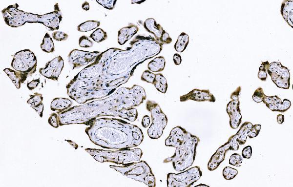
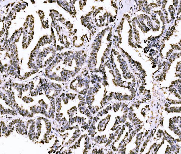
A06797-1 has IHC images from paraffin-embedded human placenta and ovarian serous adenocarcinoma (catalog image captions). No IF/ICC validation or image is listed (catalog applications and IF images).

Real IHC data IHC analysis of PIWIL4/PIWI using anti-PIWIL4/PIWI antibody (A06797-1). PIWIL4/PIWI was detected in a paraffin-embedded section of human placenta tissue. Heat mediated antigen retrieval was performed in EDTA buffer (pH 8.0, epitope retrieval solution). The tissue section was blocked with 10% goat serum. The tissue section was then incubated with 2 μg/ml rabbit anti-PIWIL4/PIWI Antibody (A06797-1) overnight at 4°C. Biotinylated goat anti-rabbit IgG was used as secondary antibody and incubated for 30 minutes at 37°C. The tissue section was developed using Strepavidin-Biotin-Complex (SABC) (Catalog # SA1022) with DAB as the chromogen.
Anti-PIWIL4/PIWI Antibody ®
Cat # A06797-1

A06797-1 is a human-reactive rabbit antibody listed for IHC (catalog applications and reactivity). Its IHC images show paraffin-embedded human placenta and ovarian serous adenocarcinoma (catalog image captions).

Which to pick: Choose A06797-1 for human paraffin-section IHC; its captions report EDTA retrieval at pH 8.0 and 2 μg/ml primary antibody (catalog image captions). No IF/ICC or cross-species option is supported by this catalog entry: A06797-1 lists human reactivity and no IF/ICC application, IF image, or clone designation (catalog reactivity, applications, IF images, and clone field). The captions identify paraffin-embedded sections but do not report a fixative (catalog image captions).

Each figure is that product's own IHC / IF validation image from its datasheet.

References

  1. UniProt Consortium. UniProt entry Q7Z3Z4 (PIWL4_HUMAN, Piwi-like protein 4).
  2. Human Protein Atlas. PIWIL4 tissue IHC expression (reliability: Enhanced).
  3. Human Protein Atlas. PIWIL4 subcellular location (ICC-IF): Localized to the nucleoplasm and mitochondria..
  4. Human Protein Atlas. PIWIL4 antibody validation summary (3 antibodies).
  5. Argonaute proteins: potential biomarkers for human colon cancer. BMC cancer 2010 — PMC2843668.
  6. The topographical distribution of spermatogonial subpopulations during the cycle of seminiferous epithelium in Macaca Fascicularis. Biology of reproduction 2026 — PMC13079449.
  7. Biopathological Significance of PIWI-piRNA Pathway Deregulation in Invasive Breast Carcinomas. Cancers 2020 — PMC7600338.
  8. Expression of genome defence protein members in proliferating and quiescent rat male germ cells and the Nuage dynamics. PloS one 2019 — PMC6557511.
  9. PubMed PMID:12906857 — UniProt-cited evidence.
  10. PubMed PMID:14702039 — UniProt-cited evidence.
  11. PubMed PMID:17974005 — UniProt-cited evidence.