This website uses cookies to ensure you get the best experience on our website.


- Table of Contents
Plan paraffin-section PKM IHC around the cytoplasmic staining reported in most tissues (HPA tissue IHC). This guide covers consistent fixation, a catalog antibody starting range of 2–5 μg/mL, chromogenic detection, and interpretation across PKM isoforms (standard IHC practice; datasheet PB9379; UniProt).

Expected localisation, validated protocols, controls and antibodies — the at-a-glance facts below, then the full design guide.
| Expected localisation | Cytoplasmic staining in most tissues (HPA tissue IHC) | |
| Staining pattern | Most tissues: cytoplasmic staining; cell type varies (HPA tissue IHC) | |
| Antigen retrieval | EDTA pH 8.0 HIER, heat-mediated (datasheet PB9379) | |
| Positive control | Adipose tissue+4 more · see all | |
| Negative control | Smooth muscle |
| Fixation | Keep fixation consistent across sections (standard IHC practice; not target-specific) | |
| Caveat | Staining has low agreement with RNA expression (HPA tissue IHC) | |
| Regulation | Enriched in proliferating cells (UniProt) | |
| Isoform / epitope | Three isoforms; PKM2-directed staining may not reflect total PKM (UniProt; datasheet PB9379) |
The catalog antibody’s IHC-P protocol (datasheet: PB9379) is presented alongside three published paraffin-section IHC protocols (PMC12328876; PMC13548708; PMC5559320).
| Sample | Paraffin-embedded human colorectal adenocarcinoma tissue; fixative not specified (datasheet PB9379) |
| Fixation | Image fixative and duration unreported (datasheet PB9379); verify before use. |
| Sectioning | 4–5 µm sections on charged slides (standard) |
| Deparaffinisation | Xylene, graded ethanol series to water (standard) |
| Antigen retrieval | Heat retrieval: EDTA pH 8.0 (datasheet PB9379); 20 min, 95–100 °C (standard) |
| Peroxidase block | 3% H2O2, 10 min, room temperature (standard) |
| Blocking | 10% goat serum (datasheet PB9379) |
| Primary antibody | Rabbit anti-PKM, 2-5μg/ml (datasheet PB9379) |
| Primary incubation | Overnight at 4 °C (datasheet PB9379) |
| Detection | HRP-conjugated secondary, DAB chromogen (datasheet PB9379) |
| Counterstain | Hematoxylin, blue, dehydrate and mount (standard) |
| Expected result | PKM-positive staining in adipocytes of adipose tissue (HPA tissue IHC: Medium). HPA tissue profile: Cytoplasmic expression in most tissues. No signal in the no-primary control. |
PKM should appear mainly in the cytoplasm of cells across many tissues (HPA tissue IHC: cytoplasmic expression in most tissues). UniProt also places PKM in the nucleus after certain signals and lists no transmembrane segment (UniProt P14618: subcellular location and topology). Interpret tissue staining cautiously: HPA rates its tissue IHC profile Uncertain because staining and RNA expression show low consistency (HPA tissue IHC: reliability Uncertain).
| Cytoplasmic staining in adipocytes or breast glandular cells, with surrounding tissue structure visible. | This fits the reported medium staining in both cell types (HPA tissue IHC: adipocytes Medium; breast glandular cells Medium). Compare the stained cells with adjacent unstained areas and the counterstain; a strong color reaction alone does not establish specificity, particularly while the overall tissue profile remains Uncertain (HPA tissue IHC: reliability Uncertain). |
| Predominantly nuclear staining, with little or no cytoplasmic staining, or a sharp membrane outline. | A membrane outline conflicts with the reported cytoplasmic tissue pattern and lack of a transmembrane segment (HPA tissue IHC: cytoplasmic expression; UniProt P14618: topology); investigate artefact. Nuclear PKM can occur after specific signals, so nuclear staining alone is not proof of error (UniProt P14618: subcellular location). An unexplained nuclear-only pattern warrants independent validation. |
| Strong staining in smooth muscle cells, especially when expected positive cells stain as well. | HPA reports smooth muscle cells as Not detected, making a strong signal there a warning for cross-reactivity or endogenous detection activity (HPA tissue IHC: smooth muscle cells Not detected; general IHC practice). Check a no-primary control and compare staining with tissue structure. Because HPA tissue reliability is Uncertain, this observation is a reason to investigate, not a definitive PKM-negative control (HPA tissue IHC: reliability Uncertain). |
| Broad color across cells, extracellular spaces, or section edges that obscures cytoplasmic detail. | A haze extending beyond cells cannot be scored as the reported cytoplasmic pattern (HPA tissue IHC: cytoplasmic expression in most tissues). Review the no-primary control for detection background and inspect whether uneven reagent coverage or inadequate washing is contributing (general IHC practice). Reassess PKM only after individual cell boundaries and the counterstain are readable. |
| No cytoplasmic signal in a well-preserved section of adipose tissue or breast. | HPA reports medium staining in adipocytes and breast glandular cells, so an absent signal calls for a run-level check (HPA tissue IHC: both Medium). Verify that the antibody is suitable for IHC-P, that detection reagents worked, and that the known-positive control was processed alongside the test section (general IHC practice). A negative result alone does not establish absent PKM. |
| Cell and tissue context | HPA describes cytoplasmic expression in most tissues but reports medium staining in the listed adipocytes and breast glandular cells, low staining in small-intestinal glandular cells, and no detected staining in smooth muscle cells (HPA tissue IHC). Select and score controls by cell type rather than assuming every cell in a section should have the same intensity; the HPA profile has Uncertain reliability (HPA tissue IHC). |
| Isoform and antibody interpretation | UniProt lists three PKM isoforms: M2, M1, and isoform 3 (UniProt P14618: isoforms). An observed pattern cannot identify an isoform unless the antibody's epitope and isoform reactivity establish that distinction (general IHC interpretation). The supplied HPA antibodies, HPA029501 and CAB019421, each have Uncertain IHC status, so their staining alone offers limited support for a disputed tissue pattern (HPA antibodies: IHC Uncertain). |
| IF/ICC Q&A: should fluorescence be only cytoplasmic? | No: HPA reports mainly cytosolic signal, with additional vesicle and principal-piece localization in its ICC-IF record (HPA subcellular: Cytosol enhanced; Vesicles and Principal piece approved). UniProt also describes signal-dependent nuclear translocation (UniProt P14618: subcellular location). These observations help interpret an IF image; they do not establish a second IHC-P protocol or confirm a nuclear signal in a particular paraffin section. |
| Situation | Likely cause | Next action |
|---|---|---|
| All sections, including a reported positive cell type, are blank. | A failed staining run or unsuitable assay conditions remain possible; adipocytes and breast glandular cells have reported medium IHC staining (HPA tissue IHC). | Check the same-run positive control, confirm IHC-P suitability and detection steps, and review the antibody's documented conditions before changing them (general IHC practice). |
| The no-primary control develops color. | The signal can arise from endogenous detection activity or nonspecific detection reagents rather than primary-antibody binding (general IHC practice). | Address the implicated detection chemistry and blocking controls, then repeat the run and compare the no-primary and antibody-stained sections (general IHC practice). |
| Signal is diffuse and hides cell boundaries. | Excess background, uneven reagent coverage, or inadequate washing can obscure a cytoplasmic pattern (general IHC practice; HPA tissue IHC: cytoplasmic expression). | Inspect section coverage, washing, and detection intensity; adjust one workflow variable at a time and retain a same-run control (general IHC practice). |
| A membrane outline dominates the stained cells. | This conflicts with PKM's reported cytoplasmic distribution and lack of a transmembrane segment (HPA tissue IHC; UniProt P14618: topology). | Inspect the no-primary control and repeat with independent specificity evidence before assigning the outline to PKM (general IHC practice; HPA tissue IHC: reliability Uncertain). |
| Nuclei stain more strongly than cytoplasm. | UniProt describes signal-dependent nuclear translocation, but the supplied tissue IHC summary describes cytoplasmic expression in most tissues (UniProt P14618; HPA tissue IHC). | Score nuclear and cytoplasmic staining separately; seek independent validation before treating a nuclear-only result as expected for this tissue (general IHC interpretation). |
| Smooth muscle cells stain strongly while other cells show plausible cytoplasm. | HPA lists smooth muscle cells as Not detected, raising concern about cross-reactivity or endogenous detection activity (HPA tissue IHC; general IHC practice). | Compare a no-primary control and an independently supported antibody result; report the discrepancy without treating HPA's Uncertain tissue profile as an absolute exclusion (HPA tissue IHC: reliability Uncertain; general IHC practice). |





Comprehensive Human Protein Atlas IHC scoring per tissue (reliability: Uncertain — Low consistency between antibody staining and RNA expression data.). Rows are taken directly from the HPA tissue chart — click any row's HPA link to view the source.
| Tissue | Cell type | Level | Evidence | Source |
|---|---|---|---|---|
| Adipose tissue | Adipocytes | Medium | Protein (IHC) | HPA → |
| Adrenal gland | Glandular cells | Medium | Protein (IHC) | HPA → |
| Bone marrow | Hematopoietic cells | Medium | Protein (IHC) | HPA → |
| Breast | Glandular cells | Medium | Protein (IHC) | HPA → |
| Bronchus | Respiratory epithelial cells | Medium | Protein (IHC) | HPA → |
| Tissue | Cell type | Level | Evidence | Source |
|---|---|---|---|---|
| Smooth muscle | Smooth muscle cells | Not detected | Protein (IHC) | HPA → |
Troubleshoot PKM staining in paraffin sections by checking retrieval, controls, compartment patterns and scoring before interpreting biological differences.
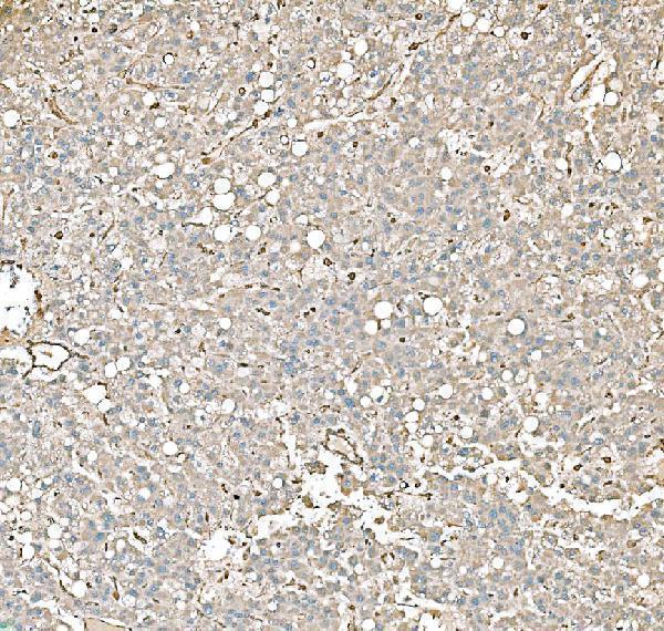
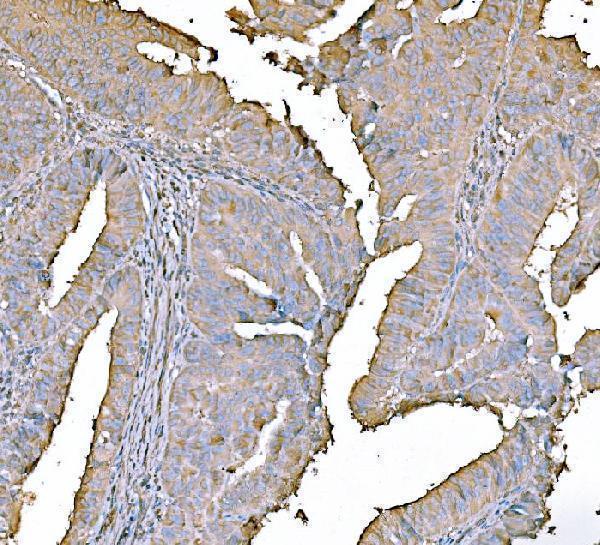
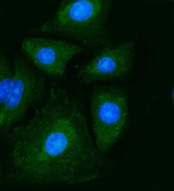
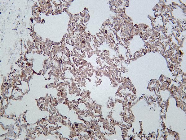
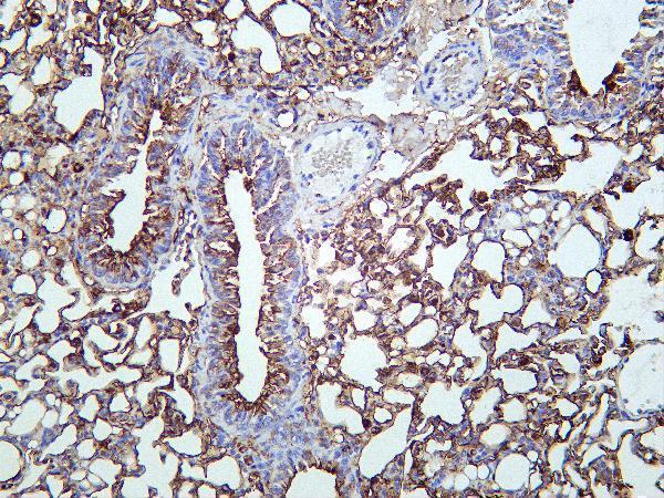
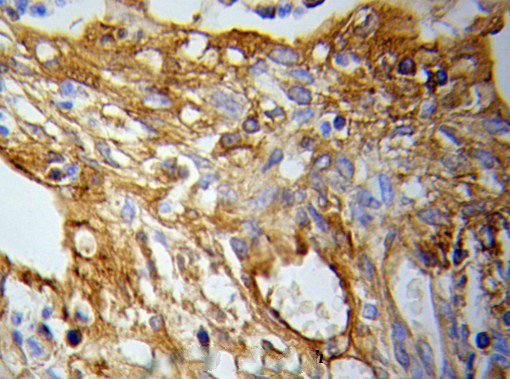
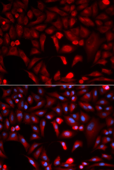
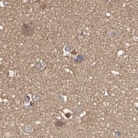
Catalog anti-PKM antibodies have IHC images from human, mouse and rat tissues, plus IF/ICC images from HeLa and A549 cells (catalog image captions).
PB9379 is shown on human colorectal adenocarcinoma, M01173-1 on human liver cancer, and M01173-2 on rat lung (respective IHC image captions). A01173-1 is shown on paraffin-embedded lung cancer tissue, and M01173 on paraffin-embedded human cervix cancer tissue (respective IHC image captions).
Which to pick: For paraffin-section IHC, pick PB9379: its human colorectal adenocarcinoma caption documents EDTA retrieval at pH 8.0 and a 2 μg/ml primary incubation overnight at 4°C; the fixative is unreported (PB9379 IHC caption). For IF/ICC, PB9379 has an A549-cell image using 5 μg/ml primary antibody (PB9379 IF caption). For cross-species IHC, M01173-2 is a rabbit monoclonal listed for human, mouse and rat, with IHC images in all three species; its rat lung caption does not report processing or fixative (M01173-2 catalog description, reactivity and IHC image captions).