This website uses cookies to ensure you get the best experience on our website.


- Table of Contents
Plan PKP2 staining in paraffin sections using its reported cytoplasmic and cardiac intercalated-disc pattern (HPA tissue IHC). The guide covers control selection and interpretation alongside the catalog antibody’s IHC conditions (datasheet A02146-2).

Expected localisation, validated protocols, controls and antibodies — the at-a-glance facts below, then the full design guide.
| Expected localisation | Cytoplasm; cardiac intercalated discs (HPA tissue IHC) | |
| Staining pattern | Cytoplasmic staining in several tissues; strong cardiac discs (HPA tissue IHC) | |
| Antigen retrieval | EDTA pH 8.0 HIER, heat-mediated (datasheet A02146-2) | |
| Positive control | Cerebellum+4 more · see all | |
| Negative control | Adipose tissue+4 more · see all |
| Fixation | Keep paraffin-section fixation consistent. (standard IHC practice; not target-specific) | |
| Caveat | Staining and RNA expression show medium consistency (HPA tissue IHC) | |
| Regulation | Heart muscle–enhanced RNA expression (HPA tissue RNA) | |
| Isoform / epitope | 2 isoforms; epitope coverage is unspecified (UniProt; datasheet A02146-2) |
Compare the catalog antibody’s IHC-P protocol (datasheet: A02146-2) with four published PKP2 IHC methods (PMC7568375; PMC3025932; PMC7837596; PMC10948840).
| Sample | Paraffin-embedded human lung cancer tissue; fixative not specified (datasheet A02146-2) |
| Fixation | Image fixative and duration unreported (datasheet A02146-2); verify before use. |
| Sectioning | 4–5 µm sections on charged slides (standard) |
| Deparaffinisation | Xylene, graded ethanol series to water (standard) |
| Antigen retrieval | Heat retrieval: EDTA pH 8.0 (datasheet A02146-2); 20 min, 95–100 °C (standard) |
| Peroxidase block | 3% H2O2, 10 min, room temperature (standard) |
| Blocking | 10% goat serum (datasheet A02146-2) |
| Primary antibody | Rabbit anti-PKP2, 2-5 μg/ml (datasheet A02146-2) |
| Primary incubation | Overnight at 4 °C (datasheet A02146-2) |
| Detection | HRP-conjugated secondary, DAB chromogen (datasheet A02146-2) |
| Counterstain | Hematoxylin, blue, dehydrate and mount (standard) |
| Expected result | PKP2-positive staining in processes in granular layer of cerebellum (HPA tissue IHC: High). HPA tissue profile: Cytoplasmic expression in several tissues, highly expressed in fallopian tube and intercalated discs in cardiac muscle. No signal in the no-primary control. |
PKP2 should mark cell junctions, especially cardiomyocyte intercalated discs, with cytoplasmic staining possible across several tissues (UniProt Q99959 localization; HPA tissue IHC). Nuclear staining can also occur (UniProt Q99959 localization). HPA rates the tissue IHC profile Enhanced, while reporting medium consistency between staining and RNA expression (HPA tissue IHC). PKP2 has no transmembrane segment (UniProt Q99959 topology).
| Sharp staining at cardiomyocyte intercalated discs, with signal in cardiomyocytes (HPA tissue IHC). | This is the strongest anatomical check: UniProt places PKP2 at cardiac intercalated discs, and HPA reports High cardiomyocyte staining (UniProt Q99959 localization; HPA tissue IHC). Judge the junctional pattern together with the stained cell type; intensity alone cannot establish specificity. |
| Signal in fallopian tube ciliated cell bodies or nasopharyngeal ciliary rootlets (HPA tissue IHC). | Both are reported High by HPA, so they offer additional positive patterns when those tissues are available (HPA tissue IHC). Their appearance need not resemble cardiac discs: compare staining with the expected cells and structures in each tissue, rather than requiring one pattern everywhere. |
| Predominantly diffuse extracellular staining, or signal confined to an unexpected compartment in a known-positive cell. | Treat this as suspect because PKP2 is reported at junctions, in cytoplasm, and in nuclei, with no transmembrane segment (UniProt Q99959 localization and topology). Nuclear signal alone is not automatically artefact: nucleoplasmic localization is supported by HPA ICC-IF (HPA subcellular). |
| Strong staining in cells reported as undetected, with little signal in adjacent expected-positive cells. | Cross-reactivity or endogenous detection activity is possible, but a single unexpected field is not proof. HPA reports adipocytes and bone-marrow hematopoietic cells as Not detected; it also reports some other cell populations in those organs separately (HPA tissue IHC). Compare the exact cell type before interpreting a negative control. |
| A uniform haze across cells and surrounding tissue, or no staining in a known-positive section. | Uniform haze weakens compartment-level interpretation; absent signal in heart cardiomyocytes or fallopian tube ciliated cells warrants a technical check (HPA tissue IHC). Neither pattern alone proves PKP2 absence. Compare morphology, the detection blank, and a matched positive section before scoring the sample. |
| Tissue and cell selection (HPA tissue IHC) | HPA reports High signal in heart cardiomyocytes and fallopian tube ciliated cells, but Not detected in adipocytes and bone-marrow hematopoietic cells (HPA tissue IHC). Use named cell populations as comparators; an organ label alone can obscure different cell types. |
| Localization and molecular topology (UniProt Q99959) | PKP2 is junctional, cytoplasmic and nuclear, and has no transmembrane segment or signal peptide (UniProt Q99959 localization, topology and processing). Evaluate intracellular and junctional signal; do not interpret extracellular deposits as a predicted PKP2 compartment. |
| Antibody validation and molecular variation (HPA antibodies; UniProt Q99959) | HPA rates IHC for HPA014314, HPA056908 and CAB037336 Enhanced (HPA antibodies). UniProt lists two isoforms and modified residues, but the supplied record gives no epitope map; it cannot predict isoform-specific staining or a retrieval response (UniProt Q99959 isoforms and modified residues). |
| IF/ICC Q&A: where should fluorescence appear? (HPA subcellular) | HPA reports enhanced cell-junction and supported nucleoplasmic localization, with images from A-431, CACO-2 and U2OS (HPA subcellular). That is an IF/ICC interpretation cue; this section supplies no IF/ICC protocol or dilution. |
| Situation | Likely cause | Next action |
|---|---|---|
| No chromogenic signal in heart cardiomyocytes or fallopian tube ciliated cells (HPA tissue IHC). | Target-specific fixation effects are not established by the supplied assay evidence. Verify with a matched IHC source before attributing a result to fixation. | Check section integrity, primary-antibody application, reagent activity and a matched positive section. Review antigen retrieval as a general IHC workflow variable; do not assume a PKP2-specific retrieval requirement. |
| Cardiac tissue stains, but intercalated discs are hard to distinguish (UniProt Q99959 localization). | Diffuse chromogen or poor structural preservation can obscure a junctional pattern. HPA's High cardiomyocyte category describes staining level; it does not guarantee every field will display clearly resolved discs (HPA tissue IHC). | Inspect well-preserved cardiomyocyte borders and counterstained morphology. Compare signal at intercalated discs with surrounding cytoplasm before calling the expected pattern (UniProt Q99959 localization). |
| Apparent positive staining in adipocytes or bone-marrow hematopoietic cells (HPA tissue IHC). | These named populations are Not detected in HPA; mistaken cell identification, nonspecific primary binding or endogenous detection activity could explain the result (HPA tissue IHC). | Confirm the cell identity and compare a no-primary detection control with a positive section. If staining persists only with primary antibody, reassess specificity using an independently validated antibody. |
| Broad brown haze or staining beyond recognizable cellular structures. | Background from detection reagents, inadequate blocking or overly concentrated primary antibody can obscure a cellular readout; this is a general IHC diagnostic, not a reported PKP2 property. | Compare the no-primary control, inspect reagent background and optimize blocking, washing and antibody dilution according to the chosen assay. Score PKP2 only where the expected cells and compartments remain discernible. |
| Nuclear signal appears alongside cell-border staining. | Nuclear localization is annotated by UniProt, and HPA ICC-IF supports nucleoplasmic signal (UniProt Q99959 localization; HPA subcellular). Its presence alone is therefore insufficient to label the stain nonspecific. | Check whether junctional staining also fits the tissue and cell type. Use morphology and the detection blank to distinguish cellular nuclear signal from generalized background. |
| A negative control tissue contains focal positive cells. | HPA's Not detected calls apply to specified cell types, such as lymph-node germinal-center cells, rather than proving every cell in that tissue lacks PKP2 (HPA tissue IHC). | Identify the stained cells before rejecting the control. Compare the exact HPA-listed population and an expected-positive section, then record cell type, compartment and intensity separately. |
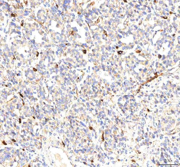
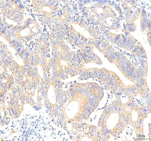
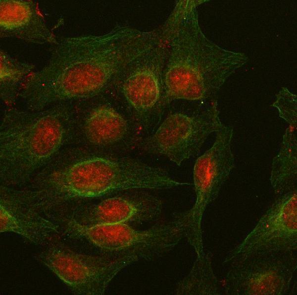


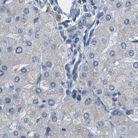


Comprehensive Human Protein Atlas IHC scoring per tissue (reliability: Enhanced — Medium consistency between antibody staining and RNA expression data.). Rows are taken directly from the HPA tissue chart — click any row's HPA link to view the source.
| Tissue | Cell type | Level | Evidence | Source |
|---|---|---|---|---|
| Cerebellum | Processes in granular layer | High | Protein (IHC) | HPA → |
| Fallopian tube | Ciliated cells (cell body) | High | Protein (IHC) | HPA → |
| Heart muscle | Cardiomyocytes | High | Protein (IHC) | HPA → |
| Nasopharynx | Ciliated cells (ciliary rootlets) | High | Protein (IHC) | HPA → |
| Appendix | Glandular cells | Medium | Protein (IHC) | HPA → |
| Tissue | Cell type | Level | Evidence | Source |
|---|---|---|---|---|
| Adipose tissue | Adipocytes | Not detected | Protein (IHC) | HPA → |
| Bone marrow | Hematopoietic cells | Not detected | Protein (IHC) | HPA → |
| Cervix | Glandular cells | Not detected | Protein (IHC) | HPA → |
| Liver | Cholangiocytes | Not detected | Protein (IHC) | HPA → |
| Lymph node | Germinal center cells | Not detected | Protein (IHC) | HPA → |
Use compartment, cell type, and matched controls to troubleshoot PKP2 staining in paraffin sections (UniProt Q99959; HPA tissue IHC).
A02146-2 has human paraffin-section IHC images and human U2OS cell IF data (catalog image captions); the catalog lists human, mouse and rat reactivity (catalog: reactivity).
A02146-2 will render with its own human lung cancer paraffin-section IHC figure (catalog: IHC image caption). The catalog also documents IHC images from human colon adenocarcinoma, liver cancer and tonsil sections, plus IF in U2OS cells (catalog: image captions).
Which to pick: Choose A02146-2 for paraffin-section IHC: its human lung cancer caption reports EDTA retrieval at pH 8.0 and 2 μg/ml primary antibody (catalog: IHC image caption); the fixative is unreported. For IF/ICC, A02146-2 has a human U2OS cell IF image and a listed concentration of 5 μg/ml (catalog: IF image caption and dilution). For cross-species planning, A02146-2 lists human, mouse and rat reactivity, while its IHC dilution entry specifies human and mouse; the supplied IHC images show human tissue (catalog: reactivity, dilution entry and IHC image captions).