This website uses cookies to ensure you get the best experience on our website.


- Table of Contents
Plan PLA2G1B paraffin IHC using exocrine pancreas as a positive tissue and cytoplasmic glandular staining as the expected pattern (HPA tissue IHC). Compare consistently fixed sections and account for secretion when interpreting staining beyond the producing cells (UniProt).

Expected localisation, validated protocols, controls and antibodies — the at-a-glance facts below, then the full design guide.
| Expected localisation | Cytoplasmic staining in exocrine pancreas (HPA tissue IHC) | |
| Staining pattern | Exocrine glandular cells show cytoplasmic staining (HPA tissue IHC) | |
| Antigen retrieval | Citrate pH 6.0 HIER, 95–98 °C, 20 min (rule: cytoplasmic / membrane antigen) | |
| Positive control | Pancreas | |
| Negative control | Adipose tissue+4 more · see all |
| Fixation | Keep fixation consistent across sections (standard IHC practice; not target-specific) | |
| Caveat | Secretion can separate protein from its source cells (HPA tissue IHC) | |
| Regulation | Pancreas-enriched expression (HPA tissue IHC) | |
| Isoform / epitope | No annotated isoforms; residues 1–22 precede the mature chain (UniProt) |
Compare the catalog antibody’s IHC-P protocol with the published mouse lung IHC workflow (PMC12571731).
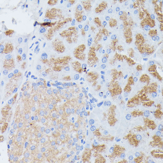
| Sample | Paraffin-embedded mouse pancreas tissue; fixative not specified (datasheet A06738-1) |
| Fixation | Image fixative and duration unreported (datasheet A06738-1); verify before use. |
| Sectioning | 4–5 µm sections on charged slides (standard) |
| Deparaffinisation | Xylene, graded ethanol series to water (standard) |
| Antigen retrieval | Heat-induced epitope retrieval in citrate buffer, pH 6.0, 20 min at 95–98 °C (standard rule: cytoplasmic / membrane antigen) |
| Peroxidase block | 3% H2O2, 10 min, room temperature (standard) |
| Blocking | 10% normal serum of the secondary host, 30 min, room temperature (standard) |
| Primary antibody | Rabbit anti-PLA2G1B, 1:100-1:200 (datasheet A06738-1) |
| Primary incubation | Overnight at 4 °C (standard) |
| Detection | HRP-polymer secondary, DAB chromogen 5–10 min (standard) |
| Counterstain | Hematoxylin, blue, dehydrate and mount (standard) |
| Expected result | PLA2G1B-positive staining in exocrine glandular cells of pancreas (HPA tissue IHC: High). HPA tissue profile: Selective cytoplasmic expression in exocrine pancreas. No signal in the no-primary control. |
PLA2G1B is secreted from pancreatic acinar cells in an inactive form and has no transmembrane segment (UniProt P04054). In tissue IHC, expect selective cytoplasmic staining in pancreatic exocrine glandular cells (HPA: High in exocrine glandular cells). HPA rates this tissue pattern Enhanced, while cautioning that secretion can separate the locations of RNA and protein (HPA: Enhanced reliability).
| Strong cytoplasmic staining in pancreatic exocrine glandular cells, with little staining elsewhere on the section. | This matches the reported selective pancreatic pattern and High staining level (HPA: tissue IHC). Interpret the stained cells and compartment together: the HPA finding supports a cytoplasmic exocrine pattern, while UniProt identifies the protein as secreted (UniProt P04054). |
| Prominent nuclear staining, especially when cytoplasmic exocrine staining is weak or absent. | A dominant nuclear pattern conflicts with HPA's selective cytoplasmic observation and UniProt's secreted annotation (HPA: tissue IHC; UniProt P04054). Treat it as suspect staining; compare a no-primary control and reassess detection and image interpretation before assigning it to PLA2G1B (general IHC practice). |
| Strong staining in unrelated cell types, or in adipocytes or adrenal glandular cells. | HPA reports PLA2G1B as Not detected in those two listed cell types, so staining there warrants a specificity check (HPA: tissue IHC). Cross-reactivity or endogenous chromogenic activity are possible explanations, not diagnoses from appearance alone; compare controls and the expected pancreatic pattern (general IHC practice). |
| Diffuse color across multiple compartments, including areas without a recognizable exocrine cell pattern. | Uniform haze does not reproduce HPA's selective cytoplasmic exocrine result (HPA: tissue IHC). Check no-primary controls, endogenous detection activity, blocking and chromogen development before scoring cells; widespread color can obscure a genuine localized signal (general IHC practice). |
| No convincing cytoplasmic signal in a pancreatic exocrine region. | That conflicts with the High pancreatic exocrine staining reported by HPA, but one negative section does not establish biological absence (HPA: tissue IHC). Confirm that exocrine tissue is present, then assess the assay with a suitable positive control and review antibody and detection conditions (general IHC practice). |
| Secreted protein and processing | UniProt describes secretion from pancreatic acinar cells, a signal peptide at residues 1–15, a propeptide at 16–22, and a mature chain at 23–148 (UniProt P04054). An extracellular signal should therefore be assessed in context; secretion alone does not make diffuse slide background specific staining. |
| Tissue context | HPA calls pancreatic RNA tissue enriched and pancreatic exocrine IHC High, while its listed samples include several Not detected cell types (HPA: tissue IHC). UniProt also reports expression in lung, liver and kidney (UniProt P04054). Do not infer that every nonpancreatic tissue must be IHC negative. |
| Antibody evidence | Two rabbit polyclonal antibodies, HPA047822 and HPA060803, have Enhanced IHC status (HPA: antibodies). That supports the reported tissue pattern; it does not establish that every new antibody, staining run or unexpected compartment is specific (general IHC practice). |
| Can the IHC pattern be applied to ICC/IF? | Use the pancreatic IHC pattern as context, not as an established cellular IF image: HPA lists no main subcellular location or ICC/IF image cell lines for PLA2G1B (HPA: subcellular). UniProt calls it secreted (UniProt P04054). Interpret any ICC/IF signal with its own controls. |
| Situation | Likely cause | Next action |
|---|---|---|
| Pancreatic exocrine cells are unstained. | The result conflicts with HPA's High exocrine signal; the reason cannot be assigned from the section alone (HPA: tissue IHC). | Verify exocrine tissue, positive control performance, antibody application and detection; review retrieval conditions without assuming PLA2G1B-specific fixation sensitivity (general IHC practice). |
| The whole section has weak, even color. | Diffuse color lacks the selective cytoplasmic distribution reported by HPA; detection background is possible (HPA: tissue IHC; general IHC practice). | Examine a no-primary control; review blocking, washes, endogenous activity control and chromogen development before scoring (general IHC practice). |
| Nuclei dominate the signal. | A dominant nuclear pattern disagrees with the HPA cytoplasmic observation and UniProt secreted annotation (HPA: tissue IHC; UniProt P04054). | Check counterstain separation and no-primary controls, then confirm whether cytoplasmic exocrine staining is independently visible (general IHC practice). |
| Adipocytes or adrenal glandular cells stain strongly. | HPA lists both cell types as Not detected; cross-reactivity or endogenous detection activity should be investigated (HPA: tissue IHC; general IHC practice). | Compare negative and no-primary controls with pancreatic positive tissue; do not score the unexpected color as PLA2G1B without corroboration (general IHC practice). |
| Extracellular color is present near otherwise plausible pancreatic staining. | Secretion makes extracellular protein plausible, but its exact histologic distribution is not established by the supplied HPA pattern (UniProt P04054; HPA: tissue IHC). | Score the documented cytoplasmic exocrine pattern separately; use controls to distinguish localized signal from diffuse chromogenic background (HPA: tissue IHC; general IHC practice). |
| An ICC/IF image appears unlike the pancreatic IHC section. | HPA provides no PLA2G1B ICC/IF image cell lines or main subcellular location for a direct comparison (HPA: subcellular). | Interpret ICC/IF with application-specific positive and negative controls; avoid transferring an IHC staining claim to a cellular IF result (general IHC/IF practice). |


Comprehensive Human Protein Atlas IHC scoring per tissue (reliability: Enhanced — High consistency between antibody staining and RNA expression data. At least one protein variant secreted, tissue location of RNA and protein might differ and correlation is complex.). Rows are taken directly from the HPA tissue chart — click any row's HPA link to view the source.
| Tissue | Cell type | Level | Evidence | Source |
|---|---|---|---|---|
| Pancreas | Exocrine glandular cells | High | Protein (IHC) | HPA → |
| Tissue | Cell type | Level | Evidence | Source |
|---|---|---|---|---|
| Adipose tissue | Adipocytes | Not detected | Protein (IHC) | HPA → |
| Adrenal gland | Glandular cells | Not detected | Protein (IHC) | HPA → |
| Appendix | Glandular cells | Not detected | Protein (IHC) | HPA → |
| Bone marrow | Hematopoietic cells | Not detected | Protein (IHC) | HPA → |
| Breast | Adipocytes | Not detected | Protein (IHC) | HPA → |
Troubleshoot PLA2G1B staining in chromogenic IHC using the documented pancreas pattern, secreted topology, and antibody-specific processing evidence.
PLA2G1B antibodies list human and mouse reactivity (catalog: both SKUs). A06738-1 has a mouse pancreas paraffin IHC figure; A06738-2 lists IF, with no IF figure provided (catalog image captions; applications).
A06738-1 lists human and mouse IHC reactivity and shows staining of mouse pancreas paraffin sections at 1:100 after microwave retrieval in 10 mM PBS, pH 7.2 (catalog applications/reactivity; A06738-1 image caption). A06738-2 lists human and mouse IHC and IF reactivity, with no IHC or IF image caption provided (catalog applications/reactivity; image captions).
Which to pick: For tissue IHC, choose A06738-1 when a pictured mouse pancreas paraffin example is useful; its caption does not report the fixative (A06738-1 image caption). For IF, choose A06738-2 because IF is listed, while ICC is unlisted and no IF figure is provided (catalog applications; image captions). Both SKUs list human and mouse reactivity, but the pictured tissue result is mouse only (catalog reactivity; A06738-1 image caption).