This website uses cookies to ensure you get the best experience on our website.


- Table of Contents
Plan PLA2G4A IHC-P around cytoplasmic tissue staining, with high staining in parathyroid and seminal-vesicle glandular cells (HPA tissue IHC). Start with the catalog antibody’s 1:10–1:50 IHC-P dilution range (datasheet), and consider calcium-dependent membrane translocation when interpreting compartment changes (UniProt).

Expected localisation, validated protocols, controls and antibodies — the at-a-glance facts below, then the full design guide.
| Expected localisation | Cytoplasmic tissue staining (HPA tissue IHC); Golgi and nuclear-envelope association possible (UniProt) | |
| Staining pattern | Cytoplasmic; high in parathyroid and seminal-vesicle glandular cells (HPA tissue IHC) | |
| Antigen retrieval | Tris-EDTA pH 9.0 HIER, 95–98 °C, 20 min (rule: nuclear antigen) | |
| Positive control | Parathyroid gland+4 more · see all | |
| Negative control | Ovary+2 more · see all |
| Fixation | Keep formalin fixation consistent across sections. (standard IHC practice; not target-specific) | |
| Caveat | Low antibody–RNA concordance limits tissue predictions (HPA tissue IHC) | |
| Regulation | Intensity regulation not specified (UniProt) | |
| Isoform / epitope | No listed isoforms; one 1–749 chain with no transmembrane segment (UniProt) |
The catalog antibody protocol is paired with a published chromogenic IHC protocol for mouse testis (PMC4372093).
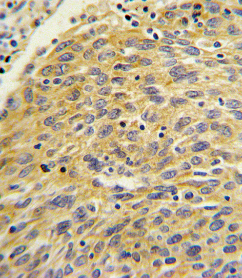
| Sample | FFPE human lung carcinoma tissue (datasheet A00854-1) |
| Fixation | Image formalin-fixed; duration unreported (datasheet A00854-1); verify before use. |
| Sectioning | 4–5 µm sections on charged slides (standard) |
| Deparaffinisation | Xylene, graded ethanol series to water (standard) |
| Antigen retrieval | Heat-induced epitope retrieval in Tris-EDTA buffer, pH 9.0, 20 min at 95–98 °C (standard rule: nuclear antigen) |
| Peroxidase block | 3% H2O2, 10 min, room temperature (standard) |
| Blocking | 10% normal serum of the secondary host, 30 min, room temperature (standard) |
| Primary antibody | Rabbit anti-PLA2G4A, 1:10-1:50 (datasheet A00854-1) |
| Primary incubation | Overnight at 4 °C (standard) |
| Detection | HRP-polymer secondary, DAB chromogen 5–10 min (standard) |
| Counterstain | Hematoxylin, blue, dehydrate and mount (standard) |
| Expected result | PLA2G4A-positive staining in glandular cells of parathyroid gland (HPA tissue IHC: High). HPA tissue profile: General cytoplasmic expression, most abundant in parathyroid gland and seminal vesicle. No signal in the no-primary control. |
PLA2G4A should stain mainly in the cytoplasm, with possible staining at intracellular membranes and the nuclear envelope; it has no transmembrane segment (UniProt P47712). In tissue IHC, glandular cells of parathyroid gland and seminal vesicle show high staining (HPA tissue IHC). HPA rates its tissue staining Approved but reports low consistency with RNA expression, so interpret unexpected patterns cautiously (HPA tissue IHC).
| Cytoplasmic staining in parathyroid or seminal vesicle glandular cells. | This matches the high staining reported for those cells (HPA tissue IHC) and the principal cytoplasmic localization of PLA2G4A (UniProt P47712). Assess the named cells rather than assigning the same intensity to every cell in the section; HPA reports a cell-specific tissue pattern (HPA tissue IHC). |
| Predominantly nuclear staining, with little cytoplasmic signal. | A nuclear envelope component is possible, but predominantly intranuclear staining is outside the reported main cytoplasmic pattern (UniProt P47712; HPA subcellular ICC-IF). Check morphology and counterstain before assigning a compartment (standard IHC practice). An unusual compartment alone does not establish target specificity, especially given HPA's low staining-to-RNA consistency (HPA tissue IHC). |
| Strong signal in ovarian stroma, salivary glandular cells, or vaginal squamous epithelium. | HPA reports PLA2G4A as not detected in those respective cells (HPA tissue IHC). Consider cross-reactivity or endogenous chromogen-generating activity, and inspect a detection-only control (standard IHC practice). Treat these tissues as comparison areas, not absolute biological negatives: HPA notes low consistency between its antibody staining and RNA expression data (HPA tissue IHC). |
| Uniform color across tissue, including empty spaces or unrelated structures. | That distribution is difficult to reconcile with the reported cell-associated cytoplasmic pattern (HPA tissue IHC; UniProt P47712). Diffuse deposition can reflect incomplete blocking, residual detection activity, or overly strong detection chemistry (standard IHC practice). Compare with a detection-only control before scoring cells as positive (standard IHC practice). |
| No signal in parathyroid or seminal vesicle glandular cells. | These are high-staining reference cells in the HPA tissue survey (HPA tissue IHC). First verify that the expected cells are present and that counterstain and detection worked (standard IHC practice). Then review the antibody's documented IHC conditions and the run controls; absence of color in one section does not establish absence of PLA2G4A (standard IHC practice). |
| Compartment and cell state | PLA2G4A is cytoplasmic and can translocate to intracellular membranes in a calcium-dependent way (UniProt P47712). Membrane-associated accentuation can therefore coexist with cytoplasmic signal; a single fixed section does not reveal when translocation occurred (UniProt P47712; standard IHC interpretation). |
| Tissue and cell selection | HPA reports high glandular staining in parathyroid gland and seminal vesicle, medium staining in bronchial respiratory epithelium, and no detected staining in ovarian stroma (HPA tissue IHC). Compare matched cell types when judging intensity; whole-tissue color can obscure that distinction (standard IHC practice). |
| Evidence strength | The HPA tissue profile is Approved but has low consistency between antibody staining and RNA expression (HPA tissue IHC). Its listed antibodies have Approved IHC status, which supports use of the reported pattern while leaving an unexpected result in need of controls (HPA antibodies; standard IHC practice). |
| Protein architecture | UniProt lists no signal peptide, propeptide, transmembrane segment, glycosylation site, or isoform for the supplied record (UniProt P47712). These annotations give no basis here for expecting an extracellular or shed-protein staining pattern (UniProt P47712). The antibody epitope is unspecified, so epitope-specific retrieval predictions are unsupported. |
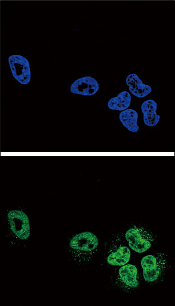
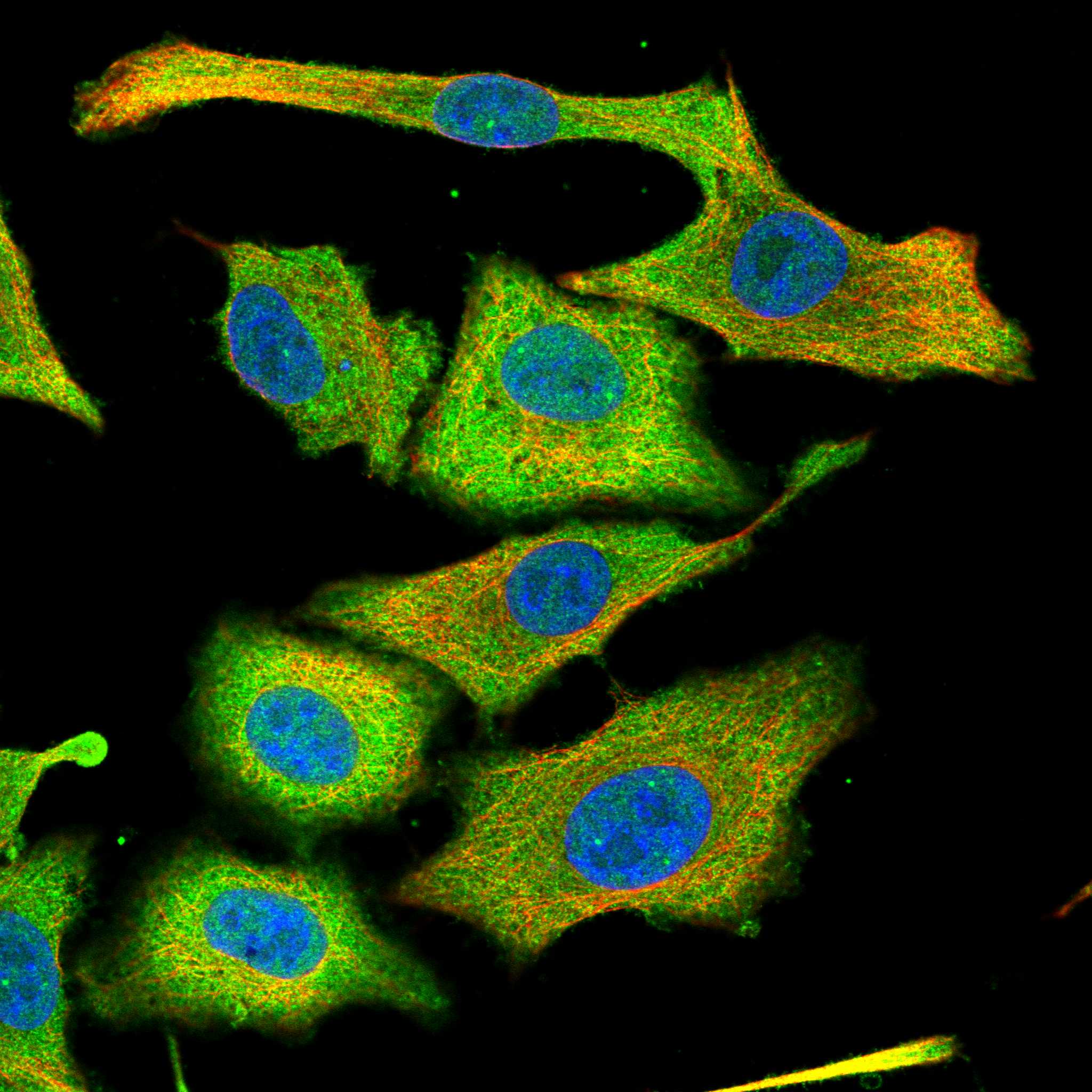
| IF/ICC Q&A: where should signal appear? | Mainly in the cytosol, with additional vesicular signal (HPA subcellular ICC-IF). UniProt also lists Golgi membrane and nuclear envelope localization (UniProt P47712). This answers the localization question only; the supplied evidence gives no IF/ICC protocol conditions. |
| Situation | Likely cause | Next action |
|---|---|---|
| High-reference glandular cells are blank. | The expected cells may be absent from the section, or an IHC run step may have failed (HPA tissue IHC; standard IHC practice). | Locate glandular cells, check a run control, and review the antibody's documented IHC conditions before interpreting the blank section (standard IHC practice). |
| Color is strongest in nuclei. | The compartment differs from the mainly cytoplasmic pattern; nuclear envelope localization should not be read as diffuse intranuclear staining (UniProt P47712; HPA subcellular ICC-IF). | Recheck focus and counterstain, then compare the signal with a detection-only control (standard IHC practice). |
| Reported negative cells stain strongly. | Cross-reactivity or endogenous detection activity is possible; HPA reports no detection in ovarian stroma, salivary glandular cells, and vaginal squamous epithelium (HPA tissue IHC; standard IHC practice). | Inspect cell identity and a detection-only control; do not assign target-specific positivity from color alone (standard IHC practice). |
| The entire section has diffuse chromogen. | Nonspecific binding, incomplete blocking, or excessive detection signal can obscure cell boundaries (standard IHC practice). | Compare the detection-only control and assess blocking, washing, and detection conditions using the documented workflow (standard IHC practice). |
| Cytoplasm stains, but intensity differs among tissues. | HPA records high, medium, low, and undetected cell-specific levels across tissues, with low staining-to-RNA consistency (HPA tissue IHC). | Score the named cell population and compartment against appropriate controls; avoid treating one tissue's intensity as a universal threshold (HPA tissue IHC; standard IHC practice). |
| Puncta or membrane-associated color accompany cytoplasmic signal. | Calcium-dependent movement to intracellular membranes is reported for PLA2G4A, while ICC-IF also supports vesicular localization (UniProt P47712; HPA subcellular ICC-IF). | Record the distribution alongside cytoplasmic staining and morphology; use controls before deciding whether the puncta are specific (standard IHC practice). |
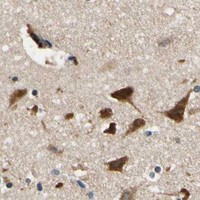
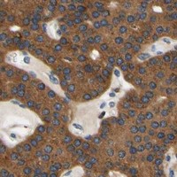
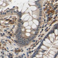
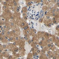

Comprehensive Human Protein Atlas IHC scoring per tissue (reliability: Approved — Low consistency between antibody staining and RNA expression data.). Rows are taken directly from the HPA tissue chart — click any row's HPA link to view the source.
| Tissue | Cell type | Level | Evidence | Source |
|---|---|---|---|---|
| Parathyroid gland | Glandular cells | High | Protein (IHC) | HPA → |
| Seminal vesicle | Glandular cells | High | Protein (IHC) | HPA → |
| Adipose tissue | Adipocytes | Medium | Protein (IHC) | HPA → |
| Adrenal gland | Glandular cells | Medium | Protein (IHC) | HPA → |
| Breast | Glandular cells | Medium | Protein (IHC) | HPA → |
Troubleshoot PLA2G4A staining in paraffin sections by checking retrieval, cell type, and compartment before comparing chromogenic IHC results (UniProt P47712; HPA tissue IHC).
Human-reactive A00854-1 has IHC data from formalin-fixed, paraffin-embedded human lung carcinoma and IF data from NCI-H460 cells (catalog: reactivity; IHC and IF image captions).
A00854-1 is listed for human IHC-P, with a DAB-stained image of formalin-fixed, paraffin-embedded human lung carcinoma (catalog: applications, reactivity, IHC image caption). The same SKU is listed for IF and has a confocal IF image of NCI-H460 cells (catalog: applications, IF image caption).
Which to pick: Choose A00854-1 for human tissue IHC-P: its own image documents formalin-fixed, paraffin-embedded lung carcinoma, and its listed IHC-P dilution is 1:10–1:50 (catalog: IHC image caption, reactivity, IHC-P dilution). For IF/ICC planning, A00854-1 is a rabbit polyclonal antibody with listed IF use and an NCI-H460 IF image; ICC is not separately listed (catalog: host, dilution_raw, applications, IF image caption). No cross-species option is supported because A00854-1 lists human reactivity only (catalog: reactivity).