This website uses cookies to ensure you get the best experience on our website.


- Table of Contents
Plan PLD1 IHC on paraffin sections using the observed cytoplasmic and membranous tissue pattern (HPA tissue IHC). Compare cell types within the section and interpret intensity with the reported variability in mind (HPA tissue IHC).

Expected localisation, validated protocols, controls and antibodies — the at-a-glance facts below, then the full design guide.
| Expected localisation | Cytoplasmic and membranous tissue staining (HPA tissue IHC) | |
| Staining pattern | Variable cytoplasmic and membranous staining in most tissues (HPA tissue IHC) | |
| Antigen retrieval | Citrate pH 6.0 HIER, 95–98 °C, 20 min (rule: cytoplasmic / membrane antigen) | |
| Positive control | Adrenal gland+4 more · see all | |
| Negative control | Caudate+3 more · see all |
| Fixation | Keep formalin fixation consistent across sections (standard IHC practice; not target-specific) | |
| Caveat | Staining and RNA expression show medium consistency (HPA tissue IHC) | |
| Regulation | Staining varies by tissue and cell type (HPA tissue IHC) | |
| Isoform / epitope | 4 isoforms; check epitope coverage across variants (UniProt) |
The catalog antibody’s IHC-P protocol is paired with published PLD1 staining methods for tissue sections (PMC3614861) and breast carcinoma tissue microarrays (PMC2360386).
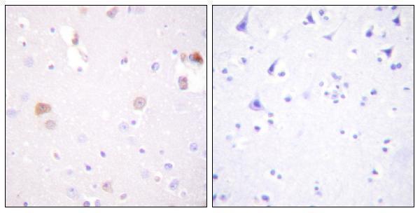
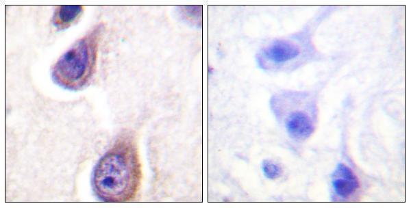
| Sample | Paraffin-embedded human brain tissue; fixative not specified (datasheet A02031) |
| Fixation | Image fixative and duration unreported (datasheet A02031); verify before use. |
| Sectioning | 4–5 µm sections on charged slides (standard) |
| Deparaffinisation | Xylene, graded ethanol series to water (standard) |
| Antigen retrieval | Heat-induced epitope retrieval in citrate buffer, pH 6.0, 20 min at 95–98 °C (standard rule: cytoplasmic / membrane antigen) |
| Peroxidase block | 3% H2O2, 10 min, room temperature (standard) |
| Blocking | 10% normal serum of the secondary host, 30 min, room temperature (standard) |
| Primary antibody | Rabbit anti-PLD1, 1:100 - 1:300 (datasheet A02031) |
| Primary incubation | Overnight at 4 °C (standard) |
| Detection | HRP-polymer secondary, DAB chromogen 5–10 min (standard) |
| Counterstain | Hematoxylin, blue, dehydrate and mount (standard) |
| Expected result | PLD1-positive staining in glandular cells of adrenal gland (HPA tissue IHC: High). HPA tissue profile: Cytoplasmic and membranous expression at variable levels in most tissues. No signal in the no-primary control. |
PLD1 staining in paraffin IHC should be cytoplasmic and membranous, with variable intensity across tissues (HPA: tissue IHC). High staining is reported in several glandular populations, cholangiocytes, keratinocytes, and fallopian tube ciliated cells at their ciliary rootlets (HPA: tissue IHC). Perinuclear, ER, Golgi, and late endosome associations are consistent with UniProt localisation; PLD1 has no transmembrane segment (UniProt Q13393 topology). HPA rates tissue IHC reliability Enhanced, with external verification pending (HPA: tissue IHC).
| Cytoplasmic and membranous staining in expected cells, with stronger staining in selected epithelia (HPA: tissue IHC). | This fits the reported IHC profile. Useful positive cells include gallbladder, duodenal, rectal, and small intestinal glandular cells; liver cholangiocytes and skin keratinocytes are also High (HPA: tissue IHC). Compare cell type and compartment together: intensity alone does not establish specificity (general IHC practice). |
| Predominantly nuclear staining, without a convincing cytoplasmic or membranous component. | A nuclear dominant pattern conflicts with the reported IHC profile and UniProt localisation (HPA: tissue IHC; UniProt Q13393 localisation). Treat it as suspect, then compare the positive control and no primary control. Atypical staining alone cannot identify whether the cause is nonspecific antibody binding or detection background (general IHC practice). |
| Strong staining in an expected low or undetected cell population, such as ovarian stromal cells or parathyroid glandular cells (HPA: tissue IHC). | HPA reports those two populations as Not detected, so strong staining warrants investigation; that rating applies to the named cells, not every cell in either tissue (HPA: tissue IHC). Compare a matched no primary control and the expected positive cells. Cross-reactivity or endogenous detection activity are possible explanations, not diagnoses from appearance alone (general IHC practice). |
| Diffuse chromogen across multiple compartments, extracellular areas, or most cells, obscuring cell boundaries. | The distribution is difficult to reconcile with the variable, cell associated cytoplasmic and membranous IHC profile (HPA: tissue IHC). Check whether the no primary control shares the background and whether expected positive cells remain distinguishable. Blocking, washing, antibody concentration, and detection background are general workflow variables (general IHC practice). |
| No staining in a selected known positive population, such as gallbladder glandular cells or liver cholangiocytes (HPA: High in these cells). | A negative test section is uninterpretable until a positive control stains under the same run conditions (general IHC practice). HPA calls these populations High, but its tissue IHC assessment has medium consistency with RNA and awaits external verification (HPA: tissue IHC). Confirm cell identity and inspect the whole staining run before inferring absence of PLD1. |
| Tissue and cell selection | PLD1 IHC levels vary across most tissues; HPA reports High glandular, cholangiocyte, keratinocyte, and ciliary rootlet staining, alongside named Not detected populations (HPA: tissue IHC). Choose a positive control by its specified cell population rather than treating a whole organ as uniformly positive or negative. |
| Compartment and topology | UniProt places PLD1 in the perinuclear cytoplasm and on ER, Golgi, and late endosome membranes, while reporting no transmembrane segment (UniProt Q13393). These associations support a cytoplasmic or membranous interpretation; they do not establish a unique organelle pattern in chromogenic sections (HPA: tissue IHC). |
| Antibody validation | HPA lists HPA042396 as IHC Enhanced and CAB004527 as IHC Approved (HPA: antibodies). The tissue profile is rated Enhanced, yet its antibody staining and RNA show medium consistency and external verification is pending (HPA: tissue IHC). Use validation status as supporting evidence, not proof that every observed signal is specific. |
| IF/ICC question: should its compartment pattern match paraffin IHC? | IF/ICC images mainly place PLD1 at the plasma membrane, with additional Golgi and vesicle localisation (HPA: subcellular ICC-IF). Paraffin tissue IHC is described more broadly as cytoplasmic and membranous (HPA: tissue IHC). Assess each preparation against its own reported pattern; this IF observation supplies no IHC protocol condition. |
| Situation | Likely cause | Next action |
|---|---|---|
| The expected positive cell population is blank. | The run may have failed, or the intended positive cells may be absent from the examined area (general IHC practice). | Check cell identity on the counterstained section, then inspect a reported High population stained in the same run, such as gallbladder glandular cells (HPA: tissue IHC). Review the run's retrieval, primary antibody, detection, and counterstain records as general workflow checks (general IHC practice). |
| The no primary control develops the same chromogen as the test section. | Antibody independent detection background, including endogenous enzyme activity in enzyme based detection, is possible (general IHC practice). | Address the detection system's endogenous activity and blocking controls, then repeat with the no primary control beside the test section (general IHC practice). Do not score shared control signal as PLD1 even if it overlaps a reported positive tissue (HPA: tissue IHC). |
| Staining is widespread and masks the reported cell pattern. | Nonspecific signal or excessive detection background may overwhelm genuine cell associated staining (general IHC practice). | Compare the no primary control, adjust general blocking and washing steps, and reassess the antibody working concentration using its documented instructions (general IHC practice). Retain a reported High cell population as the reference for interpretable cytoplasmic and membranous staining (HPA: tissue IHC). |
| Only nuclei stain strongly. | This conflicts with the reported cytoplasmic and membranous IHC pattern and listed cytoplasmic and membrane localisations (HPA: tissue IHC; UniProt Q13393). | Recheck the no primary control and a reported positive cell population in the same run. Treat nuclear dominant signal as unresolved until controls support specificity; slide appearance alone cannot assign a mechanism (general IHC practice). |
| Cells reported Not detected show strong signal. | The chosen population may be misidentified, or its signal may reflect nonspecific binding or detection background (general IHC practice). | Confirm the cell population, compare adjacent reported positive cells and the no primary control, and report the discrepancy by cell type. HPA lists ovarian stromal and parathyroid glandular cells as Not detected; that does not make their entire organs universal negative controls (HPA: tissue IHC). |
| An IF/ICC image appears more sharply membranous than the paraffin IHC section. | The reported readouts differ: ICC-IF is mainly plasma membrane with Golgi and vesicles, while tissue IHC is cytoplasmic and membranous (HPA: subcellular ICC-IF; HPA: tissue IHC). | Interpret the chromogenic section against tissue IHC cell and compartment evidence. Use the ICC-IF localisation as context, without requiring visible organelle resolution or importing an IF/ICC protocol into paraffin IHC (general IHC practice). |
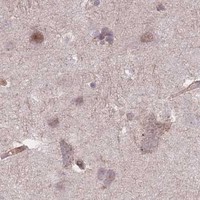


Comprehensive Human Protein Atlas IHC scoring per tissue (reliability: Enhanced — Medium consistency between antibody staining and RNA expression data. Pending external verification.). Rows are taken directly from the HPA tissue chart — click any row's HPA link to view the source.
| Tissue | Cell type | Level | Evidence | Source |
|---|---|---|---|---|
| Adrenal gland | Glandular cells | High | Protein (IHC) | HPA → |
| Duodenum | Glandular cells | High | Protein (IHC) | HPA → |
| Fallopian tube | Ciliated cells (ciliary rootlets) | High | Protein (IHC) | HPA → |
| Gallbladder | Glandular cells | High | Protein (IHC) | HPA → |
| Liver | Cholangiocytes | High | Protein (IHC) | HPA → |
| Tissue | Cell type | Level | Evidence | Source |
|---|---|---|---|---|
| Caudate | Glial cells | Not detected | Protein (IHC) | HPA → |
| Hippocampus | Glial cells | Not detected | Protein (IHC) | HPA → |
| Ovary | Ovarian stroma cells | Not detected | Protein (IHC) | HPA → |
| Parathyroid gland | Glandular cells | Not detected | Protein (IHC) | HPA → |
Troubleshoot PLD1 staining in paraffin sections by checking retrieval, controls, cellular compartment, and scoring before interpreting chromogenic signal.
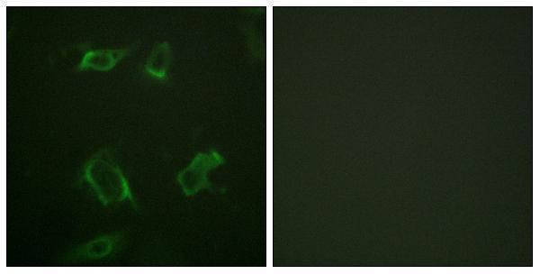
Two PLD1 antibodies have IHC images from paraffin-embedded human brain; the phospho-Ser561 antibody also has a HepG2 IF image. Both list human, mouse, and rat reactivity (catalog captions/reactivity).
A02031 lists IHC and IF/ICC applications and shows paraffin-embedded human brain IHC with peptide blocking (A02031 applications/IHC caption). P02031 lists IHC and IF/ICC applications, shows paraffin-embedded human brain IHC with phospho-peptide blocking, and has a HepG2 IF image (P02031 applications/IHC and IF captions).
Which to pick: Choose A02031 for PLD1 tissue IHC or P02031 when the target is phospho-PLD1 Ser561; each rabbit polyclonal antibody has its own paraffin-embedded human brain IHC image (catalog titles/host/dilution_raw; A02031 and P02031 IHC captions). For IF/ICC, both list those applications, while P02031 also has a HepG2 IF image (catalog applications; P02031 IF caption). Both list human, mouse, and rat reactivity, but their IHC images show human brain only; neither IHC caption reports the fixative (catalog reactivity; A02031 and P02031 IHC captions).