PLD3 / 5'-3' exonuclease PLD3 · IHC design guide

Design Immunohistochemistry for PLD3

Plan PLD3 chromogenic IHC in paraffin sections using neuronal cells or lung macrophages as positive references (HPA tissue IHC). The guide covers the catalog antibody’s 2–5 μg/ml IHC range (datasheet A01976-1), expected cytoplasmic staining (HPA tissue IHC), and controls.

Evidence assembled Oct 2026 · For research use; verify linked source records and product datasheet before use
Immunohistochemistry protocol sheet for PLD3 (IHC for PLD3): expected localisation Cytoplasmic in most tissue cell types (HPA tissue IHC), antibody A01976-1, validated IHC image, and IHC protocol steps
Printable PLD3 IHC protocol sheet — expected localisation Cytoplasmic in most tissue cell types (HPA tissue IHC), antibody A01976-1, controls and protocol steps. Open the full PLD3 IHC guide →

PLD3 Immunohistochemistry Experimental Design Guide

Expected localisation, validated protocols, controls and antibodies — the at-a-glance facts below, then the full design guide.

Must know before staining
Expected localisation Cytoplasmic in most tissue cell types (HPA tissue IHC)
Staining pattern Variable cytoplasmic staining, strongest in neurons and immune cells (HPA tissue IHC)
Antigen retrieval EDTA pH 8.0 HIER, heat-mediated (datasheet A01976-1)
Positive control ⓘ Cerebellum+4 more · see all
Negative control ⓘ Adipose tissue+1 more · see all
Important caveats
Reasons your staining may differ from the expected pattern.
Fixation Keep fixation consistent across sections. (standard IHC practice; not target-specific)
Caveat Staining–RNA consistency is medium; retesting is pending (HPA tissue IHC)
Regulation No specific expression regulator reported (UniProt)
Isoform / epitope No listed isoforms; processing releases a lumenal form, so epitope position matters (UniProt)
Section 1

Recommended PLD3 IHC & IF Protocols

The catalog antibody has a datasheet IHC-P protocol (datasheet A01976-1). Four published PLD3 IHC protocols provide tissue-specific starting points (PMC12616045; PMC4255636; PMC9729106; PMC10117289).

Recommended immunohistochemistry (IHC-P) protocol parameters
SampleParaffin-embedded human appendiceal adenocarcinoma tissue; fixative not specified (datasheet A01976-1)
FixationImage fixative and duration unreported (datasheet A01976-1); verify before use.
Sectioning4–5 µm sections on charged slides (standard)
DeparaffinisationXylene, graded ethanol series to water (standard)
Antigen retrievalHeat retrieval: EDTA pH 8.0 (datasheet A01976-1); 20 min, 95–100 °C (standard)
Peroxidase block3% H2O2, 10 min, room temperature (standard)
Blocking10% goat serum (datasheet A01976-1)
Primary antibodyRabbit anti-PLD3, 2-5 μg/ml (datasheet A01976-1)
Primary incubationOvernight at 4 °C (datasheet A01976-1)
DetectionStreptavidin-biotin complex (SABC), DAB chromogen (datasheet A01976-1)
CounterstainHematoxylin, blue, dehydrate and mount (standard)
Expected resultPLD3-positive staining in gLUC cells - cytoplasm/membrane of cerebellum (HPA tissue IHC: High). HPA tissue profile: Cytoplasmic expression at variable levels in most cell types, most abundant in neuronal cells and in immune cells. No signal in the no-primary control.
💡Decision noteStart with heat-mediated EDTA, pH 8.0, for the catalog antibody (datasheet A01976-1). Use a paper’s reported retrieval when reproducing that paper’s protocol (PMC4255636; PMC9729106).
Section 2

What Is the Expected PLD3 Staining Pattern?

PLD3 should stain mainly in the cytoplasm of neuronal and immune cells, with variable staining in many other cell types (HPA tissue IHC: Approved; medium RNA–staining consistency, pending retesting). PLD3 has a membrane-spanning segment at residues 39–59 and a lumenal region at 60–490; UniProt places it in lysosomes and endosomal, ER and Golgi compartments (UniProt Q8IV08 topology and subcellular location).

What am I looking at on my slide?
Clear cytoplasmic stain in cerebral cortex neurons or lung macrophages, with little stain outside the expected cells.This fits the reported high staining in those cells (HPA tissue IHC: High in cerebral cortex neuronal cells and lung macrophages). A granular or perinuclear appearance can be compatible with intracellular organelles, but chromogenic IHC alone cannot assign individual granules to lysosomes or endosomes (UniProt Q8IV08 subcellular location).
Predominantly nuclear, surface-only or extracellular stain replaces the expected cytoplasmic pattern.Treat this as an unsupported IHC compartment pattern and check staining controls and antibody specificity (HPA tissue IHC: cytoplasmic profile; UniProt Q8IV08 topology). The uncertain nucleoplasmic signal reported by HPA ICC-IF does not establish a nuclear IHC pattern (HPA subcellular ICC-IF: nucleoplasm uncertain).
Strong stain appears in adipocytes or chondrocytes while expected positive cells stain weakly.Investigate nonspecific antibody binding or endogenous chromogenic detection activity before calling these cells PLD3 positive (HPA tissue IHC: adipocytes and chondrocytes not detected). A negative HPA observation is a comparison point, not proof that every specimen or assay must be negative.
Color covers much of the section without clear cell boundaries or a cytoplasmic distribution.Diffuse background cannot establish PLD3 localization (HPA tissue IHC: variable cytoplasmic expression). In a chromogenic IHC workflow, incomplete blocking, excessive antibody concentration or inadequate washing can cause broad staining (general IHC practice); compare a matched negative detection control.
No detectable stain appears in cerebral cortex neurons or lung macrophages.The result conflicts with reported high staining in those cells (HPA tissue IHC: cerebral cortex neuronal cells and lung macrophages High). Check tissue preservation, retrieval and detection controls as general IHC practice; neither HPA nor UniProt supplies a PLD3-specific fixation sensitivity or retrieval condition.
💡Expected PLD3 appearanceCall a section positive when neuronal or immune cells show convincing cytoplasmic staining, potentially strong in cerebral cortex neurons or lung macrophages; isolated dominant nuclear stain or uniform section-wide color is suspect (HPA tissue IHC: cell levels and cytoplasmic profile; UniProt Q8IV08 subcellular location).
How each factor affects the staining
Compartment and topologyThe short cytoplasmic N-terminus and lumenal residues 60–490 make antibody epitope location relevant to interpretation (UniProt Q8IV08 topology). The supplied record does not identify the catalog antibody epitope, so it cannot justify a particular retrieval or permeabilisation setting.
Proteolytic processingUniProt reports that a soluble lysosomal form arises from the membrane-bound form and that PLD3 sorts into lysosomal intralumenal vesicles (UniProt Q8IV08 subcellular location). These facts support intracellular staining; they do not establish that either form yields a distinct chromogenic pattern.
Tissue and cell contextHigh staining is reported in cerebral cortex neurons, lung macrophages, epididymal glandular cells and spleen red-pulp cells; adipocytes and chondrocytes were not detected (HPA tissue IHC). Score the named cell population within each tissue, since whole-section intensity can obscure that distinction.
Evidence strengthHPA rates tissue IHC Approved but reports medium consistency with RNA expression and pending retesting (HPA tissue IHC reliability). HPA lists two antibodies with Approved IHC status; this supports comparison with its observed pattern without making every unexpected stain a confirmed PLD3 finding (HPA antibodies).
IF/ICC question: should nuclear signal be expected?HPA supports ER localization in ICC-IF, while cytosol and nucleoplasm are uncertain (HPA subcellular ICC-IF). UniProt also lists lysosomal and endosomal locations (UniProt Q8IV08 subcellular location). Assess IF localization in its separate guide; these observations provide no IF protocol option here.
Why is my staining missing, weak or wrong?
SituationLikely causeNext action
Positive-reference tissue is blank.A technical failure is possible when cells reported as High show no stain (HPA tissue IHC: cerebral cortex neurons or lung macrophages High).Confirm the intended cells are present, then check the staining run, retrieval and detection controls (general IHC practice). No PLD3-specific retrieval condition is supplied.
Stain is mainly nuclear.Nuclear-dominant IHC disagrees with the HPA cytoplasmic tissue profile; HPA calls nucleoplasmic ICC-IF localization uncertain (HPA tissue IHC; HPA subcellular ICC-IF).Recheck controls and score cytoplasmic staining separately (general IHC practice). Do not use the uncertain ICC-IF signal to validate nuclear IHC.
Adipocytes or chondrocytes stain strongly.These cell types were not detected in HPA tissue IHC, so cross-reactivity or chromogenic detection background should be considered (HPA tissue IHC).Compare a negative detection control and the expected positive cells on the same run (general IHC practice) before assigning PLD3 positivity.
Diffuse color prevents cell-level scoring.Excess reagent concentration, incomplete blocking or inadequate washing can produce background in chromogenic IHC (general IHC practice).Review dilution, blocking, washes and negative controls (general IHC practice); retain a call only where the cytoplasmic cell pattern is interpretable (HPA tissue IHC).
A tissue scores positive, but the expected cell population does not.PLD3 staining varies across cell types, and HPA reports levels for named populations rather than uniform tissue staining (HPA tissue IHC).Identify and score the reported cells, such as lung macrophages or cerebral cortex neurons, separately from surrounding cells (HPA tissue IHC).
Granules are described as definitively lysosomal.Intracellular staining is compatible with UniProt localization, but a chromogenic PLD3 pattern alone does not identify an organelle (UniProt Q8IV08 subcellular location).Report the visible cytoplasmic distribution in IHC; reserve an organelle assignment for appropriate localization evidence (general microscopy practice).

Sample controls for PLD3 IHC & IF

🧪Run lung first: macrophages should stain strongly (HPA: High in lung macrophages). Use adipose tissue adipocytes as the negative tissue (HPA: Not detected in adipocytes); on the lung slide, expect neighboring cells without identified macrophage staining to remain near background, but do not treat them as validated PLD3-negative cells (HPA: High assignment is specific to macrophages).
Positive control tissue: Cerebellum (GLUC cells - cytoplasm/membrane, HPA High)
Negative control tissue: Adipose tissue (HPA Not detected)
ICC-IF cell lines (HPA subcellular resource): HPA ICC-IF images show PLD3 in A-431, U-251MG, U2OS, with annotated localisation: Endoplasmic reticulum (supported), Cytosol (uncertain) (HPA subcellular).
Technical controls: Include a no-primary, secondary-only control and a concentration-matched rabbit IgG isotype control appropriate to the antibody’s clonality (caption: rabbit anti-PLD3 antibody). Use PLD3-knockout material as a biological negative, or immunizing-peptide competition if the peptide is available (standard IHC specificity controls). For lung IHC, block endogenous peroxidase and check endogenous biotin background before interpreting the biotin-based DAB signal (caption: biotinylated secondary, streptavidin-biotin complex and DAB; standard IHC practice).
⚠️Feasibility: A PLD3-specific fixation window or fixation effect is unreported, and the selected A01976-1 tissue-IHC caption does not state the fixative (caption: paraffin section; fixative unreported). The demonstrated paraffin-section workflow used heat retrieval in EDTA at pH 8.0, followed by 2 μg/ml antibody overnight at 4°C; the evidence does not establish that other retrieval conditions fail (caption: retrieval and incubation). No matched comparison establishes whether frozen sections or IF are easier; lung macrophage staining needs careful interpretation because endogenous peroxidase and the biotin-based detection system can produce background (HPA: High in lung macrophages; caption: biotin-based DAB detection; standard IHC practice).

HPA tissue IHC evidence for PLD3

Comprehensive Human Protein Atlas IHC scoring per tissue (reliability: Approved — Medium consistency between antibody staining and RNA expression data. Pending retesting.). Rows are taken directly from the HPA tissue chart — click any row's HPA link to view the source.

Positive expression · recommended positive controls

TissueCell typeLevelEvidenceSource
Cerebellum GLUC cells - cytoplasm/membrane High Protein (IHC) HPA →
Cerebral cortex Neuronal cells High Protein (IHC) HPA →
Epididymis Glandular cells High Protein (IHC) HPA →
Lung Macrophages High Protein (IHC) HPA →
Spleen Cells in red pulp High Protein (IHC) HPA →

Undetected expression · recommended negative controls

TissueCell typeLevelEvidenceSource
Adipose tissue Adipocytes Not detected Protein (IHC) HPA →
Soft tissue Chondrocytes Not detected Protein (IHC) HPA →
Section 3

Advanced PLD3 IHC Tips

Use compartment-aware controls and the documented paraffin-section workflow to troubleshoot PLD3 chromogenic IHC; assess IF separately.

Which retrieval conditions should I try first for weak PLD3 staining in paraffin sections?
Start with heat-mediated antigen retrieval in EDTA, pH 8.0, for paraffin sections (datasheet A01976-1). The documented PLD3 staining used that retrieval before 2 μg/ml primary antibody overnight at 4°C, so match those conditions when comparing results (datasheet A01976-1). If staining remains weak, vary heating time on matched sections and inspect morphology alongside signal (standard IHC practice). PLD3 has a membrane anchor at residues 39–59 and a luminal region at 60–490; epitope accessibility may therefore depend on the antibody’s binding site (UniProt Q8IV08 topology). Keep retrieval conditions identical across specimens used for scoring (standard IHC practice).
How should I troubleshoot PLD3 staining when fixation history is uncertain?
The selected paraffin-section caption does not report its fixative, so PLD3-specific sensitivity to fixation is unknown for this antibody (datasheet A01976-1). Record the fixative, fixation duration and processing history for each specimen, and compare sections processed together when possible (standard IHC practice). Apply the documented EDTA, pH 8.0 heat retrieval consistently before attributing weak staining to fixation (datasheet A01976-1; standard IHC practice). Include a known staining control on the same run and check tissue preservation before changing antibody conditions (standard IHC practice). Differences between specimens with unknown fixation histories should be interpreted cautiously, particularly when estimating staining intensity (standard IHC practice).
Where should convincing PLD3 chromogenic staining appear within cells?
Evaluate PLD3 chiefly as intracellular cytoplasmic staining in tissue sections, allowing for a granular pattern consistent with its endolysosomal distribution (HPA tissue IHC profile; UniProt Q8IV08 subcellular). UniProt places PLD3 in lysosomes and on endosomal, endoplasmic reticulum and Golgi membranes (UniProt Q8IV08 subcellular). HPA reports cytoplasmic expression in most cell types, with abundant staining in neuronal and immune cells (HPA tissue IHC profile). Do not score isolated nuclear DAB deposits as expected localisation: HPA lists nucleoplasmic localisation only as uncertain in its subcellular assessment (HPA subcellular). Compare compartment patterns against adjacent tissue and a negative control before assigning weak diffuse staining to PLD3 (standard IHC practice).
How could PLD3 processing or epitope position alter its IHC pattern?
PLD3 has one annotated chain and no listed isoforms, so divergent staining should not automatically be assigned to splice variants (UniProt Q8IV08 processing; UniProt Q8IV08 isoforms). Its 39–59 transmembrane segment separates a short cytoplasmic N terminus from the 60–490 luminal region (UniProt Q8IV08 topology). Proteolytic processing produces a soluble lysosomal form from membrane-bound PLD3, which could affect staining according to epitope location (UniProt Q8IV08 subcellular). Five glycosylation sites occur in the luminal region; document the antibody’s mapped epitope before testing whether processing or glycosylation explains a discrepancy (UniProt Q8IV08 glycosylation; standard IHC practice). Compare matched sections and controls rather than inferring an epitope from appearance alone (standard IHC practice).
How should I adapt PLD3 localisation checks to a separate multiplex IF experiment?
For IF, pair PLD3 with a marker of the expected cell population, such as CD68 when assessing macrophages; lung macrophages show high PLD3 tissue staining (HPA tissue IHC; standard IF practice). Choose a far-red PLD3 fluorophore and inspect unstained tissue in every channel to distinguish signal from autofluorescence (standard IF practice). PLD3’s N-terminal 1–38 segment is cytoplasmic, whereas residues 60–490 are luminal, so establish epitope position before optimizing detergent permeabilisation for intracellular access (UniProt Q8IV08 topology; standard IF practice). HPA supports endoplasmic reticulum localisation but rates its cytosolic assignment uncertain; use compartment controls when interpreting overlap (HPA subcellular). Keep IF controls and exposure settings consistent across compared specimens (standard IF practice).
What should I change when PLD3 DAB staining looks diffuse or nonspecific?
First compare the test section with a no-primary control and inspect whether brown signal follows tissue edges, necrotic areas or endogenous pigment (standard IHC practice). For the documented method, start from 10% goat-serum blocking, 2 μg/ml primary antibody overnight at 4°C, and a biotinylated secondary for 30 minutes at 37°C (datasheet A01976-1). Check peroxidase blocking and reagent-only controls when interpreting DAB deposits; these are general chromogenic IHC safeguards (standard IHC practice). If background persists, adjust wash stringency and antibody exposure on matched sections while monitoring specific intracellular staining (standard IHC practice; UniProt Q8IV08 subcellular). Document each change before comparing staining intensity between samples (standard IHC practice).
How should I quantify PLD3 staining across heterogeneous tissue sections? ⚠ ANSWER MARKED FOR VERIFICATION
Define the cell population and compartment before scoring because PLD3 tissue staining varies by cell type and is reported as cytoplasmic (HPA tissue IHC profile). For a defined population, report the percentage of positive cells and an H-score combining intensity categories with their percentages (standard IHC practice). When counting stained cells in an area, report density per mm² and normalise to the viable tissue area or relevant cell count (standard IHC practice). Exclude necrosis, folds and section edges using the same rules for every specimen (standard IHC practice). Keep retrieval, DAB development and image thresholds consistent, and report results separately for distinct cell populations (standard IHC practice).
How can I distinguish genuine PLD3 positivity from a staining artefact?
A credible PLD3 pattern is intracellular and compatible with cytoplasmic, lysosomal or endomembrane distribution (HPA tissue IHC profile; UniProt Q8IV08 subcellular). High staining in lung macrophages or cerebral-cortex neuronal cells fits the reported tissue pattern, while unexpected cell populations require closer control review (HPA tissue IHC). Question deposits confined to section edges or necrosis, as well as staining reproduced in a no-primary control (standard IHC practice). Check peroxidase controls before treating diffuse DAB precipitate as target signal (standard IHC practice). HPA rates the tissue IHC profile Approved with medium agreement to RNA data and pending retesting, so corroborate consequential assignments independently (HPA tissue IHC reliability).
Boster reagents

Best PLD3 / 5'-3' exonuclease PLD3 IHC Antibodies

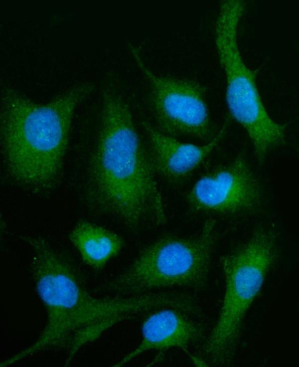
A01976-1 has IHC images from paraffin sections of human appendiceal adenocarcinoma, liver cancer and spleen, plus IF data from A549 cells (catalog image captions). Listed reactivity covers human, mouse and rat (catalog).

Real IHC data IHC analysis of PLD3 using anti-PLD3 antibody (A01976-1). PLD3 was detected in a paraffin-embedded section of human appendiceal adenocarcinoma tissue. Heat mediated antigen retrieval was performed in EDTA buffer (pH 8.0, epitope retrieval solution). The tissue section was blocked with 10% goat serum. The tissue section was then incubated with 2 μg/ml rabbit anti-PLD3 Antibody (A01976-1) overnight at 4°C. Biotinylated goat anti-rabbit IgG was used as secondary antibody and incubated for 30 minutes at 37°C. The tissue section was developed using Strepavidin-Biotin-Complex (SABC) (Catalog # SA1022) with DAB as the chromogen.
Anti-PLD3 Antibody ®
Cat # A01976-1

A01976-1 is listed for IHC, with images from paraffin sections of human appendiceal adenocarcinoma, liver cancer and spleen (catalog applications; catalog IHC captions). A01976-1 is also listed for IF/ICC, with an IF image from A549 cells (catalog applications; catalog IF caption).

Which to pick: For tissue IHC, choose A01976-1: its IHC captions document paraffin sections (catalog IHC captions). For IF/ICC, A01976-1 lists both applications and shows IF in A549 cells (catalog applications; catalog IF caption). A01976-1 lists human, mouse and rat reactivity; clone status and the fixative used for its paraffin sections are unreported (catalog reactivity; catalog clone field; catalog IHC captions).

Each figure is that product's own IHC / IF validation image from its datasheet.

References

  1. UniProt Consortium. UniProt entry Q8IV08 (PLD3_HUMAN, 5'-3' exonuclease PLD3).
  2. Human Protein Atlas. PLD3 tissue IHC expression (reliability: Approved).
  3. Human Protein Atlas. PLD3 subcellular location (ICC-IF): Mainly localized to the endoplasmic reticulum and cytosol. In addition localized to the nucleoplasm..
  4. Human Protein Atlas. PLD3 antibody validation summary (2 antibodies).
  5. Macrophage Phospholipase D3 promotes atherosclerosis via exacerbating foam cell formation and inducing inflammatory responses. International journal of cardiology. Heart & vasculature 2025 — PMC12616045.
  6. PLD3 is accumulated on neuritic plaques in Alzheimer's disease brains. Alzheimer's research & therapy 2014 — PMC4255636.
  7. PLD3 affects axonal spheroids and network defects in Alzheimer's disease. Nature 2022 — PMC9729106.
  8. Posttranslational modifications induce autoantibodies with risk prediction capability in patients with small cell lung cancer. Science translational medicine 2023 — PMC10117289.
  9. PubMed PMID:9140189 — UniProt-cited evidence.
  10. PubMed PMID:15489334 — UniProt-cited evidence.
  11. PubMed PMID:15794758 — UniProt-cited evidence.