This website uses cookies to ensure you get the best experience on our website.


- Table of Contents
Plan chromogenic PLEK IHC in paraffin sections with the IHC-validated antibody (datasheet A06656-3). This guide highlights marrow and lymph node staining and helps select tissue controls and score cell populations (HPA tissue IHC).

Expected localisation, validated protocols, controls and antibodies — the at-a-glance facts below, then the full design guide.
| Expected localisation | Cytoplasmic and extracellular in marrow and lymph node (HPA tissue IHC) | |
| Staining pattern | Marrow hematopoietic and nodal germinal center cells; cytoplasmic and extracellular staining (HPA tissue IHC) | |
| Antigen retrieval | EDTA pH 8.0 HIER, heat-mediated (datasheet A06656-3) | |
| Positive control | Lymph node+4 more · see all | |
| Negative control | Adipose tissue+4 more · see all |
| Fixation | Keep fixation consistent across sections (standard IHC practice; not target-specific) | |
| Caveat | Myeloid peroxidase may raise marrow DAB background (standard IHC practice) | |
| Regulation | Expression regulation not annotated (UniProt) | |
| Isoform / epitope |
The catalog antibody’s IHC-P protocol (datasheet: A06656-3) is followed by published PLEK IHC protocols for placenta, carotid plaque, and gingival tissue (PMC11979283; PMC10929796; PMC8787058).
| Sample | Paraffin-embedded human tonsil tissue; fixative not specified (datasheet A06656-3) |
| Fixation | Image fixative and duration unreported (datasheet A06656-3); verify before use. |
| Sectioning | 4–5 µm sections on charged slides (standard) |
| Deparaffinisation | Xylene, graded ethanol series to water (standard) |
| Antigen retrieval | Heat retrieval: EDTA pH 8.0 (datasheet A06656-3); 20 min, 95–100 °C (standard) |
| Peroxidase block | 3% H2O2, 10 min, room temperature (standard) |
| Blocking | 10% goat serum (datasheet A06656-3) |
| Primary antibody | Rabbit anti-PLEK, 2-5 μg/ml (datasheet A06656-3) |
| Primary incubation | Overnight at 4 °C (datasheet A06656-3) |
| Detection | HRP-conjugated secondary, DAB chromogen (datasheet A06656-3) |
| Counterstain | Hematoxylin, blue, dehydrate and mount (standard) |
| Expected result | PLEK-positive staining in germinal center cells of lymph node (HPA tissue IHC: High). HPA tissue profile: Cytoplasmic and extracellular expression in bone marrow and lymph node. No signal in the no-primary control. |
In paraffin-section IHC, expect PLEK in the cytoplasm of selected hematopoietic cells, with extracellular staining also reported in bone marrow and lymph node (HPA tissue IHC). Germinal center cells in lymph node and red-pulp cells in spleen show high staining; bone-marrow hematopoietic cells show medium staining (HPA tissue IHC). HPA rates tissue-IHC reliability Enhanced, with medium RNA–staining consistency (HPA tissue IHC). PLEK has no transmembrane segment (UniProt P08567 topology).
| Strong cytoplasmic signal in lymph-node germinal center cells or splenic red-pulp cells. | This fits the high-staining cell populations reported for PLEK (HPA tissue IHC). Judge the labeled cells against adjacent tissue architecture: a positive field need not imply that every cell in the section expresses PLEK (HPA tissue IHC; standard IHC interpretation). |
| Extracellular staining in bone marrow or lymph node, alongside a plausible cellular pattern. | HPA includes extracellular expression in its tissue-IHC summary, so extracellular signal alone is not grounds for rejection (HPA tissue IHC). Assess its distribution and the cellular signal together; the summary does not identify an extracellular source or mechanism (HPA tissue IHC). |
| Predominantly nuclear or sharply membranous chromogen in the expected positive cells. | That differs from HPA's cytoplasmic and extracellular tissue-IHC summary and warrants a specificity check (HPA tissue IHC). Do not equate an IHC nuclear pattern with HPA's approved nucleolar ICC-IF result: the assays report different observations (HPA subcellular ICC-IF). |
| Broad staining of cell types listed as undetected, such as adipocytes or respiratory epithelial cells. | HPA reports no detection in adipocytes of adipose tissue or respiratory epithelial cells of bronchus (HPA tissue IHC). Review morphology and controls for cross-reactivity or endogenous detection activity before interpreting widespread staining as PLEK (standard IHC practice). |
| Little or no signal in a lymph-node germinal center or splenic red-pulp control. | These are reported high-staining populations, so an absent result weakens confidence in a negative test section (HPA tissue IHC). Check section quality and the IHC detection workflow before concluding that the tested tissue lacks PLEK (standard IHC practice). |
| Tissue and cell population | HPA reports high staining in lymph-node germinal center and splenic red-pulp cells, medium staining in bone-marrow hematopoietic cells, and no detection in adipose-tissue adipocytes (HPA tissue IHC). Choose controls by cell population, not tissue name alone (standard IHC practice). |
| Evidence strength | HPA rates both HPA031838 and HPA057341 Enhanced for IHC, while describing medium consistency between antibody staining and RNA expression (HPA antibodies; HPA tissue IHC). Treat the tissue pattern as a reference that still requires appropriate slide controls (standard IHC practice). |
| Topology and processing | UniProt lists one 1–350 pleckstrin chain, no signal peptide or propeptide, and no transmembrane segment (UniProt P08567). These features do not establish the source of HPA's extracellular IHC signal or a paraffin-section retrieval condition (UniProt P08567; HPA tissue IHC). |
| IF/ICC Q: Is nucleolar signal expected? | A: HPA approves nucleolar ICC-IF localization and lists HEL, NB4 and U2OS image sets; HPA057341 is ICC Approved (HPA subcellular ICC-IF; HPA antibodies). That finding does not replace the cytoplasmic and extracellular tissue-IHC reference pattern (HPA tissue IHC). |
| Situation | Likely cause | Next action |
|---|---|---|
| No chromogen in the expected positive-control cells. | An IHC workflow failure is possible when a population reported as high staining is blank (HPA tissue IHC; standard IHC practice). | Confirm that the control contains recognizable germinal center or red-pulp cells, then check deparaffinization, the antibody's IHC-P retrieval instructions, detection reagents and counterstain (standard IHC practice). |
| Diffuse chromogen obscures cell boundaries. | Excess background can arise from inadequate blocking, excessive primary antibody or incomplete washing (standard IHC practice). HPA's extracellular summary alone cannot identify the cause (HPA tissue IHC). | Review a no-primary control, blocking and wash steps; adjust the primary-antibody condition within the validated IHC workflow, then reassess the cellular pattern (standard IHC practice). |
| Chromogen appears in many HPA-undetected cell types. | Cross-reactivity or endogenous detection activity may explain a pattern unlike HPA's listed negatives (HPA tissue IHC; standard IHC practice). | Compare the suspect section with a no-primary control and an appropriate positive tissue; check endogenous-enzyme blocking for the chosen chromogenic detection system (standard IHC practice). |
| Signal looks exclusively nuclear in a paraffin section. | Nuclear-only tissue IHC does not match HPA's cytoplasmic and extracellular summary, although HPA separately approves nucleolar ICC-IF localization (HPA tissue IHC; HPA subcellular ICC-IF). | Check nuclear morphology, counterstain and no-primary control; compare the same cell population with an appropriate positive tissue before assigning the signal to PLEK (standard IHC practice). |
| Only extracellular material stains in bone marrow or lymph node. | HPA reports extracellular expression but gives no mechanism or criterion that makes extracellular-only staining sufficient for a positive call (HPA tissue IHC). | Examine the reported hematopoietic or germinal center cell populations and a no-primary control; describe extracellular-only signal separately from cellular positivity (HPA tissue IHC; standard IHC interpretation). |
| A weak test section is difficult to score. | PLEK staining varies by cell population: HPA reports high, medium, low and undetected examples, and notes medium RNA–staining consistency (HPA tissue IHC). | Score the relevant cell type against a concurrently stained high-positive control and record intensity and distribution; avoid calling the whole tissue negative from a sparse field (HPA tissue IHC; standard IHC practice). |


Comprehensive Human Protein Atlas IHC scoring per tissue (reliability: Enhanced — Medium consistency between antibody staining and RNA expression data.). Rows are taken directly from the HPA tissue chart — click any row's HPA link to view the source.
| Tissue | Cell type | Level | Evidence | Source |
|---|---|---|---|---|
| Lymph node | Germinal center cells | High | Protein (IHC) | HPA → |
| Spleen | Cells in red pulp | High | Protein (IHC) | HPA → |
| Bone marrow | Hematopoietic cells | Medium | Protein (IHC) | HPA → |
| Duodenum | Paneth cells | Medium | Protein (IHC) | HPA → |
| Kidney | Cells in glomeruli | Medium | Protein (IHC) | HPA → |
| Tissue | Cell type | Level | Evidence | Source |
|---|---|---|---|---|
| Adipose tissue | Adipocytes | Not detected | Protein (IHC) | HPA → |
| Adrenal gland | Glandular cells | Not detected | Protein (IHC) | HPA → |
| Breast | Adipocytes | Not detected | Protein (IHC) | HPA → |
| Bronchus | Respiratory epithelial cells | Not detected | Protein (IHC) | HPA → |
| Caudate | Glial cells | Not detected | Protein (IHC) | HPA → |
Troubleshoot PLEK staining in paraffin sections by checking retrieval, cell identity, compartment, controls, and scoring before interpreting signal.
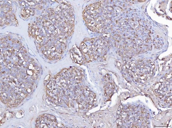
A06656-3 has real chromogenic IHC data from a paraffin-embedded human tonsil section (A06656-3 IHC image caption); human is its only listed species (catalog reactivity).
A06656-3 was demonstrated on a paraffin-embedded human tonsil section using EDTA retrieval at pH 8.0 and 2 μg/ml primary antibody (A06656-3 IHC image caption). IHC is listed as an application, while IF/ICC is not, and no IF image is supplied (catalog applications; catalog IF image alts).
Which to pick: Choose A06656-3 for human paraffin-section IHC; the catalog lists 2–5 μg/ml, and its own tonsil image used 2 μg/ml (catalog IHC dilution; A06656-3 IHC image caption). The caption does not report the fixative, and the catalog provides no IF/ICC validation for this SKU (A06656-3 IHC image caption; catalog applications and IF image alts). No cross-species choice is supported: A06656-3 lists human reactivity only (catalog reactivity).