This website uses cookies to ensure you get the best experience on our website.


- Table of Contents
Plan chromogenic PLS1 IHC on paraffin sections using colon glandular cells as a high-staining reference (HPA tissue IHC). This guide covers fixation, retrieval, a 2–5 μg/ml antibody range (datasheet A07675-1), and scoring of observed membranous and cytoplasmic staining (HPA tissue IHC).

Expected localisation, validated protocols, controls and antibodies — the at-a-glance facts below, then the full design guide.
| Expected localisation | Observed membranous and cytoplasmic staining, mainly in GI tract (HPA tissue IHC) | |
| Staining pattern | GI glandular cells show membranous and cytoplasmic staining (HPA tissue IHC) | |
| Antigen retrieval | EDTA pH 8.0 HIER, heat-mediated (datasheet A07675-1) | |
| Positive control | Appendix+4 more · see all | |
| Negative control | Adipose tissue+4 more · see all |
| Fixation | Keep fixation consistent across paraffin sections and controls. (selected-SKU IHC image A07675-1) | |
| Caveat | Antibody staining and RNA show medium consistency (HPA tissue IHC) | |
| Regulation | Intestine-enriched expression (HPA tissue IHC) | |
| Isoform / epitope | No isoforms; one cytoplasmic 1–629 chain, with no extracellular region (UniProt) |
The catalog antibody has an IHC-P protocol (datasheet: A07675-1); the published colorectal cancer tissue-core protocol below provides an additional example (PMC7419044).
| Sample | Paraffin-embedded human colon tissue; fixative not specified (datasheet A07675-1) |
| Fixation | Image fixative and duration unreported (datasheet A07675-1); verify before use. |
| Sectioning | 4–5 µm sections on charged slides (standard) |
| Deparaffinisation | Xylene, graded ethanol series to water (standard) |
| Antigen retrieval | Heat retrieval: EDTA pH 8.0 (datasheet A07675-1); 20 min, 95–100 °C (standard) |
| Peroxidase block | 3% H2O2, 10 min, room temperature (standard) |
| Blocking | 10% goat serum (datasheet A07675-1) |
| Primary antibody | Rabbit anti-PLS1, 2-5 μg/ml (datasheet A07675-1) |
| Primary incubation | Overnight at 4 °C (datasheet A07675-1) |
| Detection | HRP-conjugated secondary, DAB chromogen (datasheet A07675-1) |
| Counterstain | Hematoxylin, blue, dehydrate and mount (standard) |
| Expected result | PLS1-positive staining in glandular cells of appendix (HPA tissue IHC: High). HPA tissue profile: Membranous and cytoplasmic expression was mainly observed in gastrointestinal tract. No signal in the no-primary control. |
PLS1 is cytoplasmic and associates with cell projections; it has no transmembrane segment (UniProt Q14651). On paraffin sections, expect mainly membranous and cytoplasmic staining in gastrointestinal glandular cells (HPA tissue IHC). HPA rates the tissue pattern Enhanced, while noting only medium consistency between antibody staining and RNA expression (HPA tissue IHC). Interpret an apical rim as a staining pattern, without treating it as proof that PLS1 spans the membrane (UniProt Q14651 topology).
| Strong glandular-cell staining in small intestine, duodenum, colon, rectum, or appendix, with cytoplasmic and membranous emphasis. | This fits the reported gastrointestinal pattern and High glandular-cell staining in each listed tissue (HPA tissue IHC). Assess signal against adjacent unstained structures on the same slide; uniform chromogen over every structure is less convincing as a cell-specific result (general IHC practice). |
| Predominantly nuclear staining, with little cytoplasmic or membranous signal. | This conflicts with the reported tissue pattern (HPA tissue IHC) and cytoplasmic, cell-projection localization (UniProt Q14651). Review the no-primary control, counterstain, and detection background before assigning nuclear localization; a nuclear-only result is a warning sign, not evidence of a new compartment (general IHC practice). |
| Strong signal in a cell population reported as undetected, such as adipocytes in adipose tissue. | HPA reports PLS1 as Not detected in those adipocytes (HPA tissue IHC). Consider nonspecific antibody binding or endogenous detection activity; compare a no-primary control and an independently processed known-positive section before interpreting the unexpected cells (general IHC practice). |
| Diffuse chromogen across glandular cells, stroma, and empty spaces, without clear cellular boundaries. | That distribution does not resemble HPA's mainly gastrointestinal glandular-cell pattern (HPA tissue IHC). Broad background can reflect nonspecific detection, insufficient blocking, or overly concentrated antibody; check controls and optimize the general IHC workflow before scoring PLS1 (general IHC practice). |
| No convincing signal in small-intestinal or colonic glandular cells. | Both are High in HPA tissue IHC, so a blank section calls for a technical check before a biological negative call (HPA tissue IHC). Confirm tissue identity and section integrity, then review the antibody dilution, retrieval conditions, and detection controls (general IHC practice). |
| Cell and tissue distribution | HPA reports High glandular-cell staining in appendix, colon, duodenum, gallbladder, rectum, and small intestine; Sertoli cells in testis are also High, while bronchial respiratory epithelium is Medium (HPA tissue IHC). Choose controls by the stated cell type, since a whole tissue contains multiple populations (general IHC practice). |
| Compartment and topology | UniProt places PLS1 in cytoplasm and stereocilia and reports no transmembrane segment (UniProt Q14651). HPA describes mainly membranous and cytoplasmic gastrointestinal staining (HPA tissue IHC). A membrane-adjacent pattern is compatible with these reports, but chromogenic IHC alone cannot establish membrane insertion (general IHC practice). |
| Strength of the tissue evidence | The HPA tissue profile is Enhanced, and two listed antibodies have Enhanced IHC status: HPA055744 and HPA061936 (HPA tissue IHC; HPA antibodies). HPA also reports medium consistency between antibody staining and RNA expression, so an unexpected result deserves control-based review (HPA tissue IHC). |
| IF/ICC: what localization should be expected? | HPA reports mainly plasma-membrane localization in ICC-IF, but cautions that this assessment uses antibodies targeting proteins from multiple genes (HPA subcellular). Compare that observation with UniProt's cytoplasmic and stereocilium annotations (UniProt Q14651); the ICC-IF entry is a localization clue, not an IHC protocol (HPA subcellular). |
| Situation | Likely cause | Next action |
|---|---|---|
| Known-positive intestinal glandular cells are blank. | A failed staining or detection step is possible when a High HPA cell population lacks signal (HPA tissue IHC; general IHC practice). | Verify tissue identity, inspect the positive-control section, and check primary-antibody dilution, retrieval, and detection reagents against the selected IHC method (general IHC practice). No PLS1-specific retrieval condition is supplied here. |
| Only nuclei stain. | The compartment differs from HPA's membranous/cytoplasmic tissue profile and UniProt's cytoplasmic/cell-projection assignment (HPA tissue IHC; UniProt Q14651). | Compare the no-primary control and counterstain; repeat with controlled detection conditions before calling the nuclear signal PLS1 (general IHC practice). |
| Adipocytes in adipose tissue stain strongly. | HPA lists these cells as Not detected, so nonspecific binding or endogenous detection activity should be considered (HPA tissue IHC; general IHC practice). | Inspect a no-primary control and the chosen detection-system control, then compare with a glandular-cell positive control on the same run (general IHC practice; HPA tissue IHC). |
| Chromogen spreads across multiple unrelated tissue compartments. | Diffuse background obscures the mainly glandular-cell pattern reported by HPA (HPA tissue IHC); excess primary antibody or incomplete background suppression are general possibilities (general IHC practice). | Check the no-primary control, optimize blocking and primary-antibody dilution, and score only signal that resolves to identifiable cells (general IHC practice). |
| A low-signal tissue looks negative. | Cholangiocytes in liver and urothelial cells in urinary bladder are reported at Low levels, so a faint or absent result there is less decisive than in a High control (HPA tissue IHC). | Run a listed High glandular-cell control and compare the specified cell populations; avoid declaring assay failure from the low-level tissue alone (HPA tissue IHC; general IHC practice). |
| An unexpected cell pattern persists after basic controls pass. | HPA's Enhanced profile still has only medium antibody-to-RNA consistency, leaving some discordance unresolved by the tissue summary alone (HPA tissue IHC). | Document the cell type and compartment, compare the pattern with HPA's tissue map, and seek independent antibody or orthogonal support before reporting a new distribution (HPA tissue IHC; general IHC practice). |
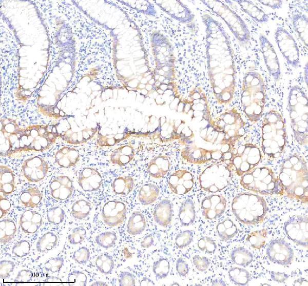
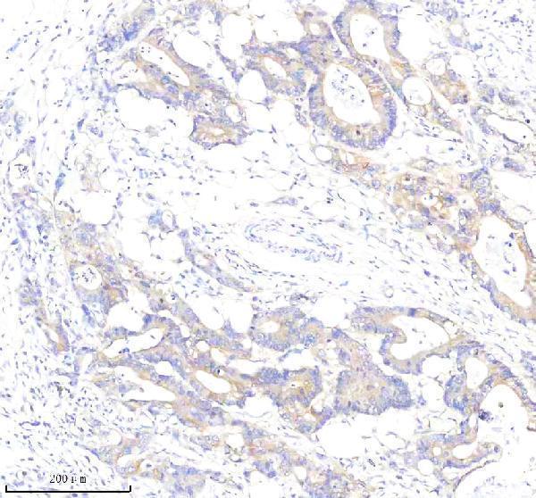
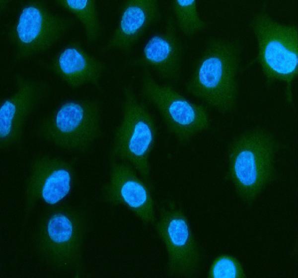


Comprehensive Human Protein Atlas IHC scoring per tissue (reliability: Enhanced — Medium consistency between antibody staining and RNA expression data.). Rows are taken directly from the HPA tissue chart — click any row's HPA link to view the source.
| Tissue | Cell type | Level | Evidence | Source |
|---|---|---|---|---|
| Appendix | Glandular cells | High | Protein (IHC) | HPA → |
| Colon | Glandular cells | High | Protein (IHC) | HPA → |
| Duodenum | Glandular cells | High | Protein (IHC) | HPA → |
| Gallbladder | Glandular cells | High | Protein (IHC) | HPA → |
| Rectum | Glandular cells | High | Protein (IHC) | HPA → |
| Tissue | Cell type | Level | Evidence | Source |
|---|---|---|---|---|
| Adipose tissue | Adipocytes | Not detected | Protein (IHC) | HPA → |
| Adrenal gland | Glandular cells | Not detected | Protein (IHC) | HPA → |
| Bone marrow | Hematopoietic cells | Not detected | Protein (IHC) | HPA → |
| Breast | Adipocytes | Not detected | Protein (IHC) | HPA → |
| Caudate | Glial cells | Not detected | Protein (IHC) | HPA → |
Troubleshoot PLS1 staining in paraffin sections by checking retrieval, cell type, compartment and controls before comparing signal intensity (UniProt Q14651; HPA tissue IHC).
A07675-1 has IHC images of paraffin-embedded human colon, colon cancer and stomach cancer, plus an IF image of A549 cells (catalog image captions).
A07675-1 is listed for IHC and IF/ICC, with images of paraffin-embedded human colon, colon cancer and stomach cancer sections (catalog applications; IHC captions). Its IF image shows A549 cells, and the catalog lists Human, Mouse and Rat reactivity (catalog IF caption; catalog reactivity).
Which to pick: For tissue IHC, choose A07675-1: its paraffin-section captions document EDTA retrieval at pH 8.0 and 2 μg/ml primary antibody; the fixative is unreported (catalog IHC captions). For IF/ICC, the same SKU is listed for both applications, with an A549 IF image using 5 μg/ml primary antibody (catalog applications; catalog IF caption). For cross-species planning, A07675-1 lists Human, Mouse and Rat reactivity, but the supplied IHC images show human tissue and the IF image shows A549 cells; clonality is unreported (catalog reactivity; catalog image captions; catalog clone field).