This website uses cookies to ensure you get the best experience on our website.


- Table of Contents
Plan PLXDC2 paraffin-section IHC around the cytoplasmic tissue pattern (HPA tissue IHC). Start the catalog antibody at 2–5 μg/mL (datasheet A08798-1), and interpret staining cautiously because its agreement with RNA expression is low (HPA tissue IHC).

Expected localisation, validated protocols, controls and antibodies — the at-a-glance facts below, then the full design guide.
| Expected localisation | Cytoplasmic staining in most tissues (HPA tissue IHC) | |
| Staining pattern | High cytoplasmic staining in colon glandular cells (HPA tissue IHC) | |
| Antigen retrieval | EDTA pH 8.0 HIER, heat-mediated (datasheet A08798-1) | |
| Positive control | Appendix+4 more · see all | |
| Negative control | Spleen |
| Fixation | Keep paraffin-section fixation consistent (standard IHC practice; not target-specific); Matched tissue-IHC evidence does not establish the fixation claim. Validate the specimen-specific method before use. (selected-SKU IHC image A08798-1) | |
| Caveat | Antibody staining has low consistency with RNA expression (HPA tissue IHC) | |
| Regulation | Stromal, but not normal colonic, endothelium (UniProt) | |
| Isoform / epitope | 3 isoforms; check extracellular vs cytoplasmic epitope (UniProt) |
Compare the catalog antibody’s IHC-P protocol (datasheet A08798-1) with the four published PLXDC2 tissue-staining protocols below (PMC12688484; PMC8194078; PMC9338914; PMC13616238).
| Sample | Paraffin-embedded human breast cancer tissue; fixative not specified (datasheet A08798-1) |
| Fixation | Image fixative and duration unreported (datasheet A08798-1); verify before use. |
| Sectioning | 4–5 µm sections on charged slides (standard) |
| Deparaffinisation | Xylene, graded ethanol series to water (standard) |
| Antigen retrieval | Heat retrieval: EDTA pH 8.0 (datasheet A08798-1); 20 min, 95–100 °C (standard) |
| Peroxidase block | 3% H2O2, 10 min, room temperature (standard) |
| Blocking | 10% goat serum (datasheet A08798-1) |
| Primary antibody | Rabbit anti-PLXDC2, 2-5 μg/ml (datasheet A08798-1) |
| Primary incubation | Overnight at 4 °C (datasheet A08798-1) |
| Detection | HRP-conjugated secondary, DAB chromogen (datasheet A08798-1) |
| Counterstain | Hematoxylin, blue, dehydrate and mount (standard) |
| Expected result | PLXDC2-positive staining in glandular cells of appendix (HPA tissue IHC: High). HPA tissue profile: Cytoplasmic expression in most tissues. No signal in the no-primary control. |
PLXDC2 is a membrane protein with an extracellular region and one transmembrane segment (UniProt Q6UX71 topology). In tissue IHC, expect predominantly cytoplasmic staining, including high staining in several glandular cell populations and selected ciliated cells (HPA tissue IHC). Treat this as a provisional pattern: HPA rates tissue IHC “Approved” but reports low agreement with RNA expression and pending external verification (HPA tissue IHC reliability).
| Strong staining in appendix or colon glandular cells, with limited surrounding signal. | This matches reported high glandular cell staining in both tissues (HPA tissue IHC). HPA describes the broader tissue pattern as cytoplasmic, even though UniProt assigns PLXDC2 to the membrane (HPA tissue IHC; UniProt Q6UX71 topology). Score the stained cell population and compartment together; neither source establishes that every positive cell must show a crisp membrane outline. |
| A sharply dominant nuclear pattern in tissue IHC, without the expected cytoplasmic pattern. | This is a compartment mismatch for the reported tissue IHC profile and merits investigation for staining artefact (HPA tissue IHC). It is not, by itself, proof of a false signal: HPA separately reports supported nuclear body localisation in ICC-IF images (HPA subcellular ICC-IF). Keep observations from the two applications distinct when interpreting a paraffin section. |
| Prominent staining in an unexpected cell population, especially spleen red pulp cells. | HPA reports PLXDC2 as not detected in spleen red pulp cells (HPA tissue IHC). Signal there raises questions about antibody cross-reactivity or endogenous chromogen-generating activity (general IHC practice). Compare the suspicious cells with an appropriate detection control and with a reported positive tissue before assigning the signal to PLXDC2; the HPA negative call is a reference observation, not an absolute biological exclusion. |
| Diffuse colour across tissue, stroma and empty areas, with little cell-specific contrast. | A widespread haze does not reproduce HPA's cell-resolved glandular, ciliary or Purkinje cell observations (HPA tissue IHC). Such background can arise from nonspecific antibody binding, detection chemistry or incomplete washing (general IHC practice). Interpret localisation only after the signal can be distinguished from background in relevant cells and a suitable control shows the detection system's contribution. |
| No signal in appendix or colon glandular cells on an otherwise interpretable section. | Both are reported as high-staining cell populations, so absent staining weakens confidence in that run (HPA tissue IHC). Check section quality, reagent performance and the chosen general IHC workflow before concluding that the specimen lacks PLXDC2 (general IHC practice). Because HPA reports low agreement with RNA expression and pending external verification, a single negative section does not resolve the target's distribution (HPA tissue IHC reliability). |
| Membrane topology and tissue IHC compartment | PLXDC2 has an extracellular region at residues 31–454 and a transmembrane segment at 455–475 (UniProt Q6UX71 topology). HPA nevertheless describes most tissue IHC staining as cytoplasmic (HPA tissue IHC). Use the observed cell and tissue context rather than requiring membrane-only chromogen. |
| Strength and limits of the tissue reference | HPA reports high staining in several glandular populations, ciliated cell sites and cerebellar Purkinje cells, and no detection in spleen red pulp cells (HPA tissue IHC). Its “Approved” rating carries low staining–RNA agreement and pending external verification; HPA017268 is IHC “Approved,” with no supplied IHC “Enhanced” designation (HPA tissue IHC reliability; HPA antibodies). |
| Application-specific localisation | HPA reports supported nuclear body localisation in ICC-IF, while its tissue IHC summary is predominantly cytoplasmic (HPA subcellular ICC-IF; HPA tissue IHC). The ICC-IF observation answers why a different compartment may appear in fluorescence images; it does not establish a nuclear-positive tissue IHC pattern. |
| Isoforms and unreported epitope details | UniProt lists 3 PLXDC2 isoforms (UniProt Q6UX71 isoforms). The supplied record does not map the catalog antibody's epitope to an isoform or domain, so it cannot predict isoform-specific staining or an antigen retrieval requirement. Treat retrieval settings as general IHC workflow choices, without assigning a PLXDC2-specific fixation effect. |
| Situation | Likely cause | Next action |
|---|---|---|
| A reported high-staining glandular tissue gives no visible chromogen. | The run may have inadequate detection or an unsuitable general workflow; appendix and colon glandular cells are reported high references (HPA tissue IHC; general IHC practice). | Check section integrity and positive-control performance, then review the documented antibody dilution, antigen retrieval and detection steps if available (general IHC practice). Do not infer a PLXDC2-specific fixation sensitivity from this result. |
| The slide has broad haze that obscures cell boundaries. | Nonspecific binding, residual detection reagent or insufficient washing may reduce contrast (general IHC practice). | Inspect a suitable detection control; review blocking, washes and detection exposure within the laboratory's IHC workflow (general IHC practice). Judge the result against HPA's cell-resolved tissue observations only after background is controlled (HPA tissue IHC). |
| Spleen red pulp cells show strong staining. | This conflicts with HPA's “not detected” observation and could reflect cross-reactivity or endogenous detection activity (HPA tissue IHC; general IHC practice). | Compare a detection control and a reported positive tissue processed in the same run (general IHC practice; HPA tissue IHC). Record the unexpected cell type rather than calling every coloured cell PLXDC2-positive. |
| Tissue IHC appears chiefly nuclear. | That compartment differs from HPA's predominantly cytoplasmic tissue IHC summary, although nuclear bodies are supported in ICC-IF (HPA tissue IHC; HPA subcellular ICC-IF). | Check whether the nuclear signal persists with appropriate detection controls and whether reported tissue cell populations also stain (general IHC practice; HPA tissue IHC). Do not transfer the ICC-IF localisation claim directly to paraffin-section scoring. |
| Only faint signal appears in a selected reference tissue. | The chosen cells may be a low-staining HPA population; parathyroid glandular cells and ovarian stroma cells are examples (HPA tissue IHC). | Identify and score the actual cell population first. If a stronger reference is needed, use a reported high-staining population such as appendix glandular cells (HPA tissue IHC). Compare runs using the same scoring criteria (general IHC practice). |
| Why do ICC-IF images show nuclear bodies when tissue IHC looks cytoplasmic? | The two HPA datasets report different application-specific observations: supported nuclear bodies in ICC-IF and predominantly cytoplasmic tissue IHC (HPA subcellular ICC-IF; HPA tissue IHC). | Report the application and observed compartment separately. For this paraffin-section IHC guide, assess tissue staining against the HPA tissue IHC cell patterns and its reliability caveat; consult the separate IF/ICC guide for fluorescence design (HPA tissue IHC reliability). |


Comprehensive Human Protein Atlas IHC scoring per tissue (reliability: Approved — Low consistency between antibody staining and RNA expression data. Pending external verification.). Rows are taken directly from the HPA tissue chart — click any row's HPA link to view the source.
| Tissue | Cell type | Level | Evidence | Source |
|---|---|---|---|---|
| Appendix | Glandular cells | High | Protein (IHC) | HPA → |
| Bronchus | Ciliated cells (tip of cilia) | High | Protein (IHC) | HPA → |
| Cerebellum | Purkinje cells | High | Protein (IHC) | HPA → |
| Cervix | Glandular cells | High | Protein (IHC) | HPA → |
| Colon | Glandular cells | High | Protein (IHC) | HPA → |
| Tissue | Cell type | Level | Evidence | Source |
|---|---|---|---|---|
| Spleen | Cells in red pulp | Not detected | Protein (IHC) | HPA → |
Troubleshoot PLXDC2 staining in paraffin sections by checking retrieval, compartment, cell identity, and controls before interpreting chromogenic signal.
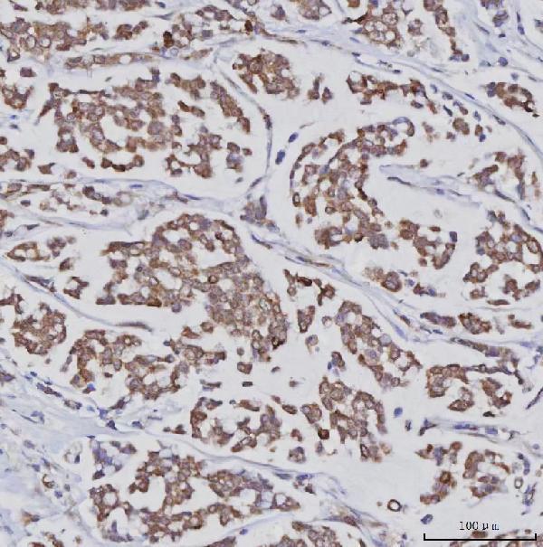
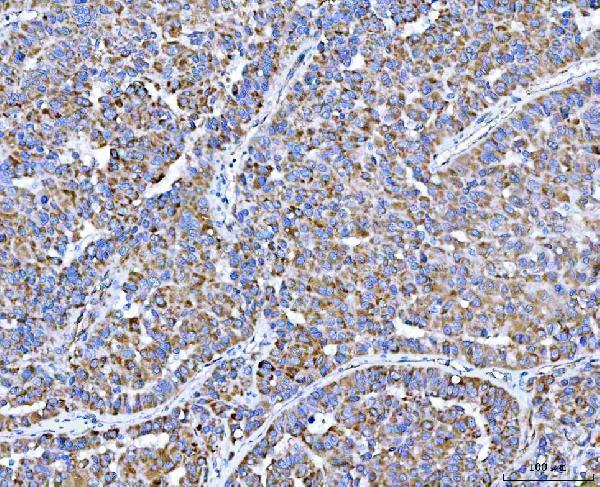
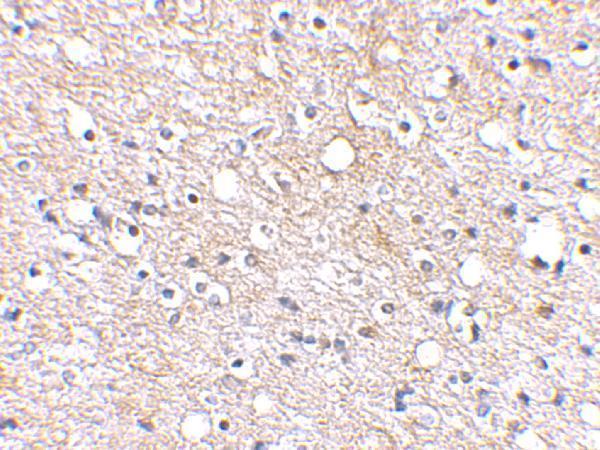
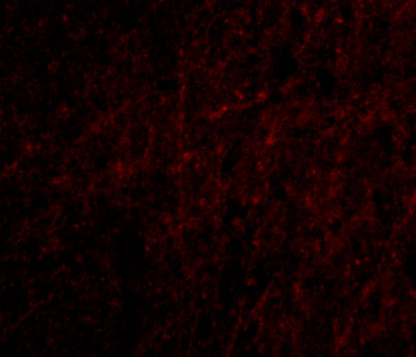
Two anti-PLXDC2 antibodies have human tissue IHC images (catalog image captions); A08798 also has an IF image of human brain cells (catalog IF image caption).
A08798-1 shows IHC staining in paraffin sections of human breast cancer, liver cancer, and lymphoma tissue (A08798-1 image captions). A08798 shows IHC staining in human brain tissue and IF staining in human brain cells (A08798 image captions).
Which to pick: For paraffin-section IHC, choose A08798-1 when its documented EDTA pH 8.0 retrieval and 2 μg/mL staining condition suit the experiment; the caption does not report the fixative (A08798-1 IHC image caption). For IF, choose A08798, which lists IF and shows human brain-cell staining at 20 μg/mL; ICC validation is not specified (A08798 applications and IF image caption). For mouse or rat work, A08798 lists those species as reactive, but its supplied IHC and IF images show human samples, so validate staining in the intended species (A08798 reactivity and image captions).