This website uses cookies to ensure you get the best experience on our website.


- Table of Contents
Plan chromogenic PMEL IHC in paraffin sections using skin melanocytes as a positive reference (HPA tissue IHC). Assess cytoplasmic staining while accounting for melanin pigment and PMEL processing when interpreting the result (HPA tissue IHC; UniProt; standard IHC practice).

Expected localisation, validated protocols, controls and antibodies — the at-a-glance facts below, then the full design guide.
| Expected localisation | Melanocyte cytoplasm (HPA tissue IHC) | |
| Staining pattern | Selective cytoplasmic staining in melanocytes (HPA tissue IHC) | |
| Antigen retrieval | Citrate pH 6.0 HIER, 95–98 °C, 20 min (rule: cytoplasmic / membrane antigen) | |
| Positive control | Skin | |
| Negative control | Adipose tissue+4 more · see all |
| Fixation | Keep fixation consistent across sections. (standard IHC practice; not target-specific) | |
| Caveat | Melanin may obscure brown chromogen (UniProt; standard IHC practice) | |
| Regulation | Higher with proliferation and in melanoma (UniProt) | |
| Isoform / epitope | 5 isoforms; cleavage separates M-alpha from the cytoplasmic tail (UniProt) |
The catalog antibody protocol is accompanied by published chromogenic PMEL IHC protocols for mouse distant organs, melanoma tissue, and a human tissue microarray (PMC13609154; PMC5587917; PMC8225232).
| Sample | Paraffin-embedded human melanoma tissue; fixative not specified (datasheet M01262) |
| Fixation | Image fixative and duration unreported (datasheet M01262); verify before use. |
| Sectioning | 4–5 µm sections on charged slides (standard) |
| Deparaffinisation | Xylene, graded ethanol series to water (standard) |
| Antigen retrieval | Heat-induced epitope retrieval in citrate buffer, pH 6.0, 20 min at 95–98 °C (standard rule: cytoplasmic / membrane antigen) |
| Peroxidase block | 3% H2O2, 10 min, room temperature (standard) |
| Blocking | 10% normal serum of the secondary host, 30 min, room temperature (standard) |
| Primary antibody | Rabbit monoclonal (clone AHH-16) anti-PMEL, 1:50 (datasheet M01262) |
| Primary incubation | Overnight at 4 °C (standard) |
| Detection | HRP-polymer secondary, DAB chromogen 5–10 min (standard) |
| Counterstain | Hematoxylin, blue, dehydrate and mount (standard) |
| Expected result | PMEL-positive staining in melanocytes of skin (HPA tissue IHC: High). HPA tissue profile: Selective cytoplasmic expression in melanocytes. No signal in the no-primary control. |
In paraffin-section IHC, expect selective cytoplasmic PMEL staining in melanocytes, with high staining reported in skin melanocytes (HPA tissue IHC: Enhanced; High in melanocytes). PMEL occupies melanosomes and their precursors after passage through the ER and Golgi; it has a membrane-spanning segment at residues 596–616 (UniProt P40967: localization; topology).
| Selective cytoplasmic staining in skin melanocytes, stronger than surrounding cells. | This matches the reported tissue pattern (HPA tissue IHC: selective cytoplasmic expression; High in melanocytes). Score identified melanocytes rather than treating all pigmented areas as positive (standard IHC practice). |
| Predominantly nuclear staining, without a convincing melanocyte cytoplasmic pattern. | A dominant nuclear pattern is unexpected given PMEL's reported membrane and melanosome localization (UniProt P40967: localization; topology). Review the negative control and antibody conditions before interpreting it as PMEL. |
| Staining predominates in cells outside the expected melanocyte population. | HPA reports selective staining in melanocytes (HPA tissue IHC). Consider cross-reactivity or endogenous detection activity; establish the cell identity and compare an appropriate negative control before scoring. |
| Broad, diffuse color obscures cell boundaries or appears across many cell types. | This does not reproduce selective cytoplasmic staining (HPA tissue IHC). Assess background with controls, then review blocking, washing and detection conditions (standard IHC practice). |
| No convincing melanocyte signal in an otherwise evaluable skin section. | This conflicts with the reported high staining in skin melanocytes (HPA tissue IHC). Confirm that melanocytes are present, then check the positive control and run conditions before calling the sample negative. |
| Tissue and cell context | HPA reports high staining in skin melanocytes, while UniProt describes low expression in quiescent adult melanocytes and higher expression during proliferation and melanoma growth (HPA tissue IHC; UniProt P40967: tissue specificity). Interpret intensity with the specimen context. |
| Processing and antibody epitope | PMEL has processed M-alpha (25–467) and M-beta (470–661) chains, and a transmembrane segment at 596–616 (UniProt P40967: processing; topology). The antibody epitope is not supplied, so fragment recognition and the effect of retrieval cannot be predicted from this record. |
| Strength of the tissue evidence | The HPA tissue profile is Enhanced, indicating high consistency between antibody staining and RNA expression; HPA031649 has Enhanced IHC validation (HPA tissue IHC: reliability; HPA antibodies). These findings support the expected pattern without validating every new specimen or run. |
| IF/ICC Q&A: where should signal localize? | HPA reports mainly ER localization with additional Golgi localization in ICC-IF, with images from SK-MEL-30 (HPA subcellular). This answers the IF localization question; the tissue IHC pattern remains selective cytoplasmic staining in melanocytes (HPA tissue IHC). |
| Situation | Likely cause | Next action |
|---|---|---|
| Skin melanocytes appear negative. | Melanocytes may be absent from the assessed area, or the staining run may have failed; HPA reports high staining in skin melanocytes (HPA tissue IHC). | Verify melanocyte identity on the section and inspect a known-positive control. If that control also fails, review retrieval, antibody incubation and detection as general IHC workflow steps (standard IHC practice). |
| The positive control stains, but the sample is weak. | Expression can vary with melanocyte state; UniProt describes low levels in quiescent adult melanocytes (UniProt P40967: tissue specificity). Weak staining alone does not identify a technical failure. | Compare like cell populations and specimen contexts, and score only localized signal above the run's background (standard IHC practice). Avoid applying a universal intensity cutoff from the HPA skin result. |
| Many unrelated cells stain. | This departs from selective melanocyte staining (HPA tissue IHC) and may reflect cross-reactivity or endogenous detection activity (standard IHC practice). | Compare the appropriate negative control, confirm cell identity, and review blocking and detection conditions before assigning PMEL positivity (standard IHC practice). |
| Brown deposits are difficult to separate from pigment. | Melanocytes contain pigment, which can complicate interpretation of a brown chromogenic signal (standard IHC practice; UniProt P40967: pigmentation function). | Compare the stained section with an appropriate control and inspect signal distribution within identified cells; do not score pigment alone as antibody staining (standard IHC practice). |
| Staining is chiefly nuclear or broadly diffuse. | That pattern differs from HPA's selective cytoplasmic tissue staining and UniProt's organelle localization (HPA tissue IHC; UniProt P40967: localization). | Check the negative control and review antibody and detection conditions. Reassess whether any discrete melanocyte cytoplasmic signal remains (standard IHC practice). |
| Retrieval changes the apparent pattern between runs. | The supplied sources do not establish PMEL-specific fixation or retrieval sensitivity; the antibody epitope is unspecified (UniProt P40967: processing; topology). | Compare runs using the same positive tissue and controls, document retrieval conditions, and retain the condition that yields selective melanocyte cytoplasmic staining with acceptable background (HPA tissue IHC; standard IHC practice). |


Comprehensive Human Protein Atlas IHC scoring per tissue (reliability: Enhanced — High consistency between antibody staining and RNA expression data.). Rows are taken directly from the HPA tissue chart — click any row's HPA link to view the source.
| Tissue | Cell type | Level | Evidence | Source |
|---|---|---|---|---|
| Skin | Melanocytes | High | Protein (IHC) | HPA → |
| Tissue | Cell type | Level | Evidence | Source |
|---|---|---|---|---|
| Adipose tissue | Adipocytes | Not detected | Protein (IHC) | HPA → |
| Adrenal gland | Glandular cells | Not detected | Protein (IHC) | HPA → |
| Appendix | Glandular cells | Not detected | Protein (IHC) | HPA → |
| Bone marrow | Hematopoietic cells | Not detected | Protein (IHC) | HPA → |
| Breast | Adipocytes | Not detected | Protein (IHC) | HPA → |
Troubleshoot PMEL staining in paraffin sections by checking retrieval, cellular distribution, processing, and controls before interpreting chromogenic signal.
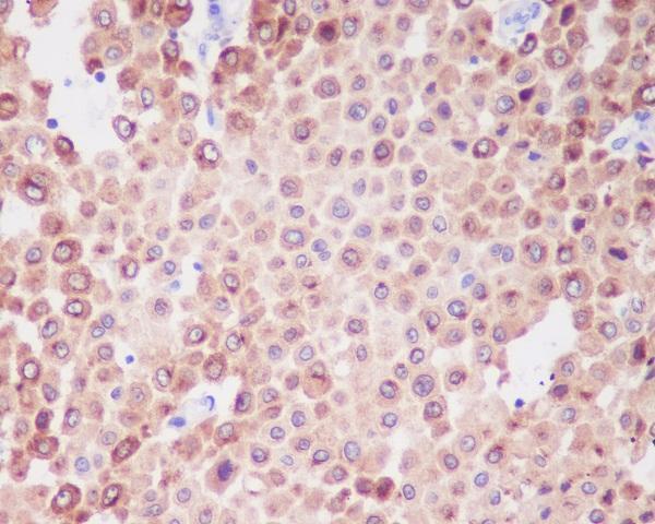
M01262 has real IHC data from paraffin-embedded human melanoma (catalog image caption); IF data are absent (catalog).
M01262 is listed for human IHC (catalog applications and reactivity). Its IHC image shows paraffin-embedded human melanoma (catalog image caption); no IF image is supplied (catalog).
Which to pick: For tissue IHC, choose M01262, a human-reactive rabbit monoclonal with an IHC image from paraffin-embedded human melanoma (catalog; catalog image caption). The caption does not report the fixative (catalog image caption). No listed SKU is supported here for IF/ICC or other species (catalog applications, IF images and reactivity).