This website uses cookies to ensure you get the best experience on our website.


- Table of Contents
Plan PMS2 staining in paraffin sections around the nuclear pattern reported in most tissues (HPA tissue IHC). This guide identifies cell-specific control choices (HPA tissue IHC) and flags MLH1-dependent stability when interpreting weak signal (UniProt).

Expected localisation, validated protocols, controls and antibodies — the at-a-glance facts below, then the full design guide.
| Expected localisation | Nuclear staining in most tissues (HPA tissue IHC) | |
| Staining pattern | Nuclear staining across most tissue cell types (HPA tissue IHC) | |
| Antigen retrieval | Tris-EDTA pH 9.0 HIER, 95–98 °C, 20 min (rule: nuclear antigen) | |
| Positive control | Cerebellum+4 more · see all | |
| Negative control | Adipose tissue+3 more · see all |
| Fixation | Keep fixation consistent across paraffin sections. (standard IHC practice; not target-specific) | |
| Caveat | MLH1 loss may reduce PMS2 signal through reduced stability (UniProt) | |
| Regulation | Stability depends on MLH1 binding (UniProt) | |
| Isoform / epitope | 4 isoforms; epitope coverage is unspecified (UniProt) |
The catalog antibody protocol is followed by published PMS2 IHC methods for colorectal carcinoma and tumor tissue (PMC13018393; PMC3273660; PMC8011511).
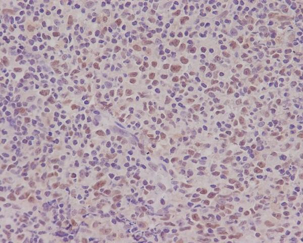
| Sample | Paraffin-embedded human tonsil tissue; fixative not specified (datasheet M01028-1) |
| Fixation | Image fixative and duration unreported (datasheet M01028-1); verify before use. |
| Sectioning | 4–5 µm sections on charged slides (standard) |
| Deparaffinisation | Xylene, graded ethanol series to water (standard) |
| Antigen retrieval | Heat-induced epitope retrieval in Tris-EDTA buffer, pH 9.0, 20 min at 95–98 °C (standard rule: nuclear antigen) |
| Peroxidase block | 3% H2O2, 10 min, room temperature (standard) |
| Blocking | 10% normal serum of the secondary host, 30 min, room temperature (standard) |
| Primary antibody | Rabbit monoclonal (clone AEH-16) anti-PMS2, 1:50 (datasheet M01028-1) |
| Primary incubation | Overnight at 4 °C (standard) |
| Detection | HRP-polymer secondary, DAB chromogen 5–10 min (standard) |
| Counterstain | Hematoxylin, blue, dehydrate and mount (standard) |
| Expected result | PMS2-positive staining in purkinje cells of cerebellum (HPA tissue IHC: High). HPA tissue profile: Nuclear expression in most tissues. No signal in the no-primary control. |
PMS2 should appear predominantly in nuclei across most tissues (UniProt P54278: nucleus; HPA tissue IHC: nuclear expression in most tissues). Purkinje cells, cerebral cortical neurons, and spermatogonia show high staining; several epithelial and glandular populations show medium staining (HPA tissue IHC). HPA rates the tissue profile Approved, with medium consistency between staining and RNA data (HPA tissue IHC). PMS2 has no transmembrane segment (UniProt P54278: topology).
| Distinct nuclear chromogen in Purkinje cells, cerebral cortical neurons, or spermatogonia. | This matches the expected compartment and HPA's high staining levels in those cells (UniProt P54278: nucleus; HPA tissue IHC: High). Assess individual nuclei against adjacent cells; a uniformly dark section is harder to interpret (general IHC practice). |
| Nuclear staining in appendix or breast glandular cells, bronchial respiratory epithelium, or caudate glia. | Medium staining is consistent with the reported cell-specific pattern (HPA tissue IHC: Medium). Compare the relevant cell population, since intensity across an entire tissue section can obscure which cells carry the signal (general IHC practice). |
| Predominantly cytoplasmic staining, with little convincing nuclear signal. | This conflicts with the expected IHC pattern (UniProt P54278: nucleus; HPA tissue IHC: nuclear expression in most tissues). Review background and antibody specificity before calling the section positive. HPA's uncertain additional cytosolic ICC-IF location does not establish a cytoplasmic IHC pattern (HPA subcellular ICC-IF). |
| Strong staining in adipocytes, cholangiocytes, alveolar cells, or soft-tissue fibroblasts. | Those specific populations are reported as not detected, making prominent signal a reason to investigate cross-reactivity or endogenous detection activity (HPA tissue IHC: Not detected; general IHC practice). The HPA result is a reference pattern, not proof that every specimen must be negative. |
| Diffuse staining across nuclei, cytoplasm, and surrounding tissue, or no staining in a known-positive control. | Diffuse signal limits compartment-based interpretation; absent control signal leaves a negative result uninterpretable (general IHC practice). A section containing an HPA high-staining cell population can provide a reference, provided that population is present and identifiable (HPA tissue IHC: High). |
| Tissue and cell selection | HPA reports High staining in Purkinje cells, cerebral cortical neurons, and spermatogonia, but Not detected in adipocytes, cholangiocytes, alveolar cells, and soft-tissue fibroblasts (HPA tissue IHC). Match the comparison to the named cell type, since a tissue label alone does not describe every cell in a section. |
| Strength of the tissue reference | The profile is Approved, with medium consistency between antibody staining and RNA expression data (HPA tissue IHC). Use its reported levels as interpretation guides rather than absolute thresholds for an individual specimen. |
| Antibody validation | CAB010235 has Approved IHC status; HPA070310 has no IHC status in the supplied antibody record (HPA antibodies). ICC support for either antibody does not establish IHC performance (HPA antibodies). |
| Isoforms and epitope coverage | Four isoforms are listed, but the payload does not map an IHC antibody's epitope to them (UniProt P54278: isoforms 1–4). A difference in staining cannot be assigned to isoform recognition from these records alone. |
| Does the IF/ICC result change the IHC call? | No: HPA supports nucleoplasmic localization in ICC-IF and marks additional cytosolic localization uncertain (HPA subcellular ICC-IF). That observation informs compartment review, while this paraffin-section guide relies on the IHC tissue pattern for its positive call (HPA tissue IHC). |
| Situation | Likely cause | Next action |
|---|---|---|
| No nuclear signal in an expected high-staining cell population. | The staining run may have failed, or the chosen section may lack identifiable target cells (general IHC practice; HPA tissue IHC: High in the named populations). | Confirm the target cells on the counterstained section and inspect a known-positive control run with the same reagents. Review the catalog antibody's IHC-P instructions and the run's retrieval, antibody incubation, and detection steps (general IHC practice). |
| Signal appears mainly cytoplasmic. | Background or nonspecific binding is possible; the predominant compartment conflicts with nuclear IHC expression (general IHC practice; HPA tissue IHC: nuclear expression in most tissues). | Compare nuclear and cytoplasmic staining in the same cells, then review blocking, antibody concentration, and detection controls (general IHC practice). Do not use the uncertain cytosolic ICC-IF observation as confirmation of cytoplasmic IHC staining (HPA subcellular ICC-IF). |
| Strong color appears in HPA not-detected cell populations. | Cross-reactivity or endogenous detection activity may contribute, although tissue differences and assay conditions also require review (HPA tissue IHC: Not detected; general IHC practice). | Verify the cell identity and run a control that omits the primary antibody to assess detection-related signal. Compare localization with a known-positive nuclear control before assigning PMS2 positivity (general IHC practice; UniProt P54278: nucleus). |
| Diffuse color obscures nuclei throughout the section. | Excess background can arise from nonspecific binding, detection chemistry, or insufficient washing (general IHC practice). It prevents a reliable comparison with the nuclear reference pattern (HPA tissue IHC). | Inspect the no-primary control, then review blocking, wash steps, antibody concentration, and chromogen development for the assay (general IHC practice). Reassess only when individual nuclei and cell types can be distinguished. |
| A weakly stained section is called negative. | Some named populations are reported Low, while others are Medium; a single intensity cutoff can misclassify a cell-specific result (HPA tissue IHC: Low and Medium). | Identify the exact cell population and compare it with its HPA level. Use a high-staining control to check that the run worked, then score nuclear localization alongside intensity (HPA tissue IHC: High; general IHC practice). |
| An ICC-IF image shows cytosolic signal that seems inconsistent with IHC. | HPA supports the nucleoplasm as the main ICC-IF location but labels its additional cytosolic location uncertain (HPA subcellular ICC-IF). The two assays also present tissue and cells differently (general IHC/IF practice). | Interpret the paraffin IHC section against its nuclear tissue pattern and controls (HPA tissue IHC; general IHC practice). Address ICC-IF assay settings and images in the separate IF/ICC guide. |


Comprehensive Human Protein Atlas IHC scoring per tissue (reliability: Approved — Medium consistency between antibody staining and RNA expression data.). Rows are taken directly from the HPA tissue chart — click any row's HPA link to view the source.
| Tissue | Cell type | Level | Evidence | Source |
|---|---|---|---|---|
| Cerebellum | Purkinje cells | High | Protein (IHC) | HPA → |
| Cerebral cortex | Neuronal cells | High | Protein (IHC) | HPA → |
| Testis | Spermatogonia cells | High | Protein (IHC) | HPA → |
| Adrenal gland | Glandular cells | Medium | Protein (IHC) | HPA → |
| Appendix | Glandular cells | Medium | Protein (IHC) | HPA → |
| Tissue | Cell type | Level | Evidence | Source |
|---|---|---|---|---|
| Adipose tissue | Adipocytes | Not detected | Protein (IHC) | HPA → |
| Liver | Cholangiocytes | Not detected | Protein (IHC) | HPA → |
| Lung | Alveolar cells | Not detected | Protein (IHC) | HPA → |
| Soft tissue | Fibroblasts | Not detected | Protein (IHC) | HPA → |
Troubleshoot PMS2 staining by checking nuclear signal, tissue controls, and processing variables before interpreting apparent loss of expression (UniProt P54278; HPA tissue IHC).
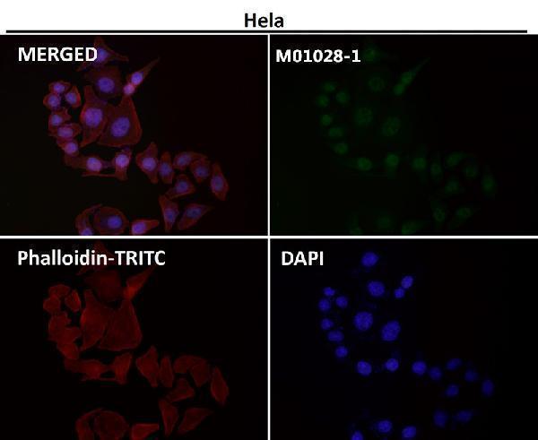
Both antibodies list human IHC and IF applications (catalog applications/reactivity); both have IHC images, while only M01028-1 has an IF image (catalog image captions).
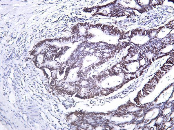
M01028-1 lists human IHC and IF applications, with IHC shown on paraffin-embedded human tonsil (catalog applications/reactivity; M01028-1 IHC caption). M01028-3 lists human IHC and IF applications, with IHC shown on human rectal carcinoma tissue; its processing and fixative are unreported (catalog applications/reactivity; M01028-3 IHC caption).
Which to pick: For paraffin-section IHC, start with M01028-1 at 1:50, matching its tonsil image; the fixative is unreported (M01028-1 IHC caption; datasheet: 1:50). For IF/ICC, choose M01028-1 because it lists both applications and has an IF image, though the IF sample type is unreported; M01028-3 lists IF but has no IF image (catalog applications; catalog IF captions). Neither SKU has documented cross-species reactivity: both list Human only (catalog reactivity).