PNCK / Calcium/calmodulin-dependent protein kinase type 1B · IHC design guide

Design Immunohistochemistry for PNCK

Plan PNCK staining in paraffin sections using the reported cytoplasmic and nuclear tissue pattern (HPA tissue IHC). Compare glandular-cell staining with reference tissues while accounting for low consistency between antibody staining and RNA expression (HPA tissue IHC).

Evidence assembled Oct 2026 · For research use; verify linked source records and product datasheet before use
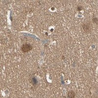
Immunohistochemistry protocol sheet for PNCK (IHC for PNCK): expected localisation Cytoplasmic and nuclear tissue staining (HPA tissue IHC), antibody A11515, validated IHC image, and IHC protocol steps
Printable PNCK IHC protocol sheet — expected localisation Cytoplasmic and nuclear tissue staining (HPA tissue IHC), antibody A11515, controls and protocol steps. Open the full PNCK IHC guide →

PNCK Immunohistochemistry Experimental Design Guide

Expected localisation, validated protocols, controls and antibodies — the at-a-glance facts below, then the full design guide.

Must know before staining
Expected localisation Cytoplasmic and nuclear tissue staining (HPA tissue IHC)
Staining pattern Glandular cells show cytoplasmic and nuclear staining (HPA tissue IHC)
Antigen retrieval Tris-EDTA pH 9.0 HIER, 95–98 °C, 20 min (rule: nuclear antigen)
Positive control ⓘ Appendix+4 more · see all
Negative control ⓘ None in HPA (detected in all 45 tissues); use no-primary + isotype controls
Important caveats
Reasons your staining may differ from the expected pattern.
Fixation Keep fixation conditions consistent across sections. (standard IHC practice; not target-specific)
Caveat Staining has low consistency with RNA expression (HPA tissue IHC)
Regulation Expression regulation is not annotated (UniProt)
Isoform / epitope 5 isoforms; epitope coverage is unspecified (UniProt)
Section 1

Recommended PNCK IHC & IF Protocols

The catalog antibody's IHC-P protocol is accompanied by three published PNCK staining protocols (PMC3636239; PMC9078829; PMC6909947).

Recommended immunohistochemistry (IHC-P) protocol parameters
SampleParaffin-embedded human brain tissue; fixative not specified (datasheet A11515)
FixationImage fixative and duration unreported (datasheet A11515); verify before use.
Sectioning4–5 µm sections on charged slides (standard)
DeparaffinisationXylene, graded ethanol series to water (standard)
Antigen retrievalHeat-induced epitope retrieval in Tris-EDTA buffer, pH 9.0, 20 min at 95–98 °C (standard rule: nuclear antigen)
Peroxidase block3% H2O2, 10 min, room temperature (standard)
Blocking10% normal serum of the secondary host, 30 min, room temperature (standard)
Primary antibodyRabbit anti-PNCK, 1:100 - 1:300 (datasheet A11515)
Primary incubationOvernight at 4 °C (standard)
DetectionHRP-polymer secondary, DAB chromogen 5–10 min (standard)
CounterstainHematoxylin, blue, dehydrate and mount (standard)
Expected resultPNCK-positive staining in glandular cells of appendix (HPA tissue IHC: High). HPA tissue profile: General cytoplasmic and nuclear expression. No signal in the no-primary control.
💡Decision noteStart with Tris-EDTA pH 9.0, 95–98 °C for 20 min for the catalog antibody (page retrieval); use each article's stated retrieval when reproducing its protocol.
Section 2

What Is the Expected PNCK Staining Pattern?

In paraffin tissue sections, expect PNCK mainly in the cytoplasm and nuclei of selected cells, including appendix and stomach glandular cells, pancreatic exocrine glandular cells, placental decidual cells, and skin fibroblasts (HPA tissue IHC: High; general cytoplasmic and nuclear expression). UniProt also lists cytoplasm and nucleus and reports no transmembrane segment (UniProt Q6P2M8). Treat intensity as a guide: HPA rates the tissue staining Approved but notes low consistency with RNA expression (HPA tissue IHC).

What am I looking at on my slide?
Distinct cytoplasmic or nuclear stain in glandular cells or other listed positive cells.This fits the reported tissue pattern; compare the same cell population across sections before scoring intensity (HPA tissue IHC: general cytoplasmic and nuclear expression). A high HPA level describes observed staining, not a guaranteed result in every specimen (HPA tissue IHC: High).
Stain sits mainly in gland lumens, extracellular material, or a compartment unrelated to the cells.Treat it as possible deposited chromogen or nonspecific signal and inspect morphology and controls. Tissue IHC supports cellular cytoplasmic and nuclear staining (HPA tissue IHC); membrane signal alone needs care because ICC-IF also reports plasma membrane localization (HPA subcellular: approved).
Strong stain appears in an unexpected cell population, such as vaginal squamous epithelium.Investigate cross-reactivity or endogenous detection activity before calling PNCK positive. HPA reports low staining in vaginal squamous epithelial cells, but this is not a validated negative control (HPA tissue IHC: Low). Cell identity and control staining matter.
Color spreads diffusely across tissue, including areas without plausible positive cells.A weak boundary between cells and background limits compartment scoring. Check the no-primary control and detection chemistry as general chromogenic IHC practice. HPA describes a cellular pattern, so diffuse section-wide color should not be counted as PNCK expression (HPA tissue IHC: general expression).
No detectable stain in a listed high-staining tissue.First check that the relevant cells are present: pancreatic exocrine cells, for example, are listed High (HPA tissue IHC). Then review the control and IHC workflow. An absent signal in one section cannot establish PNCK absence, given HPA's reported antibody–RNA inconsistency (HPA tissue IHC: Approved, low consistency).
💡Expected PNCK appearanceCall a convincing positive when identifiable listed cells show discrete cytoplasmic and/or nuclear staining, potentially strong in HPA High populations; diffuse extracellular color or strong stain in an unexpected cell type needs control review before being scored (HPA tissue IHC: profile, levels, and low consistency).
How each factor affects the staining
Tissue and cell identityExpected intensity is cell specific: glandular cells in appendix, epididymis, gallbladder, and stomach; pancreatic exocrine cells; placental decidual cells; and skin fibroblasts are listed High (HPA tissue IHC). Adipocytes are Medium, while vaginal squamous cells and soft-tissue fibroblasts are Low (HPA tissue IHC).
Compartment and topologyUniProt assigns PNCK to cytoplasm and nucleus and reports no transmembrane segment or signal peptide (UniProt Q6P2M8). These annotations help assess a cellular IHC pattern; they do not prove that every observed membrane edge is nonspecific, because HPA ICC-IF reports an approved plasma membrane location (HPA subcellular).
Validation strengthHPA lists the tissue IHC pattern as Approved, with low consistency between antibody staining and RNA expression (HPA tissue IHC). The listed rabbit polyclonal HPA007458 is IHC Approved, while the supplied record does not label its IHC validation Enhanced (HPA antibody record). Interpret isolated unexpected staining conservatively.
Isoform coverageUniProt lists five PNCK isoforms and a protein kinase domain at residues 15–270 (UniProt Q6P2M8). No antibody epitope or isoform coverage is supplied, so a missing or altered pattern cannot be assigned to a particular isoform from these sources.
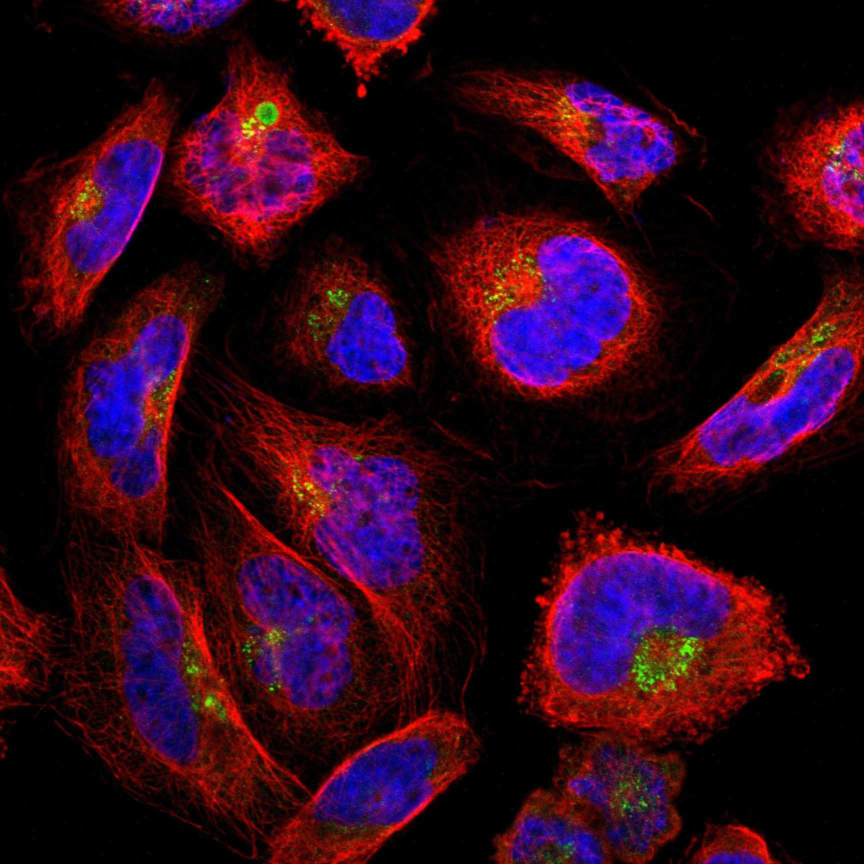
IF/ICC Q: What localization is reported?A: HPA reports approved plasma membrane and cytosol locations, with an additional approved Golgi location in ICC-IF (HPA subcellular). That cell-image summary does not set the expected chromogenic pattern for every paraffin tissue; use the tissue IHC profile for that decision (HPA tissue IHC).
Why is my staining missing, weak or wrong?
SituationLikely causeNext action
No signal in a section chosen as a positive comparison.The section may lack the named positive cell population, or an IHC step may have failed; HPA levels refer to specific cells (HPA tissue IHC).Confirm cell morphology, then check the IHC-validated antibody's supplied procedure and a working detection control. Review antigen retrieval as general IHC practice; PNCK-specific retrieval or fixation sensitivity is unreported here.
Uniform brown color or stain in vessel contents and unrelated areas.Background or endogenous enzyme activity can mimic chromogenic staining in general IHC practice; the reported PNCK tissue pattern is cellular (HPA tissue IHC).Inspect a no-primary control, review blocking and washes, and check the detection system's endogenous-activity control. Score cells only after background is separated from specific staining.
Strong signal in a population reported Low.Unexpected signal may reflect cross-reactivity or detection background; HPA lists vaginal squamous cells and soft-tissue fibroblasts as Low, not definitively negative (HPA tissue IHC).Verify cell identity and compare controls on the same tissue. Avoid using the Low label alone to declare a false positive, especially because HPA reports low antibody–RNA consistency (HPA tissue IHC).
Only a sharp membrane outline is visible in tissue IHC.HPA tissue IHC describes general cytoplasmic and nuclear expression, while ICC-IF also reports plasma membrane localization (HPA tissue IHC; HPA subcellular).Review cellular morphology, controls, and accompanying cytoplasmic or nuclear stain. Record the membrane-predominant finding separately rather than treating the ICC-IF pattern as proof of the tissue result.
Neighboring sections give different apparent intensities.The proportions of the named cell populations may differ between sections; HPA reports levels by tissue and cell type (HPA tissue IHC).Compare the same identifiable cell type and compartment, with matched scoring and detection conditions. Record heterogeneity instead of assigning a whole-tissue score from a small positive area.
Signal is weak after a change to the IHC workflow.The supplied sources give no PNCK-specific retrieval, fixation, or dilution effect, so the reason for the change is undetermined.Return to the IHC-validated antibody's documented conditions and check a known working section alongside the changed run. Adjust one general IHC step at a time and judge the result by cellular pattern and controls.

Sample controls for PNCK IHC & IF

🧪Run placenta first and look for staining in decidual cells (HPA: High in placenta decidual cells). HPA detects PNCK in all 45 scored tissues, so there is no supported negative tissue; use no-primary and isotype controls for the negative comparison, and expect cells without specific signal on the positive slide to lack chromogen above local background without assigning a cell type as PNCK-negative (HPA: no negative rows; standard IHC practice).
Positive control tissue: Appendix (Glandular cells, HPA High)
Negative control tissue: None in HPA: PNCK is detected in all 45 scored tissues. Use a no-primary (secondary-only) and an isotype control instead.
ICC-IF cell lines (HPA subcellular resource): HPA ICC-IF images show PNCK in A-431, U-251MG, U2OS, hTERT-RPE1 (serum starved), NIH 3T3, with annotated localisation: Plasma membrane (approved), Cytosol (approved) (HPA subcellular).
Technical controls: Include a no-primary (secondary-only) control and an irrelevant antibody matched to the primary antibody’s host species, class, and concentration; use a PNCK-knockout specimen as a biological negative if available, or assess peptide competition as shown for A11515 (standard IHC practice; A11515 tissue-IHC caption: peptide block). For placenta, quench endogenous peroxidase and check blood-associated chromogenic background (standard IHC practice).
⚠️Feasibility: A target-specific fixation window and retrieval dependence are unreported in the supplied PNCK evidence; the A11515 paraffin-section caption does not state a fixative (A11515 tissue-IHC caption). Paraffin IHC is documented by that caption, and HPA has ICC-IF images, but the supplied evidence does not establish that frozen sections or IF are easier (A11515 tissue-IHC caption; HPA: PNCK subcellular ICC-IF images). In placenta, blood-associated peroxidase can complicate chromogenic interpretation (standard IHC practice).

HPA tissue IHC evidence for PNCK

Comprehensive Human Protein Atlas IHC scoring per tissue (reliability: Approved — Low consistency between antibody staining and RNA expression data.). Rows are taken directly from the HPA tissue chart — click any row's HPA link to view the source.

Positive expression · recommended positive controls

TissueCell typeLevelEvidenceSource
Appendix Glandular cells High Protein (IHC) HPA →
Epididymis Glandular cells High Protein (IHC) HPA →
Gallbladder Glandular cells High Protein (IHC) HPA →
Pancreas Exocrine glandular cells High Protein (IHC) HPA →
Placenta Decidual cells High Protein (IHC) HPA →

Undetected expression · recommended negative controls

TissueCell typeLevelEvidenceSource
None in HPA: PNCK is detected in all 45 scored tissues. Use a no-primary (secondary-only) and an isotype control instead.
Section 3

Advanced PNCK IHC Tips

These PNCK questions focus on interpreting chromogenic IHC in paraffin sections, with one IF/ICC question for a complementary localisation check.

How should I optimize retrieval if PNCK staining is weak or uneven?
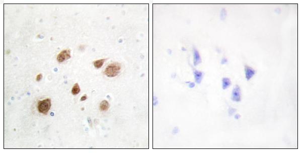
Start with Tris-EDTA pH 9.0 HIER at 95–98 °C for 20 min (page retrieval setting: nuclear antigen). If staining remains weak, compare a shorter heating period or citrate pH 6.0 on matched sections while holding antibody dilution and chromogen development constant (standard IHC optimization). Include a positive reference section and a no-primary control in each comparison (standard IHC practice). Score nuclear and cytoplasmic signal separately because both are reported for PNCK in tissue (HPA tissue IHC; UniProt Q6P2M8 subcellular). The catalog image shows peptide-blocked staining in paraffin-embedded human brain, but its caption does not identify a retrieval method (A11515 tissue-IHC caption).
Can the available evidence tell me whether fixation has masked PNCK?
PNCK-specific fixation sensitivity is unknown; the selected paraffin-embedded human brain caption does not state a fixative (A11515 tissue-IHC caption). Record the fixative, fixation duration, tissue thickness and processing history for each section so staining differences can be traced to preparation (standard IHC practice). If archived samples vary, compare cases within matched processing groups before assigning biological meaning to intensity differences (standard IHC practice). Examine morphology and include a consistently processed positive reference section in each run (standard IHC practice). Do not infer fixation tolerance from PNCK’s cytoplasmic and nuclear annotation or from tissue staining patterns (UniProt Q6P2M8 subcellular; HPA tissue IHC).
Which compartments should count as plausible PNCK staining in tissue?
Assess cytoplasmic and nuclear staining as plausible tissue patterns because both are reported for PNCK (UniProt Q6P2M8 subcellular; HPA tissue IHC). HPA IF/ICC additionally places PNCK mainly at the plasma membrane and cytosol, with Golgi staining, so a membrane rim alone needs independent confirmation in the tissue context (HPA subcellular). PNCK has no annotated transmembrane segment, which does not by itself exclude membrane-associated localisation (UniProt Q6P2M8 topology; standard cell biology). Record nuclear, diffuse cytoplasmic and sharply membranous staining as separate observations rather than combining their intensities (standard IHC scoring practice). Compare each pattern with morphology, a no-primary control and the same compartment in a reference section (standard IHC practice).
Could isoforms or an unknown epitope explain discordant PNCK staining?
PNCK has 5 annotated isoforms, while the supplied caption does not identify the catalog antibody’s epitope or isoform coverage (UniProt Q6P2M8 isoforms; A11515 tissue-IHC caption). Its annotated kinase domain spans residues 15–270, but that range cannot establish where this antibody binds (UniProt Q6P2M8 domains). Ask for an epitope map before treating staining as evidence for a particular isoform (standard antibody validation practice). The selected image includes staining blocked by a synthesized peptide, supporting recognition of the immunizing peptide under that image condition without establishing isoform specificity (A11515 tissue-IHC caption). Compare compartment patterns with an independently validated reagent or orthogonal expression measurement when the distinction matters (standard IHC validation practice).
How can IF/ICC help resolve a disputed PNCK compartment pattern?
Use IF/ICC as a complementary localisation check: HPA reports approved cytosol and plasma membrane localisation, plus Golgi staining (HPA subcellular). For tissue multiplexing, pair PNCK with a marker identifying the cell population under study; HPA reports high staining in appendix glandular cells, for example (HPA tissue IHC: High in appendix glandular cells). Choose fluorophores after inspecting tissue autofluorescence and reserve a spectrally distinct channel for the weaker signal (standard IF practice). Use permeabilisation when testing intracellular PNCK epitopes, while assessing an accessible surface epitope without it if epitope mapping supports that placement (UniProt Q6P2M8 topology; standard IF practice). Verify each channel with single-label controls before interpreting overlap (standard IF practice).
How do I distinguish PNCK signal from chromogenic background?
Inspect a no-primary section for endogenous enzyme activity, pigment and nonspecific chromogen deposition before interpreting the PNCK-stained section (standard chromogenic IHC practice). Apply a peroxidase block when using peroxidase detection, and compare chromogen development times across matched sections (standard chromogenic IHC practice). If widespread staining persists, optimize protein blocking and antibody dilution with the same retrieval and detection conditions (standard IHC practice). The selected A11515 image includes a synthesized-peptide block in paraffin-embedded human brain; that comparison is specific to the depicted antibody and condition (A11515 tissue-IHC caption). Treat edge staining or deposits that ignore cell boundaries as technical concerns requiring repeat assessment (standard IHC interpretation practice).
What scoring method makes PNCK IHC comparable across samples? ⚠ ANSWER MARKED FOR VERIFICATION
Define the cell population and compartment before scoring, then report nuclear and cytoplasmic PNCK separately (HPA tissue IHC; standard IHC scoring practice). For each compartment, record the percentage of viable positive cells and an intensity grade from 0–3; an H-score sums percentage at each intensity multiplied by that intensity, yielding 0–300 (standard IHC scoring practice). For spatial comparisons, report positive cells per mm² of evaluable tissue as a separate measure (standard image-analysis practice). Normalize cell counts to viable cells of the prespecified type, and area measurements to evaluable tissue area (standard IHC scoring practice). Apply identical thresholds, section selection and chromogen settings across cases (standard IHC scoring practice).
What would make a PNCK-positive IHC result convincing?
A convincing pattern identifies intact cells, respects their boundaries and recurs in a consistently processed reference section (standard IHC interpretation practice). Cytoplasmic and nuclear signal is compatible with reported PNCK tissue staining, while a sharply isolated membrane pattern warrants comparison with HPA IF/ICC localisation and an independent control (HPA tissue IHC; HPA subcellular). Check cell identity against morphology because HPA reports high staining in specified glandular cells, decidual cells, skin fibroblasts and adipocytes at medium intensity, rather than uniform positivity across all cells (HPA tissue IHC). Exclude edge effects, necrotic regions and no-primary staining before calling a positive result (standard IHC practice). Interpret RNA agreement cautiously because HPA rates tissue staining approved but reports low consistency with RNA expression (HPA tissue IHC reliability).
Boster reagents

Best PNCK / Calcium/calmodulin-dependent protein kinase type 1B IHC Antibodies

A11515 has real IHC data from paraffin-embedded human brain tissue (catalog IHC image caption). IF/ICC applications and Human, Mouse, and Rat reactivity are listed (catalog applications/reactivity).

Real IHC data Immunohistochemistry analysis of paraffin-embedded human brain tissue, using CaMK1-beta Antibody. The picture on the right is blocked with the synthesized peptide.
Anti-CaMK1-beta PNCK Antibody
Cat # A11515

A11515 is listed for IHC, IF, and ICC with Human, Mouse, and Rat reactivity (catalog applications/reactivity). Its IHC image shows paraffin-embedded human brain tissue and a peptide-blocked comparison (catalog IHC image caption); no IF image is supplied (catalog IF image alts).

Which to pick: Choose A11515 for tissue IHC based on its paraffin-embedded human brain image (catalog IHC image caption); the fixative is unreported (catalog IHC image caption). For IF/ICC, A11515 lists both applications, with an IF dilution of 1:50 (catalog applications/IF dilution). For cross-species work, A11515 lists Human, Mouse, and Rat reactivity and is rabbit polyclonal (catalog reactivity/host/dilution raw).

Each figure is that product's own IHC / IF validation image from its datasheet.

References

  1. UniProt Consortium. UniProt entry Q6P2M8 (KCC1B_HUMAN, Calcium/calmodulin-dependent protein kinase type 1B).
  2. Human Protein Atlas. PNCK tissue IHC expression (reliability: Approved).
  3. Human Protein Atlas. PNCK subcellular location (ICC-IF): Mainly localized to the plasma membrane and cytosol. In addition localized to the Golgi apparatus..
  4. Human Protein Atlas. PNCK antibody validation summary (1 antibodies).
  5. Increased expression of pregnancy up-regulated non-ubiquitous calmodulin kinase is associated with poor prognosis in clear cell renal cell carcinoma. PloS one 2013 — PMC3636239.
  6. Upregulation of PNCK Promotes Metastasis and Angiogenesis via Activating NF-κB/VEGF Pathway in Nasopharyngeal Carcinoma. Journal of oncology 2022 — PMC9078829.
  7. PNCK depletion inhibits proliferation and induces apoptosis of human nasopharyngeal carcinoma cells in vitro and in vivo. Journal of Cancer 2019 — PMC6909947.
  8. PubMed PMID:14702039 — UniProt-cited evidence.
  9. PubMed PMID:15772651 — UniProt-cited evidence.
  10. PubMed PMID:15489334 — UniProt-cited evidence.