This website uses cookies to ensure you get the best experience on our website.


- Table of Contents
Plan paraffin-section POC1A IHC with 2–5 μg/mL catalog antibody and chromogenic detection (datasheet A08694-1). Use high-staining glandular tissue or testis Leydig cells as positive controls, with adipocytes as a negative comparator (HPA tissue IHC).

Expected localisation, validated protocols, controls and antibodies — the at-a-glance facts below, then the full design guide.
| Expected localisation | Granular cytoplasm in tissue (HPA tissue IHC) | |
| Staining pattern | Granular cytoplasm in glandular and Leydig cells (HPA tissue IHC) | |
| Antigen retrieval | EDTA pH 8.0 HIER, heat-mediated (datasheet A08694-1) | |
| Positive control | Adrenal gland+4 more · see all | |
| Negative control | Adipose tissue+2 more · see all |
| Fixation | Keep paraffin-section fixation consistent across samples. (standard IHC practice; not target-specific) | |
| Caveat | Tissue staining has medium RNA concordance (HPA tissue IHC) | |
| Regulation | No expression regulator annotated (UniProt) | |
| Isoform / epitope | 3 isoforms; no TM segment; epitope coverage unknown (UniProt) |
The catalog antibody’s IHC-P protocol (datasheet: A08694-1) is accompanied by four published POC1A IHC protocols (PMC13286842; PMC10907829; PMC7732308; PMC12366406).
| Sample | Paraffin-embedded human prostate cancer tissue; fixative not specified (datasheet A08694-1) |
| Fixation | Image fixative and duration unreported (datasheet A08694-1); verify before use. |
| Sectioning | 4–5 µm sections on charged slides (standard) |
| Deparaffinisation | Xylene, graded ethanol series to water (standard) |
| Antigen retrieval | Heat retrieval: EDTA pH 8.0 (datasheet A08694-1); 20 min, 95–100 °C (standard) |
| Peroxidase block | 3% H2O2, 10 min, room temperature (standard) |
| Blocking | 10% goat serum (datasheet A08694-1) |
| Primary antibody | Rabbit anti-POC1A, 2-5 μg/ml (datasheet A08694-1) |
| Primary incubation | Overnight at 4 °C (datasheet A08694-1) |
| Detection | HRP-conjugated secondary, DAB chromogen (datasheet A08694-1) |
| Counterstain | Hematoxylin, blue, dehydrate and mount (standard) |
| Expected result | POC1A-positive staining in glandular cells of adrenal gland (HPA tissue IHC: High). HPA tissue profile: General granular cytoplasmic expression. No signal in the no-primary control. |
POC1A is a centriolar and basal body protein with no transmembrane segment (UniProt Q8NBT0). In paraffin IHC, expect granular cytoplasmic staining, especially in adrenal, appendix, duodenal, gallbladder, rectal and small-intestinal glandular cells and testicular Leydig cells (HPA tissue IHC). HPA rates its tissue staining “Approved,” with medium consistency between antibody staining and RNA expression; interpret the pattern with that limitation (HPA tissue IHC).
| Granular cytoplasmic staining is strong in glandular cells of the listed positive tissues or in testicular Leydig cells (HPA tissue IHC). | This matches the reported tissue IHC pattern and high staining levels (HPA tissue IHC). Granularity is the practical paraffin-section readout; individual centrioles need not be resolved to call a tissue pattern consistent with POC1A (UniProt Q8NBT0; general IHC practice). |
| A strong, predominantly nuclear or cell-surface pattern replaces granular cytoplasmic staining (HPA tissue IHC). | That compartment is discordant with the reported tissue pattern and centriolar localization (HPA tissue IHC; UniProt Q8NBT0). Check antibody specificity and detection controls before interpreting it as POC1A; a discordant pattern alone does not identify its cause (general IHC practice). |
| Prominent staining appears in adipocytes, ovarian stromal cells or skeletal myocytes (HPA tissue IHC). | HPA reports POC1A as not detected in those cell types (HPA tissue IHC). Treat conspicuous staining there as unexpected and assess cross-reactivity or endogenous detection activity with appropriate controls (general IHC practice). A negative HPA call does not prove absolute absence in every specimen. |
| Diffuse stain covers cells and surrounding section without a readable granular cytoplasmic pattern (HPA tissue IHC). | The distribution cannot support a POC1A localization call (HPA tissue IHC; general IHC practice). Nonspecific reagent binding or detection background are possibilities; compare a no-primary control and review blocking, washing and chromogen development (general IHC practice). |
| No convincing signal appears in adrenal or intestinal glandular cells, or in testicular Leydig cells (HPA tissue IHC). | These are reported high-staining cell populations, so an entirely negative run needs technical review (HPA tissue IHC). Check tissue identity and assay controls, then evaluate retrieval, antibody dilution and detection performance as general IHC variables; no POC1A-specific retrieval sensitivity is supplied (general IHC practice). |
| Tissue and cell selection | Reported high staining occurs in adrenal, appendix, duodenal, gallbladder, rectal and small-intestinal glandular cells and testicular Leydig cells (HPA tissue IHC). Stomach glandular cells are reported at medium level; adipocytes, ovarian stromal cells and skeletal myocytes are reported as not detected (HPA tissue IHC). |
| Compartment and image resolution | POC1A is assigned to centrioles, centrosomes, cilium basal bodies and spindle poles (UniProt Q8NBT0). Tissue IHC is described as generally granular cytoplasmic (HPA tissue IHC). Interpret the paraffin-section pattern at the resolution actually obtained, rather than requiring visible individual centrioles (general IHC practice). |
| Strength of tissue evidence | The tissue IHC profile is “Approved,” with medium consistency between antibody staining and RNA expression (HPA tissue IHC). HPA040600 is listed as IHC “Approved” and ICC “Approved” (HPA antibodies). These classifications support a reference pattern but do not make every stained structure specific. |
| Molecular forms and topology | UniProt lists three POC1A isoforms, no transmembrane segment, no signal peptide or propeptide, and a chain spanning residues 1–407 (UniProt Q8NBT0). Without an epitope map, these annotations cannot establish which isoforms an antibody detects or predict retrieval behavior; avoid isoform claims from staining alone. |
| IF/ICC interpretation | HPA's ICC-IF images are from sperm; the supported main location is the flagellar centriole, with approved additional mid-piece and principal-piece localization (HPA subcellular). This is a distinct imaging context from the granular cytoplasmic tissue IHC profile and does not set a paraffin-IHC scoring threshold (HPA tissue IHC). |
| Situation | Likely cause | Next action |
|---|---|---|
| A reported high-staining glandular or Leydig-cell population is blank (HPA tissue IHC). | Possible assay failure, unsuitable tissue identification or inadequate signal development (general IHC practice); POC1A-specific fixation or retrieval sensitivity is unreported in the supplied evidence. | Confirm the cell population and a working positive control; review retrieval conditions, antibody dilution and detection reagents using the validated assay instructions (general IHC practice). Do not infer target absence from one blank section. |
| The whole section has brown haze or extracellular deposit instead of readable granular cytoplasm (HPA tissue IHC). | Excess detection background, incomplete blocking or washing, or excessive chromogen development can obscure localization (general IHC practice). | Compare the no-primary control, then adjust blocking, washing or development within the assay workflow (general IHC practice). Score POC1A only where cell-associated granular staining is distinguishable from background (HPA tissue IHC). |
| Unexpected adipocyte, ovarian stromal-cell or myocyte staining is conspicuous (HPA tissue IHC). | These populations are reported as not detected (HPA tissue IHC). Cross-reactivity or endogenous enzyme activity is possible in chromogenic IHC (general IHC practice). | Check a no-primary control and the relevant endogenous-activity control; reassess the antibody pattern against a reported positive tissue (general IHC practice; HPA tissue IHC). Do not score unexpected color as POC1A solely because it is strong. |
| Nuclear or membrane-like staining dominates a reported positive cell population (HPA tissue IHC). | The localization differs from granular cytoplasmic tissue IHC and centriolar assignment (HPA tissue IHC; UniProt Q8NBT0). The appearance alone cannot distinguish cross-reactivity from detection artefact. | Review control slides, staining distribution and cell morphology; repeat with an independently validated specificity control if available (general IHC practice). Record the result as discordant until the compartment assignment is supported. |
| Only faint staining is visible in stomach glandular cells (HPA tissue IHC). | HPA reports stomach glandular cells at medium level, whereas several other glandular populations are high (HPA tissue IHC); intensity need not match across tissues. | Compare like-for-like processed sections and confirm the pattern in a reported high-staining population before treating the stomach result as assay failure (HPA tissue IHC; general IHC practice). |
| Q: What should an IF/ICC image show? | HPA's subcellular ICC-IF evidence comes from sperm, while the primary tissue IHC profile is granular cytoplasmic (HPA subcellular; HPA tissue IHC). | A: In sperm ICC-IF, assess a flagellar centriole signal; mid-piece and principal-piece signals are also reported (HPA subcellular). Use the separate IF/ICC guide for assay setup; do not apply that sperm image pattern as a paraffin-IHC requirement. |


Comprehensive Human Protein Atlas IHC scoring per tissue (reliability: Approved — Medium consistency between antibody staining and RNA expression data.). Rows are taken directly from the HPA tissue chart — click any row's HPA link to view the source.
| Tissue | Cell type | Level | Evidence | Source |
|---|---|---|---|---|
| Adrenal gland | Glandular cells | High | Protein (IHC) | HPA → |
| Appendix | Glandular cells | High | Protein (IHC) | HPA → |
| Duodenum | Glandular cells | High | Protein (IHC) | HPA → |
| Gallbladder | Glandular cells | High | Protein (IHC) | HPA → |
| Rectum | Glandular cells | High | Protein (IHC) | HPA → |
Troubleshoot POC1A staining in paraffin sections by comparing matched controls, documented processing conditions, and the expected cellular pattern.
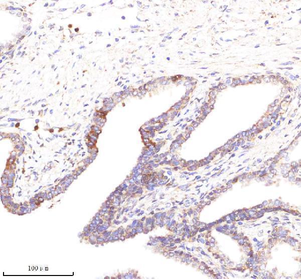
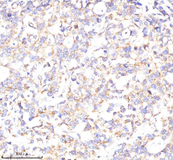
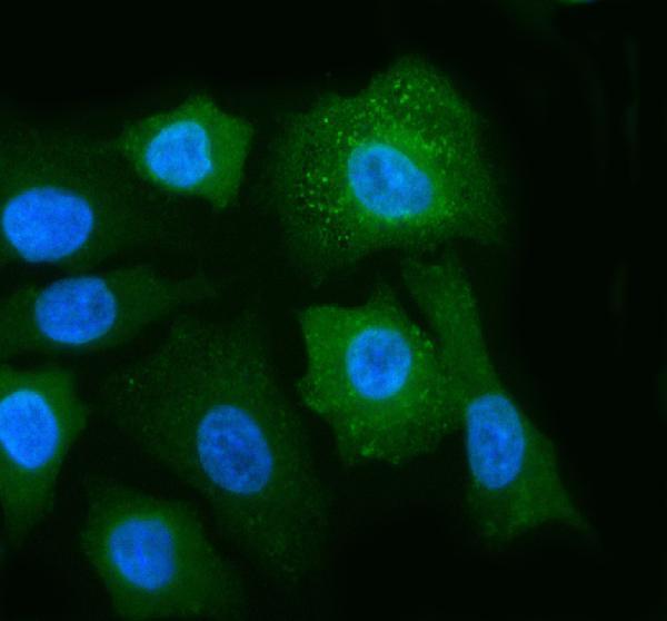
Anti-POC1A A08694-1 has IHC images from human prostate and stomach cancer sections and mouse and rat testis sections, plus an IF image from A549 cells (catalog image captions).
A08694-1 is listed for IHC and IF/ICC and is reactive with human, mouse, and rat samples (catalog applications and reactivity). Its IHC captions show paraffin sections of human prostate and stomach cancer and mouse and rat testis; its IF caption shows A549 cells (catalog image captions).
Which to pick: Choose A08694-1 for tissue IHC: its images show staining in paraffin sections, with an IHC working range of 2–5 μg/ml (catalog IHC captions; datasheet: 2–5 μg/ml); the fixative is unreported (catalog IHC captions). For IF/ICC, A08694-1 has an A549 cell image and a listed dilution of 5 μg/ml (catalog IF caption; datasheet: 5 μg/ml). For cross-species IHC, A08694-1 lists human, mouse, and rat reactivity and has IHC images for each; its host is rabbit, and clonality is unreported (catalog reactivity, IHC captions, and antibody details).