This website uses cookies to ensure you get the best experience on our website.


- Table of Contents
Plan chromogenic POLB IHC in paraffin sections around the general nuclear tissue pattern (HPA tissue IHC). Use high-staining glandular cells as positive references and adipocytes as a low-signal comparison (HPA tissue IHC).

Expected localisation, validated protocols, controls and antibodies — the at-a-glance facts below, then the full design guide.
| Expected localisation | Nuclear staining in tissue; cytoplasmic POLB is also annotated (HPA tissue IHC; UniProt) | |
| Staining pattern | General nuclear staining across tissue cell types (HPA tissue IHC) | |
| Antigen retrieval | Tris-EDTA pH 9.0 HIER, 95–98 °C, 20 min (rule: nuclear antigen) | |
| Positive control | Breast+4 more · see all | |
| Negative control | Adipose tissue+2 more · see all |
| Fixation | Keep fixation consistent across paraffin sections (standard IHC practice; not target-specific) | |
| Caveat | Adipocytes may lack detectable staining despite the general pattern (HPA tissue IHC) | |
| Regulation | DNA damage drives nuclear translocation (UniProt) | |
| Isoform / epitope | No annotated isoforms; one 1–335 chain (UniProt) |
The catalog antibody’s IHC-P protocol (datasheet) is followed by four published POLB chromogenic IHC methods (PMC9869049; PMC7957714; PMC13232957; PMC4964131).
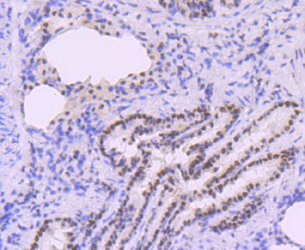
| Sample | Paraffin-embedded rat lung tissue; fixative not specified (datasheet A01946-2) |
| Fixation | Image fixative and duration unreported (datasheet A01946-2); verify before use. |
| Sectioning | 4–5 µm sections on charged slides (standard) |
| Deparaffinisation | Xylene, graded ethanol series to water (standard) |
| Antigen retrieval | Heat-induced epitope retrieval in Tris-EDTA buffer, pH 9.0, 20 min at 95–98 °C (standard rule: nuclear antigen) |
| Peroxidase block | 3% H2O2, 10 min, room temperature (standard) |
| Blocking | 10% normal serum of the secondary host, 30 min, room temperature (standard) |
| Primary antibody | Rabbit anti-POLB, 1:50-1:200 (datasheet A01946-2) |
| Primary incubation | Overnight at 4 °C (standard) |
| Detection | HRP-polymer secondary, DAB chromogen 5–10 min (standard) |
| Counterstain | Hematoxylin, blue, dehydrate and mount (standard) |
| Expected result | POLB-positive staining in glandular cells of breast (HPA tissue IHC: High). HPA tissue profile: General nuclear expression. No signal in the no-primary control. |
POLB IHC in paraffin sections should show a predominantly nuclear pattern in appropriate cells, including breast glandular cells and bladder urothelial cells (HPA: general nuclear expression; High in both cell types). The tissue IHC profile has Enhanced reliability, reflecting agreement between antibody staining and RNA data (HPA: Enhanced). POLB can also be cytoplasmic under normal conditions and has no transmembrane segment (UniProt P06746: subcellular location; topology).
| Distinct nuclear staining in glandular, urothelial, or squamous epithelial cells. | This fits the reported tissue IHC pattern; breast glandular cells, bladder urothelial cells, and tonsil squamous epithelial cells are High (HPA: tissue IHC). Compare stained cells with neighboring cells rather than treating every nucleus as equally positive (standard IHC practice). |
| Predominantly membrane-edge or extracellular staining, with little nuclear signal. | That distribution does not fit the reported general nuclear IHC pattern or POLB's lack of a transmembrane segment (HPA: tissue IHC; UniProt P06746: topology). Review morphology and controls before calling it specific; cytoplasmic signal alone needs more caution because UniProt also places POLB in the cytoplasm (UniProt P06746: subcellular location). |
| Strong staining in adipocytes, cardiomyocytes, or smooth muscle cells. | Those cell types are reported as Not detected by tissue IHC (HPA: adipose tissue, heart muscle, smooth muscle). Unexpected staining can indicate cross-reactivity or detection activity; compare a no-primary control and the expected nuclear pattern (standard IHC practice). |
| Diffuse color across cells and surrounding tissue, obscuring boundaries. | A uniform haze cannot be scored confidently as cell-specific POLB staining. Check the no-primary control, blocking and detection steps, and whether the counterstain permits nuclear assessment (standard IHC practice); the expected tissue pattern is general nuclear expression (HPA: tissue IHC). |
| No staining in breast glandular cells or bladder urothelial cells. | Both are reported High and can serve as reference-positive cell populations (HPA: breast; urinary bladder). First verify that the relevant cells are present and the detection control worked; then review antibody use and retrieval against its IHC-P instructions (standard IHC practice). |
| Tissue and cell selection | HPA reports High staining in breast, gallbladder, and salivary gland glandular cells, plus testis round or early spermatids (HPA: tissue IHC). Its Not detected examples include adipocytes and cardiomyocytes (HPA: tissue IHC). Select and interpret controls by cell type, not tissue name alone. |
| Antibody-specific IHC evidence | HPA049104 and CAB011616 have Enhanced IHC status; HPA069013 is Supported (HPA: antibody validation). Validation status helps prioritize an IHC antibody, but a result still needs interpretable morphology and appropriate slide controls (standard IHC practice). |
| Compartment and damage context | UniProt places POLB in both cytoplasm and nucleus and reports nuclear translocation after DNA damage (UniProt P06746: subcellular location). HPA describes general nuclear expression in tissue IHC (HPA: tissue IHC). Do not infer DNA damage from localization in an uncontrolled section. |
| Processing and topology | The annotated POLB chain spans residues 1–335, with no signal peptide, propeptide, or transmembrane segment (UniProt P06746: processing; topology). These annotations support scrutiny of membrane-only staining; they do not establish an antibody epitope or a retrieval condition. |
| Does IF/ICC require the same localization call? | No: HPA reports vesicles as Approved and cytosol as Supported in ICC-IF, while tissue IHC shows general nuclear expression (HPA: subcellular ICC-IF; tissue IHC). Interpret each assay against its own evidence; this IHC section supplies no IF protocol. |
| Retrieval and detection choices | Follow the catalog antibody's IHC-P instructions for retrieval and detection, then assess no-primary and known-positive controls (standard IHC practice). Neither HPA staining levels nor UniProt annotations establish POLB-specific fixation sensitivity or an optimal retrieval setting. |
| Situation | Likely cause | Next action |
|---|---|---|
| Known-positive cells are blank. | The target cells may be absent from the section, or the IHC workflow may have failed (standard IHC practice); breast glandular cells and bladder urothelial cells are reported High (HPA: tissue IHC). | Confirm cell identity on the counterstained section, check a working positive control, and compare retrieval and antibody use with the IHC-P instructions (standard IHC practice). |
| Signal is mainly cytoplasmic, with weak nuclei. | UniProt reports cytoplasmic POLB under normal conditions, whereas HPA's tissue IHC profile is generally nuclear (UniProt P06746: subcellular location; HPA: tissue IHC). The discrepancy alone cannot identify its cause. | Compare high-staining reference cells, inspect controls, and report the observed compartment explicitly before assigning the result to POLB (standard IHC practice). |
| Unexpected cells stain strongly. | Adipocytes, cardiomyocytes, and smooth muscle cells are Not detected in HPA tissue IHC (HPA: tissue IHC); cross-reactivity or endogenous detection activity may mimic positivity (standard IHC practice). | Check a no-primary control and whether staining is nuclear and cell-associated; treat unexpected staining as unconfirmed until controls support it (standard IHC practice). |
| Brown signal spreads beyond recognizable cells. | Diffuse background can obscure compartment and cell-type assessment (standard IHC practice); it does not match the reported general nuclear pattern (HPA: tissue IHC). | Review blocking, washes, detection exposure, and counterstain with a no-primary control, then score only distinguishable cells (standard IHC practice). |
| A low-staining tissue appears negative. | Neuronal cells in hippocampus and glial cells in caudate are reported Low (HPA: tissue IHC); absence of an obvious chromogenic signal there is less informative than failure in a High reference population. | Assess a reported High population on a comparable run before interpreting a negative result; avoid calling a low-expression sample a failed assay by itself (HPA: tissue IHC; standard IHC practice). |
| IF/ICC images disagree with the IHC slide. | HPA reports vesicle and cytosol localization by ICC-IF but general nuclear expression by tissue IHC (HPA: subcellular ICC-IF; tissue IHC). | Use the assay-specific HPA observations and controls when interpreting each image; do not transfer an ICC-IF localization call directly to paraffin IHC (standard IHC practice). |

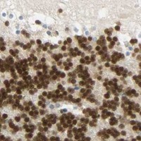


Comprehensive Human Protein Atlas IHC scoring per tissue (reliability: Enhanced — High consistency between antibody staining and RNA expression data.). Rows are taken directly from the HPA tissue chart — click any row's HPA link to view the source.
| Tissue | Cell type | Level | Evidence | Source |
|---|---|---|---|---|
| Breast | Glandular cells | High | Protein (IHC) | HPA → |
| Gallbladder | Glandular cells | High | Protein (IHC) | HPA → |
| Salivary gland | Glandular cells | High | Protein (IHC) | HPA → |
| Testis | Round or early spermatids | High | Protein (IHC) | HPA → |
| Tonsil | Squamous epithelial cells | High | Protein (IHC) | HPA → |
Troubleshoot POLB chromogenic IHC in paraffin sections by checking retrieval, compartment, cell type and controls before interpreting staining intensity.
The catalog includes human, mouse and rat reactive anti-POLB antibodies; A01946-2 also lists zebrafish reactivity (catalog: reactivity). A01946-2 has a rat lung paraffin-section IHC figure (catalog: image caption).
A01946-2 will render with IHC and ICC listed, human, mouse, rat and zebrafish reactivity, and a rat lung paraffin-section IHC figure (catalog: applications, reactivity and image caption). M01946 will render with IHC and IF/ICC listed and human, mouse and rat reactivity; no IHC or IF figure is supplied (catalog: applications, reactivity and image captions).
Which to pick: Choose A01946-2 for tissue IHC when a matching example matters: its own figure shows a hematoxylin-counterstained rat lung paraffin section, but the fixative is unreported (catalog: A01946-2 image caption). Choose monoclonal M01946 for IF/ICC because IF is explicitly listed, although no IF figure is supplied (catalog: M01946 title, applications and image captions). Choose A01946-2 when zebrafish reactivity is needed; both SKUs list human, mouse and rat reactivity (catalog: reactivity).