This website uses cookies to ensure you get the best experience on our website.


- Table of Contents
Use the general nuclear tissue pattern to assess POLD1 staining in paraffin sections (HPA tissue IHC). This guide covers fixation consistency, chromogenic detection and nuclear scoring, using 2 μg/ml as a catalog antibody starting concentration (datasheet: M03720).

Expected localisation, validated protocols, controls and antibodies — the at-a-glance facts below, then the full design guide.
| Expected localisation | General nuclear staining (HPA tissue IHC) | |
| Staining pattern | Nuclear staining; high in colon glandular cells (HPA tissue IHC) | |
| Antigen retrieval | EDTA pH 8.0 HIER, heat-mediated (datasheet M03720) | |
| Positive control | Appendix+4 more · see all | |
| Negative control | None in HPA (detected in all 45 tissues); use no-primary + isotype controls |
| Fixation | Keep fixation consistent across paraffin sections (standard IHC practice; not target-specific) | |
| Caveat | Low staining in cholangiocytes and adipocytes (HPA tissue IHC) | |
| Regulation | Abundance regulation not specified (UniProt) | |
| Isoform / epitope | One 1–1107 chain; no isoforms annotated (UniProt) |
The catalog antibody uses an EDTA pH 8.0 retrieval protocol (datasheet: M03720). Three published POLD1 IHC protocols provide additional tissue and detection details (PMC7583138; PMC6907229; PMC10095303).
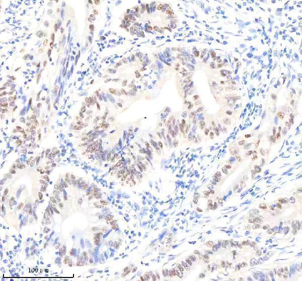
| Sample | Paraffin-embedded human colon cancer tissue; fixative not specified (datasheet M03720) |
| Fixation | Image fixative and duration unreported (datasheet M03720); verify before use. |
| Sectioning | 4–5 µm sections on charged slides (standard) |
| Deparaffinisation | Xylene, graded ethanol series to water (standard) |
| Antigen retrieval | Heat retrieval: EDTA pH 8.0 (datasheet M03720); 20 min, 95–100 °C (standard) |
| Peroxidase block | 3% H2O2, 10 min, room temperature (standard) |
| Blocking | 10% goat serum (datasheet M03720) |
| Primary antibody | Rabbit monoclonal (clone 21P14) anti-POLD1, 1:50 recommended; image 2 μg/ml (datasheet M03720) |
| Primary incubation | Overnight at 4 °C (datasheet M03720) |
| Detection | HRP-conjugated secondary, DAB chromogen (datasheet M03720) |
| Counterstain | Hematoxylin, blue, dehydrate and mount (standard) |
| Expected result | POLD1-positive staining in glandular cells of appendix (HPA tissue IHC: High). HPA tissue profile: General nuclear expression. No signal in the no-primary control. |
POLD1 is a nuclear DNA polymerase with no transmembrane segment (UniProt P28340: nucleus; topology). In paraffin-section IHC, expect staining mainly in nuclei across multiple cell types (HPA: general nuclear expression; low tissue specificity). HPA reports high staining in appendix and colon glandular cells, cervical squamous cells, and kidney tubular cells (HPA: tissue IHC). Its tissue IHC profile is supported by consistency between antibody staining and RNA expression (HPA: reliability Supported).
| Nuclear staining in glandular cells of appendix or colon, squamous epithelial cells of cervix, or kidney tubular cells. | This matches the reported compartment and high-staining cell populations (HPA: general nuclear expression; High in these cells). Compare nuclei with adjacent tissue structures on the same section; high is HPA's reported category, not a required score for every specimen. |
| Predominantly membranous staining, or cytoplasmic staining that obscures the nuclei. | This conflicts with the expected IHC pattern (HPA: general nuclear expression; UniProt P28340: nucleus, no transmembrane segment). Review localisation before scoring it as POLD1. HPA separately reports uncertain additional cytosolic localisation by ICC-IF, so a minor cytosolic signal alone is inconclusive (HPA: subcellular). |
| Strong staining in an unexpected cell population while the reported positive cells lack nuclear signal. | Consider antibody cross-reactivity or endogenous chromogenic detection activity (standard IHC practice). Check the staining against tissue morphology and the reported cell-level pattern; HPA's low tissue specificity does not make every cell type an equally strong positive (HPA: tissue IHC). |
| Diffuse colour across nuclei, cytoplasm, and tissue spaces, with little distinction between cells. | Treat this as background until controls support a cellular pattern (standard IHC practice). Broad POLD1 expression does not explain colour in tissue spaces (HPA: low tissue specificity; general nuclear expression). Reassess blocking, washes, and detection controls before interpreting intensity. |
| No nuclear staining in an appendix or colon section with identifiable glandular cells. | This misses a reported high-staining population (HPA: High in appendix and colon glandular cells). Check the section and assay controls first (standard IHC practice). A single negative result cannot establish absence of POLD1, especially without a working positive control. |
| Cell population and comparator tissue | HPA reports High staining in appendix, colon, duodenum, and fallopian tube glandular cells; cervical and esophageal squamous cells; cerebellar Purkinje cells; and kidney tubular cells (HPA: tissue IHC). Liver cholangiocytes and adipocytes are Low, so they are weaker comparators, not established negatives (HPA: tissue IHC). |
| Nuclear localisation and replication context | POLD1 is nuclear and colocalises with PCNA and POLD3 at S-phase replication sites (UniProt P28340: subcellular). That supports nuclear interpretation but does not require visible replication foci in routine chromogenic sections; HPA describes the tissue-level appearance as general nuclear expression (HPA: tissue IHC). |
| Antibody evidence | HPA lists HPA046524 and CAB004375 as IHC Supported; HPA046524 is also ICC Supported (HPA: antibodies). These statuses support use of the observed patterns as comparators, but they do not validate an unspecified catalog antibody, dilution, or retrieval condition. |
| Topology and processing | UniProt reports no transmembrane segment, signal peptide, propeptide, or alternative isoforms, and describes one chain spanning residues 1–1107 (UniProt P28340: topology; processing; isoforms). These annotations give no basis to expect a membrane-restricted or secreted IHC pattern; they do not identify an antibody epitope. |
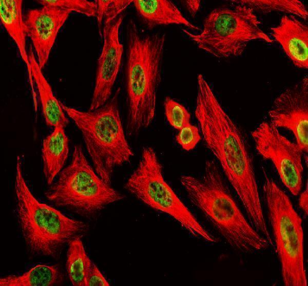
| IF/ICC Q&A | Q: Should IF/ICC show the same compartment? A: Predominantly nucleoplasmic signal is supported; additional cytosolic localisation is uncertain (HPA: subcellular ICC-IF). Treat that as a localisation cross-check, not an IHC protocol option. HPA lists ICC images for A-431, HEK293, U2OS, and NIH 3T3 (HPA: subcellular). |
| Situation | Likely cause | Next action |
|---|---|---|
| Reported high-staining cells show no nuclear signal. | The assay may have failed, or section conditions may be unsuitable; the slide alone cannot distinguish these possibilities (standard IHC practice). | Confirm tissue identity and cell preservation, then inspect positive and detection controls; review the antibody's IHC validation and the chosen retrieval and dilution settings (standard IHC practice; HPA: High in the selected cells). |
| Signal is mainly at cell borders or membranes. | This differs from the nuclear pattern and lacks support from POLD1 topology (HPA: general nuclear expression; UniProt P28340: no transmembrane segment). | Check whether nuclear counterstain and cell boundaries are being read correctly; compare with a known positive tissue and a detection control before attributing the border signal to POLD1 (standard IHC practice). |
| Unexpected cells stain strongly while expected nuclei are faint. | Cross-reactivity or endogenous detection activity is possible; HPA's tissue profile does not establish the identity of this signal (standard IHC practice; HPA: tissue IHC). | Check staining without primary antibody for detection background, and compare the suspicious cells with reported high-staining populations on a positive section (standard IHC practice; HPA: tissue IHC). |
| Brown colour is diffuse and obscures nuclear boundaries. | Background from blocking, washing, or detection can prevent localisation assessment (standard IHC practice). | Review blocking and wash steps, then assess a detection control and the nuclear counterstain. Score POLD1 only where cellular nuclear signal remains distinguishable (standard IHC practice; HPA: general nuclear expression). |
| Low signal in liver cholangiocytes or adipocytes is called assay failure. | Both populations are reported Low rather than negative (HPA: tissue IHC). Their weak staining is a poor standalone test of assay performance. | Use a reported High population, such as colon glandular cells or kidney tubular cells, as a positive comparator and assess its nuclear pattern (HPA: tissue IHC; general nuclear expression). |
| IF/ICC shows nucleoplasmic signal plus some cytosolic fluorescence. | HPA supports nucleoplasmic localisation but marks additional cytosolic localisation uncertain (HPA: subcellular ICC-IF). Fluorescence alone does not settle whether that component is specific. | Record the nucleoplasmic pattern as the supported localisation and interpret the cytosolic component cautiously against controls (HPA: subcellular ICC-IF; standard IF practice). Keep IF/ICC protocol decisions in its separate guide. |
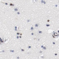


Comprehensive Human Protein Atlas IHC scoring per tissue (reliability: Supported — High consistency between antibody staining and RNA expression data.). Rows are taken directly from the HPA tissue chart — click any row's HPA link to view the source.
| Tissue | Cell type | Level | Evidence | Source |
|---|---|---|---|---|
| Appendix | Glandular cells | High | Protein (IHC) | HPA → |
| Cerebellum | Purkinje cells | High | Protein (IHC) | HPA → |
| Cervix | Squamous epithelial cells | High | Protein (IHC) | HPA → |
| Colon | Glandular cells | High | Protein (IHC) | HPA → |
| Duodenum | Glandular cells | High | Protein (IHC) | HPA → |
| Tissue | Cell type | Level | Evidence | Source |
|---|---|---|---|---|
| None in HPA: POLD1 is detected in all 45 scored tissues. Use a no-primary (secondary-only) and an isotype control instead. | ||||
Troubleshoot POLD1 staining in paraffin sections by checking retrieval, nuclear localisation and controls before comparing chromogenic signal across samples.
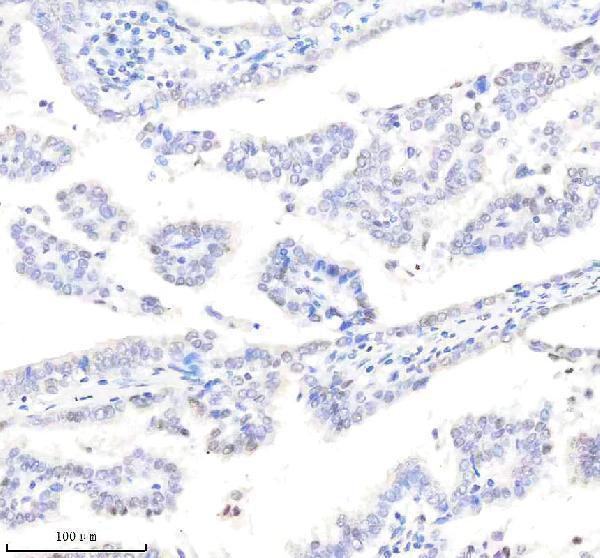
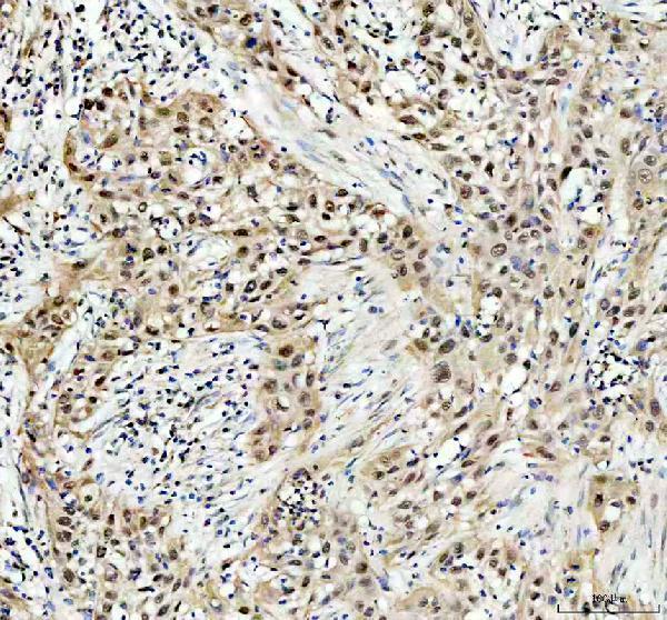
POLD1 antibodies have human paraffin-section IHC images (catalog IHC captions); A03720-2 also has a U2OS cell IF image (catalog IF caption). Both list human, mouse, and rat reactivity (catalog applications/reactivity).
M03720 has IHC images from human colon cancer, lung adenocarcinoma, ovarian cancer, and tonsil paraffin sections (M03720 IHC captions). A03720-2 has a human bladder cancer paraffin-section IHC image and a U2OS cell IF image (A03720-2 IHC/IF captions).
Which to pick: For tissue IHC, choose M03720 if its broader set of human paraffin-section images is useful; its caption specifies EDTA retrieval at pH 8.0, while the fixative is unreported (M03720 IHC captions). For IF/ICC, choose A03720-2 because it has a U2OS cell IF image; its own IHC caption also supports paraffin-section use with EDTA retrieval at pH 8.0, with fixative unreported (A03720-2 IF/IHC captions). Both list human, mouse, and rat reactivity, but the supplied IHC images show human samples only; M03720 is monoclonal and A03720-2 is polyclonal (catalog applications/reactivity; catalog antibody descriptions; IHC captions).