This website uses cookies to ensure you get the best experience on our website.


- Table of Contents
Plan POLR3G chromogenic IHC in paraffin sections using the observed nuclear tissue pattern as a guide (HPA tissue IHC). Compare testis and squamous epithelia with an appropriate negative control, and interpret weak staining cautiously because tissue staining awaits external verification (HPA tissue IHC).

Expected localisation, validated protocols, controls and antibodies — the at-a-glance facts below, then the full design guide.
| Expected localisation | Nuclear staining observed in tissue (HPA tissue IHC) | |
| Staining pattern | Nuclei in seminiferous ducts and squamous epithelia (HPA tissue IHC) | |
| Antigen retrieval | Tris-EDTA pH 9.0 HIER, 95–98 °C, 20 min (rule: nuclear antigen) | |
| Positive control | Skin+4 more · see all | |
| Negative control | Adipose tissue+4 more · see all |
| Fixation | Keep fixation consistent across sections (standard IHC practice; not target-specific); Matched tissue-IHC evidence does not establish the fixation claim. Validate the specimen-specific method before use. (selected-SKU IHC image A10669) | |
| Caveat | Tissue staining awaits external verification (HPA tissue IHC) | |
| Regulation | Expressed in dividing cells (UniProt) | |
| Isoform / epitope | No annotated isoforms or processing changes (UniProt) |
Compare the catalog antibody’s IHC-P protocol (datasheet) with one published POLR3G IHC protocol in bladder cancer tissue (PMC13092746).
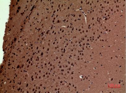
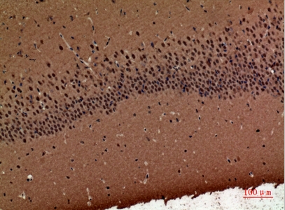
| Sample | Paraffin-embedded Mouse Brain tissue; fixative not specified (datasheet A10669) |
| Fixation | Image fixative and duration unreported (datasheet A10669); verify before use. |
| Sectioning | 4–5 µm sections on charged slides (standard) |
| Deparaffinisation | Xylene, graded ethanol series to water (standard) |
| Antigen retrieval | Heat-induced epitope retrieval in Tris-EDTA buffer, pH 9.0, 20 min at 95–98 °C (standard rule: nuclear antigen) |
| Peroxidase block | 3% H2O2, 10 min, room temperature (standard) |
| Blocking | 10% normal serum of the secondary host, 30 min, room temperature (standard) |
| Primary antibody | Rabbit anti-POLR3G, 1:100-1:300 (datasheet A10669) |
| Primary incubation | Overnight at 4 °C (standard) |
| Detection | HRP-polymer secondary, DAB chromogen 5–10 min (standard) |
| Counterstain | Hematoxylin, blue, dehydrate and mount (standard) |
| Expected result | POLR3G-positive staining in keratinocytes of skin (HPA tissue IHC: Medium). HPA tissue profile: Nuclear expression in several different tissue types, most abundant in seminiferous ducts of testis and squamous epithelia. No signal in the no-primary control. |
POLR3G should stain mainly in nuclei, with nucleolar exclusion (UniProt O15318; HPA: nuclear tissue expression). Expect the clearest tissue signal in testis preleptotene spermatocytes and several squamous epithelia (HPA: Medium). POLR3G has no transmembrane segment (UniProt O15318 topology). HPA rates the tissue IHC profile Enhanced, while describing medium agreement with RNA data and pending external verification (HPA: tissue reliability).
| Nuclear staining in testis preleptotene spermatocytes or squamous epithelial cells; nucleoli remain unstained. | This fits the reported tissue distribution and nuclear pattern (HPA: Medium in these cells; UniProt O15318: excluded from nucleoli). Judge the cells and compartment together: nuclear color elsewhere alone is less informative because HPA reports nuclear expression across several tissue types (HPA: tissue profile). |
| Prominent nucleolar staining, or predominantly cytoplasmic staining in the listed adult positive tissues. | This conflicts with the expected dominant nuclear pattern and nucleolar exclusion (HPA: tissue profile; UniProt O15318). Check morphology, counterstain and detection controls before calling it POLR3G. Cytoplasmic localisation is recorded by UniProt, so its presence alone is not proof of an artefact (UniProt O15318: cytoplasm). |
| Strong staining in HPA-listed negative cells, such as adipocytes or adrenal glandular cells. | HPA reports POLR3G as Not detected in those cells (HPA: adipose tissue and adrenal gland IHC). Consider cross-reactivity or endogenous detection activity, especially if the positive cells lack the expected nuclear pattern (general IHC practice). A single discrepant field does not identify the cause. |
| Diffuse color over tissue and empty spaces obscures the nuclei. | That distribution cannot be scored as a specific nuclear result (HPA: nuclear tissue profile). Review background in the no-primary and detection controls, then assess blocking, washing and detection conditions (general IHC practice). Interpret any remaining cell signal only after the background is sufficiently low. |
| No nuclear signal in an interpretable testis section containing preleptotene spermatocytes. | HPA reports Medium staining in these cells, making this a useful check of the run (HPA: testis IHC). Confirm the cells are present and the section and detection controls worked (general IHC practice). Absence in one run cannot by itself establish that POLR3G is absent from the sample. |
| Cell population and tissue | HPA reports Medium staining in testis preleptotene spermatocytes, keratinocytes, squamous epithelial cells, ovarian follicle cells and splenic red-pulp cells (HPA: tissue IHC). Its Not detected calls apply to the named cells, not every cell in those organs (HPA: tissue IHC). |
| Compartment and nucleoli | The tissue profile is nuclear, while UniProt records both nucleus and cytoplasm and specifically excludes nucleoli (HPA: tissue IHC; UniProt O15318). Score the nuclear pattern first; interpret cytoplasmic color in the context of the sample and its controls. |
| Strength and validation limits | HPA labels the tissue profile Enhanced and reports Medium staining in its listed positive cells (HPA: tissue IHC). It also notes medium agreement with RNA data and pending external verification; neither designation guarantees that every section or antibody run will reproduce the profile (HPA: reliability description). |
| Antibody-specific evidence | Among the listed antibodies, CAB010109 has Enhanced IHC validation; HPA052658 and HPA074330 have no IHC status supplied (HPA: antibody records). Treat ICC support for all three as evidence for that application only (HPA: antibody records). |
| IF/ICC Q: Where should signal appear? | A: Mainly in the nucleoplasm; nuclear bodies are additionally supported, while cytosol is uncertain (HPA: subcellular ICC-IF). This is an interpretation cue for IF/ICC, not an IHC-P protocol or a reason to score diffuse cytoplasmic chromogen as positive. |
| Situation | Likely cause | Next action |
|---|---|---|
| Expected nuclei are blank in testis preleptotene spermatocytes. | The positive cells may be absent from the viewed field, or the IHC run may have failed (HPA: Medium in preleptotene spermatocytes; general IHC practice). | Find the relevant cells on the counterstained section; check a working positive control and detection reagents, then review the catalog antibody's IHC-P conditions (general IHC practice). Do not infer target-specific fixation sensitivity: none is supplied here. |
| Nucleoli stain more strongly than surrounding nuclei. | That reverses the reported nucleolar exclusion (UniProt O15318). Misread nuclear detail or nonspecific staining are possibilities (general IHC practice). | Recheck nuclear boundaries against the counterstain and compare a no-primary control (general IHC practice). Score only a reproducible pattern consistent with nuclear staining and nucleolar sparing (HPA: tissue IHC; UniProt O15318). |
| Cytoplasm dominates the signal in an adult tissue section. | HPA's tissue profile is nuclear, although UniProt also lists cytoplasm (HPA: tissue IHC; UniProt O15318). Location alone cannot establish whether the color is specific. | Compare nuclear signal in the expected positive cells with the no-primary control (HPA: tissue IHC; general IHC practice). Record cytoplasmic staining separately until its specificity is supported; do not convert an IF/ICC observation into an IHC-P expectation. |
| HPA-listed negative cells stain strongly. | The result disagrees with the cell-specific Not detected calls (HPA: tissue IHC). Cross-reactivity or endogenous detection activity may contribute (general IHC practice). | Check a no-primary control and the relevant endogenous-activity control for the chromogenic system (general IHC practice). Reassess cell identity and nuclear localisation before treating the staining as POLR3G (HPA: tissue IHC). |
| Broad, uneven chromogen makes nuclear scoring difficult. | Background from blocking, washing or detection conditions can obscure the expected nuclear distribution (general IHC practice; HPA: tissue profile). | Compare tissue and blank areas with a no-primary control; optimize blocking and washing within the established IHC workflow (general IHC practice). Score cell-specific nuclear signal only when background permits a clear distinction. |
| Signal is weak in a cell population reported as Medium. | HPA levels describe its observed samples and do not set a guaranteed intensity for each section (HPA: tissue IHC). Weak detection or a poorly represented positive cell population are also possible (general IHC practice). | Confirm the named cells are present, compare a validated positive section, and review the catalog antibody's IHC-P dilution and retrieval instructions (general IHC practice). No POLR3G-specific retrieval or fixation effect is established by the supplied sources. |
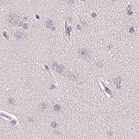


Comprehensive Human Protein Atlas IHC scoring per tissue (reliability: Enhanced — Medium consistency between antibody staining and RNA expression data. Pending external verification.). Rows are taken directly from the HPA tissue chart — click any row's HPA link to view the source.
| Tissue | Cell type | Level | Evidence | Source |
|---|---|---|---|---|
| Skin | Keratinocytes | Medium | Protein (IHC) | HPA → |
| Cervix | Squamous epithelial cells | Medium | Protein (IHC) | HPA → |
| Esophagus | Squamous epithelial cells | Medium | Protein (IHC) | HPA → |
| Oral mucosa | Squamous epithelial cells | Medium | Protein (IHC) | HPA → |
| Ovary | Follicle cells | Medium | Protein (IHC) | HPA → |
| Tissue | Cell type | Level | Evidence | Source |
|---|---|---|---|---|
| Adipose tissue | Adipocytes | Not detected | Protein (IHC) | HPA → |
| Adrenal gland | Glandular cells | Not detected | Protein (IHC) | HPA → |
| Bone marrow | Hematopoietic cells | Not detected | Protein (IHC) | HPA → |
| Breast | Adipocytes | Not detected | Protein (IHC) | HPA → |
| Caudate | Glial cells | Not detected | Protein (IHC) | HPA → |
Troubleshoot POLR3G staining by checking nuclear localisation, tissue controls, and the processing details documented for the antibody.
A10669 lists IHC and IF applications (catalog: applications) and human and mouse reactivity (catalog: reactivity). Its IHC figure shows paraffin-embedded mouse brain at 1:100 (IHC image caption).
A10669 will render with an IHC figure of paraffin-embedded mouse brain stained at 1:100 (IHC image caption). IF is listed as an application, and human and mouse are listed as reactive species (catalog: applications and reactivity); no IF figure is provided (catalog: IF image alts).
Which to pick: Choose A10669 for paraffin-section tissue IHC based on its mouse brain figure (IHC image caption); the fixative is unreported (IHC image caption). For IF, A10669 is listed (catalog: applications), but ICC validation is unreported (catalog: applications). For work across species, it lists human and mouse reactivity (catalog: reactivity), while the IHC figure documents mouse brain only (IHC image caption).