This website uses cookies to ensure you get the best experience on our website.


- Table of Contents
Plan PON2 paraffin-section IHC around its generally cytoplasmic tissue staining (HPA tissue IHC) and membrane annotation (UniProt). Use high-staining glandular or glial cells and tissues with undetected staining as reference controls (HPA tissue IHC).

Expected localisation, validated protocols, controls and antibodies — the at-a-glance facts below, then the full design guide.
| Expected localisation | General cytoplasmic tissue staining (HPA tissue IHC); membrane annotated (UniProt) | |
| Staining pattern | High staining in glandular and glial cells; generally cytoplasmic (HPA tissue IHC) | |
| Antigen retrieval | Tris-EDTA pH 8.0 HIER, heat-mediated (datasheet M02027-1) | |
| Positive control | Adrenal gland+4 more · see all | |
| Negative control | Esophagus+2 more · see all |
| Fixation | Keep fixation consistent across sections. (standard IHC practice; not target-specific) | |
| Caveat | A secreted variant may shift protein from RNA-defined sites (HPA tissue IHC) | |
| Regulation | Widely expressed; tissue levels vary (UniProt) | |
| Isoform / epitope | 3 isoforms; verify which forms the epitope covers (UniProt) |
The catalog antibody protocol is paired with 3 published PON2 chromogenic IHC protocols for lymphoma, glioblastoma and pancreatic tissue (PMC9559328; PMC5362434; PMC5567863).
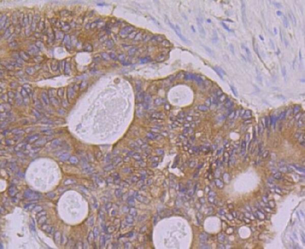
| Sample | Paraffin-embedded human colon carcinoma tissue; fixative not specified (datasheet M02027-1) |
| Fixation | Image fixative and duration unreported (datasheet M02027-1); verify before use. |
| Sectioning | 4–5 µm sections on charged slides (standard) |
| Deparaffinisation | Xylene, graded ethanol series to water (standard) |
| Antigen retrieval | Heat retrieval: Tris-EDTA pH 8.0, 20 minutes (datasheet M02027-1) |
| Peroxidase block | 3% H2O2, 10 min, room temperature (standard) |
| Blocking | 10% normal serum of the secondary host, 30 min, room temperature (standard) |
| Primary antibody | Rabbit monoclonal (clone 19P05) anti-PON2, 1:50 (datasheet M02027-1) |
| Primary incubation | Overnight at 4 °C (standard) |
| Detection | HRP-polymer secondary, DAB chromogen 5–10 min (standard) |
| Counterstain | Hematoxylin, blue, dehydrate and mount (standard) |
| Expected result | PON2-positive staining in glandular cells of adrenal gland (HPA tissue IHC: High). HPA tissue profile: General cytoplasmic expression. No signal in the no-primary control. |
PON2 should show predominantly cytoplasmic staining, with possible membrane-associated signal, in glandular cells and glia (HPA: general cytoplasmic expression; HPA: high glandular and glial staining; UniProt Q15165: membrane, no transmembrane segment). HPA rates tissue IHC reliability Enhanced, while noting only medium consistency with RNA data (HPA: tissue IHC reliability description). Judge the pattern by cell type and compartment together, not by overall tissue darkness.
| Clear cytoplasmic staining in adrenal gland or duodenal glandular cells, or in cerebral cortex glia. | This fits the reported high cell-specific signal (HPA: High in these cells). Compare stained cells with adjacent structures; a uniformly dark section does not establish the reported pattern (HPA: general cytoplasmic expression). |
| Predominantly nuclear or other isolated compartment staining in otherwise positive cells. | This is discordant with the reported cytoplasmic pattern (HPA: general cytoplasmic expression). Review morphology, counterstain and detection controls before calling it PON2; UniProt's membrane annotation does not establish a nuclear pattern (UniProt Q15165: membrane). |
| Strong signal confined to esophageal or vaginal squamous epithelial cells, or salivary glandular cells. | Those cells were reported as not detected (HPA: tissue IHC). Consider antibody cross-reactivity or endogenous detection activity, then check a positive tissue alongside them; an unexpected stain alone does not identify its cause. |
| Color spread across stroma, lumina and many unrelated cell types without distinct cellular outlines. | Treat this as diffuse background rather than a cell-specific positive result (HPA: general cytoplasmic expression). Check a control without primary antibody and inspect whether the background follows tissue structures or the detection chemistry (general IHC practice). |
| No convincing signal in adrenal glandular cells or cerebellar Bergmann glia. | Both have reported high staining (HPA: tissue IHC). First verify tissue identity, cell preservation and a working positive control; a negative slide may reflect assay performance and should not establish absence of PON2 by itself. |
| Cell type within the section | HPA reports high staining in selected glandular cells and glia, but not detected staining in specified squamous or salivary cells (HPA: tissue IHC). Score the named cells rather than assigning one intensity to a whole organ. |
| Compartment assignment | HPA describes general cytoplasmic expression, while UniProt annotates membrane association without a transmembrane segment (HPA: tissue IHC; UniProt Q15165 topology). This supports a cytoplasmic or membrane-associated reading, not a requirement for a sharp surface rim. |
| Antibody validation | HPA029193 has Enhanced IHC validation and CAB025430 has Supported IHC validation (HPA: antibody validation). These grades help prioritize concordant patterns; they do not prove that every colored structure in a new specimen is PON2. |
| Isoforms and processing | Three isoforms and three glycosylation sites are annotated (UniProt Q15165: isoforms; glycosylation at 254, 269, 323). No supplied epitope map links these features to IHC staining, so do not predict a compartment or tissue difference from them. |
| IF/ICC evidence | HPA labels the subcellular summary Secreted but provides no main location or ICC-IF image-bearing cell lines (HPA: ICC-IF). Its tissue IHC description remains the direct basis for this paraffin-section call; an IF/ICC pattern needs its own validation. |
| Situation | Likely cause | Next action |
|---|---|---|
| Known high-staining glandular or glial cells look negative. | Tissue sampling, detection failure or assay conditions may be responsible; HPA reports these cells as High (HPA: tissue IHC). | Confirm cell identity and run a known-positive section with the same stain; review the IHC-validated antibody's documented IHC-P conditions (general IHC practice). |
| Every compartment is weak and poorly defined. | Diffuse background can obscure the expected cellular pattern (HPA: general cytoplasmic expression); its source cannot be assigned from the image alone. | Compare a no-primary control, then review blocking, washes and detection exposure for the entire run (general IHC practice). |
| Unexpected color appears in HPA not-detected cell types. | Cross-reactivity or endogenous detection activity is possible; HPA reports no detection in esophageal squamous and salivary glandular cells (HPA: tissue IHC). | Check a no-primary control and an expected positive tissue; interpret residual cell-specific staining cautiously (general IHC practice). |
| Only nuclei appear positive. | Nuclear-only staining conflicts with HPA's general cytoplasmic pattern (HPA: tissue IHC); the cause remains undetermined. | Recheck compartment boundaries against the counterstain and compare controls before scoring nuclei as PON2 (general IHC practice). |
| Two antibodies give different tissue patterns. | Their IHC validation levels differ: HPA029193 is Enhanced and CAB025430 is Supported (HPA: antibody validation). This does not by itself explain the disagreement. | Compare the same cell types on matched sections and retain the discordance in the interpretation (general IHC practice). |
| Can an IF/ICC image establish the expected IHC-P pattern? | HPA supplies no ICC-IF image-bearing cell lines or main subcellular location for PON2 (HPA: ICC-IF). | Use HPA tissue IHC to assess paraffin sections; evaluate IF/ICC separately with its own controls (HPA: tissue IHC; general IF practice). |


Comprehensive Human Protein Atlas IHC scoring per tissue (reliability: Enhanced — Medium consistency between antibody staining and RNA expression data. At least one protein variant secreted, tissue location of RNA and protein might differ and correlation is complex.). Rows are taken directly from the HPA tissue chart — click any row's HPA link to view the source.
| Tissue | Cell type | Level | Evidence | Source |
|---|---|---|---|---|
| Adrenal gland | Glandular cells | High | Protein (IHC) | HPA → |
| Appendix | Glandular cells | High | Protein (IHC) | HPA → |
| Caudate | Glial cells | High | Protein (IHC) | HPA → |
| Cerebellum | Bergmann glia - cytoplasm/membrane | High | Protein (IHC) | HPA → |
| Cerebral cortex | Glial cells | High | Protein (IHC) | HPA → |
Troubleshoot PON2 chromogenic IHC in paraffin sections with matched controls, documented processing, and cell level scoring.
M02027-1 has real IHC data from paraffin-embedded human colon carcinoma tissue (IHC image caption). Human, Mouse, and Rat reactivity is listed (catalog reactivity); IF data are absent (catalog IF images).
M02027-1 is shown on paraffin-embedded human colon carcinoma tissue at 1:50 (IHC image caption). Its listed applications include IHC, and its listed reactivity covers Human, Mouse, and Rat (catalog applications/reactivity).
Which to pick: For tissue IHC, choose M02027-1, a rabbit monoclonal (catalog host/clone) shown on paraffin-embedded human colon carcinoma tissue (IHC image caption); the fixative is unreported (IHC image caption). For IF/ICC, no SKU in this payload has a listed IF application or IF image (catalog applications/IF images). For cross-species work, M02027-1 lists Human, Mouse, and Rat reactivity (catalog reactivity), while its IHC image documents human tissue only (IHC image caption).